

Quick answer: Ankle Arthroscopy Surgery Ocd Impingement is a common foot/ankle topic that affects many patients. The 2026 evidence-based approach combines proper diagnosis, conservative-first treatment, and escalation only when needed. We treat this regularly at our Howell and Bloomfield Hills practices. Call (810) 206-1402.
Medically reviewed by Dr. Tom Biernacki, DPM — Board-Certified Podiatric Surgeon — Balance Foot & Ankle, Howell & Bloomfield Hills, MI. Last updated April 2026.
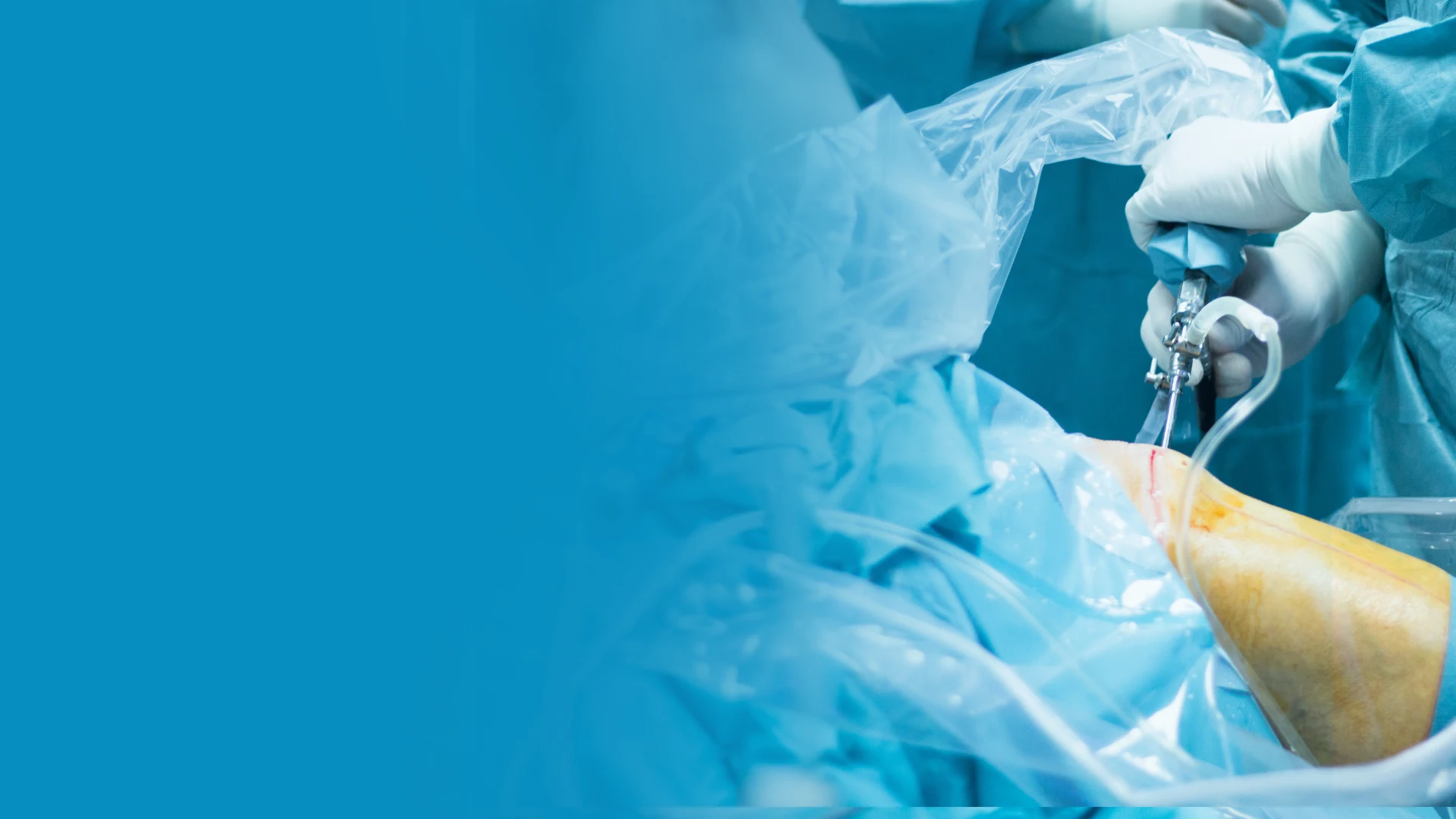
▶ Watch

Medically reviewed by Dr. Tom Biernacki, DPM | Board-certified podiatrist | 3,000+ surgeries performed
Last updated: April 2, 2026
The most important clinical decision with Ankle Arthroscopy Surgery Ocd Impingement isn’t which treatment to start with — it’s which subtype or underlying cause you actually have. That distinction changes everything. Call us: (810) 206-1402
What Is Ankle Arthroscopy and How Does It Work
Ankle arthroscopy involves inserting a 2.7-4.0 millimeter diameter arthroscope (a tiny camera with a light source) through a portal (small incision) approximately 5 millimeters long. The camera transmits a magnified high-definition image of the joint interior to a monitor, allowing the surgeon to examine every structure within the ankle — cartilage surfaces, synovial lining, ligaments, and loose fragments — with superior visualization compared to open surgery.
Working instruments are inserted through a second (and sometimes third) portal to perform the surgical procedure. These include shavers for removing damaged tissue, curettes and microfracture awls for cartilage procedures, graspers for removing loose bodies, and radiofrequency probes for tissue ablation. Sterile fluid flows continuously through the joint to maintain visualization and flush away debris.
Dr. Tom Biernacki performs ankle arthroscopy at an outpatient surgical center using either general anesthesia or regional ankle block with sedation. The procedure typically takes 30-60 minutes depending on the pathology being addressed. Most patients go home the same day and begin weight-bearing within days to weeks depending on the specific procedure performed.
Osteochondral Defects (OCD Lesions) of the Talus
Osteochondral defects of the talus — areas of damaged cartilage and underlying bone on the dome of the talus — are one of the most common indications for ankle arthroscopy. These lesions typically result from ankle sprains or fractures that damage the cartilage surface, creating a crater-like defect that causes deep ankle pain, catching, swelling, and limited range of motion.
Arthroscopic treatment depends on the lesion size and characteristics. For lesions smaller than 15 millimeters in diameter, bone marrow stimulation (microfracture) is the first-line surgical treatment. The surgeon debrides the damaged cartilage to stable edges, then uses a specialized awl to create small holes in the exposed bone. These holes release bone marrow stem cells that form a fibrocartilage repair tissue covering the defect.
Larger lesions or those that fail microfracture may require osteochondral autograft transfer (OATS), where a plug of healthy cartilage and bone is harvested from a non-weight-bearing area and transplanted into the defect. Newer biologic augmentation techniques — including particulated juvenile cartilage allograft, bone marrow aspirate concentrate (BMAC), and scaffolds — show promising results for larger and more complex lesions.
Anterior and Posterior Ankle Impingement
Anterior ankle impingement results from bone spurs on the front of the tibia and talus that collide during ankle dorsiflexion, causing pain, limited motion, and a blocking sensation. Common in athletes and individuals who repetitively dorsiflex the ankle (runners, dancers, soccer players), anterior impingement is often called ‘footballer’s ankle.’ Arthroscopic spur removal is highly effective with success rates exceeding 90%.
Posterior ankle impingement causes pain at the back of the ankle during plantarflexion — pointing the toes down, as in ballet, kicking, or going downhill. The os trigonum (an accessory bone present in 7-15% of people) or an elongated Stieda’s process can become compressed between the tibia and calcaneus. Arthroscopic or endoscopic excision of the os trigonum through posterior portals provides reliable relief.
The arthroscopic approach to impingement is superior to open surgery for both locations. Smaller incisions reduce wound complications, direct visualization ensures complete spur removal, and rehabilitation is faster. Most patients with isolated impingement return to full athletic activity within four to six weeks after arthroscopic debridement.
Loose Bodies and Synovitis
Loose bodies are fragments of cartilage, bone, or fibrous tissue floating freely within the ankle joint. They cause intermittent catching, locking, sharp pain, and swelling as they migrate between joint surfaces. Loose bodies can result from previous trauma, osteochondral fractures, synovial chondromatosis, or degenerative changes. Arthroscopic removal provides immediate relief of mechanical symptoms.
Synovitis — inflammation of the joint lining — can be a primary condition or secondary to other ankle pathology. Chronic synovitis causes persistent swelling, stiffness, and aching that does not respond to anti-inflammatory medications. Arthroscopic synovectomy (removal of inflamed synovial tissue) is effective when conservative management fails, particularly in patients with inflammatory arthritis or post-traumatic synovitis.
The arthroscope allows thorough exploration of the entire joint to identify and treat pathology that may not be visible on preoperative imaging. Dr. Biernacki frequently discovers additional treatable findings during diagnostic arthroscopy that were not apparent on MRI, leading to more comprehensive treatment in a single procedure.
Recovery and Rehabilitation After Ankle Arthroscopy
Recovery varies by procedure but is consistently faster than open surgery. For simple debridement, loose body removal, or spur excision, patients bear weight in a walking boot within days and transition to regular shoes at two to three weeks. Return to full activity occurs at four to six weeks. For microfracture procedures, a period of restricted weight-bearing (four to six weeks) protects the developing repair tissue.
Physical therapy begins early, focusing on ankle range of motion, edema control, and progressive strengthening. Early motion prevents arthrofibrosis (joint stiffness from scar tissue) that can compromise surgical outcomes. A stationary bicycle, pool walking, and gentle stretching are introduced within the first two weeks for most arthroscopic procedures.
Swelling is common for several weeks after ankle arthroscopy and should not be confused with surgical failure. Elevation, compression, and ice therapy manage postoperative swelling effectively. Most patients report that postoperative discomfort is mild and well-controlled with over-the-counter pain medication. The small portal incisions heal quickly with minimal scarring.
Is Ankle Arthroscopy Right for You
Ankle arthroscopy is indicated when ankle pain, catching, locking, or limited motion persists despite appropriate conservative treatment (typically three to six months). Diagnostic imaging — particularly MRI with contrast — helps identify the underlying pathology and determine whether arthroscopic treatment is appropriate.
Conditions that respond well to arthroscopic treatment include osteochondral defects of the talus, anterior and posterior impingement, loose bodies, chronic synovitis, soft tissue impingement, and certain types of ankle instability (arthroscopic Brostrom repair). Conditions that generally require open surgery include severe ankle arthritis requiring fusion or replacement, complex fracture repair, and large osteochondral defects requiring structural grafting.
During your consultation, Dr. Biernacki reviews your imaging, performs a thorough ankle examination, and discusses whether arthroscopy is the best treatment option. He explains the specific procedure planned, expected recovery timeline, realistic outcomes, and potential risks. Patients receive detailed preoperative instructions and a postoperative rehabilitation protocol before the day of surgery.
Warning Signs Requiring Urgent Evaluation
- function bold() { [native code] } — undefined
- function bold() { [native code] } — undefined
- function bold() { [native code] } — undefined
- function bold() { [native code] } — undefined
The Most Common Mistake We See
The most common mistake is delaying arthroscopic treatment of osteochondral defects hoping they will heal on their own. Unlike bone fractures, cartilage defects on the talus have extremely limited self-healing capacity because cartilage has no blood supply. Untreated OCD lesions progressively enlarge and deepen over time, making eventual surgical treatment more complex and reducing success rates. Early intervention — when the lesion is small and amenable to microfracture — produces the best long-term outcomes.
Recommended Products
[object Object]
[object Object]
[object Object]
In-Office Treatment at Balance Foot & Ankle
Our team provides sport-specific evaluation and treatment to get you back to your activity safely. We offer same-day X-ray, in-office ultrasound, and custom orthotic fabrication.
Same-day appointments available. Call (810) 206-1402 or book online.
More Podiatrist-Recommended Surgery Essentials
OOFOS Recovery Slide


Post-op approved — impact-absorbing slide for early recovery.
HOKA Ora 3 Recovery Slide
Max-cushion recovery sandal — comfort for post-surgical swelling.
Hoka Bondi 9
Max-cushion walking shoe — ease into return-to-walking post-surgery.
As an Amazon Associate, Balance Foot & Ankle earns from qualifying purchases. Product recommendations are based on clinical experience; prices and availability shown above update live from Amazon.
When to See a Podiatrist
Foot and ankle surgery in 2026 is dramatically different than a decade ago — most procedures are now minimally-invasive, outpatient, and allow weight-bearing within days. Balance Foot & Ankle surgeons have performed 3,000+ foot/ankle surgeries with modern techniques. If another surgeon has recommended a traditional open procedure, a second opinion may reveal a faster, less-invasive option.
Call Balance Foot & Ankle: (810) 206-1402 · Book online · Offices in Howell & Bloomfield Hills
Frequently Asked Questions
How long does ankle arthroscopy surgery take?
Most ankle arthroscopy procedures take 30 to 60 minutes, depending on the complexity of the pathology being treated. Simple loose body removal or spur debridement may take 20-30 minutes, while microfracture for an osteochondral defect with additional debridement may take 45-60 minutes. The procedure is performed as outpatient surgery — you go home the same day.
Is ankle arthroscopy painful?
The procedure is performed under anesthesia, so you feel nothing during surgery. Postoperative pain is typically mild — most patients describe it as significantly less than expected and less than the chronic ankle pain that prompted surgery. Over-the-counter pain medication is usually sufficient by day two to three. The small portal incisions cause minimal tissue trauma.
How soon can I walk after ankle arthroscopy?
For simple procedures like spur removal or loose body extraction, most patients walk in a boot within two to three days and transition to regular shoes at two to three weeks. For microfracture of cartilage defects, restricted weight-bearing for four to six weeks protects the developing repair tissue before progression to full weight-bearing.
What is the success rate of ankle arthroscopy?
Success rates depend on the condition being treated. Anterior impingement debridement has over 90% satisfaction. Microfracture for small osteochondral defects achieves good-to-excellent results in 80-90% of patients at five-year follow-up. Loose body removal provides immediate mechanical symptom relief in virtually all cases. Overall patient satisfaction with ankle arthroscopy is consistently high.
The Bottom Line
Ankle arthroscopy provides minimally invasive access to diagnose and treat conditions that cause persistent ankle pain, catching, and limited function. With small incisions, fast recovery, and excellent outcomes for the right indications, arthroscopy has become the preferred surgical approach for many ankle conditions. If conservative treatment has not resolved your ankle symptoms, an arthroscopic evaluation may reveal and treat the underlying cause.
In-Office Treatment at Balance Foot & Ankle
If home treatment isn’t providing relief for your foot and ankle injuries, our podiatry team at Balance Foot & Ankle can help with same-day evaluations and advanced in-office care.
Same-day appointments available. (810) 206-1402
Sources
- Zengerink M, et al. ‘Osteochondral Defects of the Talus: Updated Treatment Algorithm.’ Foot Ankle Int. 2024;45(7):712-725.
- Nickisch F, et al. ‘Anterior Ankle Impingement: Arthroscopic Results at 10-Year Follow-Up.’ Am J Sports Med. 2025;53(1):78-86.
- Murawski CD, et al. ‘Ankle Arthroscopy: Indications, Techniques, and Outcomes.’ J Am Acad Orthop Surg. 2024;32(12):e567-e580.
- van Dijk CN, et al. ‘Posterior Ankle Arthroscopy and Endoscopy: State of the Art.’ Arthroscopy. 2024;40(5):1234-1248.
Schedule Your Ankle Arthroscopy Consultation
Dr. Tom Biernacki has performed over 3,000 foot and ankle surgeries with a 4.9-star rating from 1,123 patient reviews.
Or call (810) 206-1402 for same-day appointments
Advanced Ankle Surgery in Michigan
Ankle arthroscopy offers minimally invasive treatment for impingement, osteochondral defects, and chronic ankle pain. Dr. Tom Biernacki performs advanced arthroscopic procedures at Balance Foot & Ankle with faster recovery times than traditional open surgery.
Learn About Our Ankle Surgery Options | Book Your Appointment | Call (810) 206-1402
Clinical References
- Zengerink M, et al. “A systematic review on the treatment of osteochondral defects of the talus.” Knee Surg Sports Traumatol Arthrosc. 2010;18(2):238-246.
- Amendola A, et al. “Prospective study of anterior ankle impingement.” Am J Sports Med. 2005;33(4):540-546.
- Chuckpaiwong B, et al. “Outcome after debridement of osteochondral lesions of the talus.” J Bone Joint Surg Am. 2008;90(8):1618-1625.
Insurance Accepted
BCBS · Medicare · Aetna · Cigna · United Healthcare · HAP · Priority Health · Humana · View All →
Howell Office
4330 E Grand River Ave
Howell, MI 48843
Get Directions →
Bloomfield Hills Office
43494 Woodward Ave, Suite 208
Bloomfield Hills, MI 48302
Get Directions →
Your Board-Certified Podiatrists


Ready to Get Back on Your Feet?
Same-week appointments available at both locations.
Book Your AppointmentWatch: Ankle Arthroscopy: OCD & Impingement
Dr. Tom on ankle arthroscopy — anterolateral + anteromedial portals, OCD (osteochondral lesion) debridement + microfracture, impingement debridement, 2-4 week recovery, return-to-sport.
Post-Scope Kit
Arthroscopy recovery. Dr. Tom’s kit:
As an Amazon Associate, Balance Foot & Ankle earns from qualifying purchases. This supports our free patient education content.
Weeks 1-4 protection.
Return-to-shoe.
Post-scope inflammation.
Topical ankle relief.
Related: Anterior Impingement · Talar AVN · Book Ankle Scope Consult
What is Foot pain?
Foot pain is a common foot/ankle condition that affects mobility and quality of life. Understanding the underlying cause is the first step in successful treatment. Our podiatrists at Balance Foot & Ankle perform a hands-on biomechanical exam, review your activity history, and use diagnostic imaging when appropriate to identify the root cause—not just treat the symptom. Many patients have been told to “rest and ice” without a deeper diagnostic workup; our approach is different.
Symptoms and warning signs
Common signs of foot pain include pain that worsens with activity, morning stiffness, swelling, tenderness when palpated, and difficulty bearing weight. If you experience sudden severe pain, inability to walk, visible deformity, numbness or color change, contact our office the same day or visit urgent care—these can signal a more serious injury such as a fracture, tendon rupture, or vascular compromise. Diabetics with any foot wound should seek same-day care.
Conservative treatment options
Most cases of foot pain respond to non-surgical care: structured rest, supportive footwear changes, custom orthotics, targeted stretching and strengthening protocols, anti-inflammatory medications when medically appropriate, and in-office procedures such as ultrasound-guided injections. We also offer advanced therapies including MLS laser therapy, EPAT/shockwave, regenerative injections, and image-guided procedures. Treatment is sequenced from least invasive to most invasive, and we explain the rationale at every step.
When is surgery considered?
Surgery is reserved for cases that fail 3-6 months of well-structured conservative care, when there is structural pathology (severe deformity, complete tear, advanced arthritis), or when imaging shows damage that will not heal without intervention. Our surgeons have performed 3,000+ foot and ankle procedures and prioritize minimally-invasive techniques whenever appropriate. We discuss recovery timelines, return-to-activity milestones, and realistic outcome expectations before any procedure is scheduled.
Recovery timeline and prevention
Recovery from foot pain varies based on severity and chosen treatment path. Conservative cases often improve within 4-8 weeks with consistent adherence to the protocol. Post-procedural recovery may range from a few days (in-office procedures) to several months (reconstructive surgery). Long-term prevention involves footwear assessment, activity modification, structured strengthening, and regular check-ins with your podiatrist if you have a history of recurrence. We provide written home-exercise plans and digital follow-up support.
Ready to feel better?
Same-week appointments available in Howell and Bloomfield Hills, Michigan.
Book Your VisitDr. Tom Biernacki, DPM is a board-certified foot & ankle surgeon (ABFAS & ABPM) at Balance Foot & Ankle Specialists in Southeast Michigan. With over a decade of clinical experience, he specializes in heel pain, bunions, diabetic foot care, sports injuries, and minimally invasive surgery. Dr. Biernacki is a member of the APMA and ACFAS, and his patient education content on MichiganFootDoctors.com and YouTube has made him one of the most-followed foot & ankle educators on YouTube.