

Medically reviewed by Dr. Tom Biernacki, DPM
Good nail habits help prevent reinfection — see our toenail care guide and, if you are diabetic, nail care for diabetics.
Treated locally at both offices: see toenail fungus treatment in Howell, MI or toenail fungus treatment in Bloomfield Township.
Ready to be seen? Book a real appointment time online, 24/7 — Howell or Bloomfield Township. Takes about a minute, 24/7.
Board-certified podiatric surgeon | Balance Foot & Ankle, Howell and Bloomfield Township, MI
Last reviewed: May 2026
Quick answer: Toenail fungus (onychomycosis) causes thickened, yellowed, brittle nails affecting 10% of adults. Treatment requires 3–12 months with oral antifungals (terbinafine) being most effective at 70–80% cure rate. Without treatment, fungus spreads and becomes harder to eliminate. Call (810) 206-1402 for same-week appointments in Howell & Bloomfield Township, MI.
Laser Toenail Fungus Treatment in Howell & Bloomfield Township, MI
At Balance Foot & Ankle, our board-certified podiatrists treat stubborn toenail fungus (onychomycosis) with in-office laser treatment for toenail fungus — a needle-free option that targets the fungus in the nail bed without the liver-function blood tests oral antifungals require. We care for patients across Livingston and Oakland County at our Howell and Bloomfield Township offices, usually within a week of calling.
Searching for laser toenail fungus treatment near me? We first confirm the nail is truly fungal (not trauma, psoriasis, or a nail-bed injury), then walk you through laser, topical, and oral options so you can pick what fits your nails and schedule.
How laser treatment for toenail fungus works
Laser devices for toenail fungus deliver focused light energy through the nail plate. That energy is absorbed by the fungal cells and the tissue immediately around them, heating the fungus enough to damage it while leaving your nail and skin intact. The treatment happens right in our office, takes roughly 15–30 minutes for both feet, and most patients describe a brief warming sensation rather than pain. No needles, no anesthesia, no downtime — you put your shoes back on and drive yourself home.
What a laser treatment course actually looks like
One session is rarely enough, and any clinic promising a one-visit cure deserves skepticism. Most plans involve a short series of sessions spaced weeks apart, and because toenails grow slowly, you measure success by clear nail growing in at the base — full cosmetic clearing usually takes 9–12 months as the old, damaged nail grows out. We typically pair laser with in-office nail debridement and a topical protocol so the new nail has the best possible chance of growing in clean.
Is laser right for you? An honest look
We tell patients the truth: published results for laser therapy vary, and its FDA clearance is for a temporary increase in clear nail — not a guaranteed cure. Where laser genuinely shines is for patients who cannot or would rather not take oral terbinafine: people with liver concerns, medication interactions, or who simply do not want months of pills and lab monitoring. For severe, thick, years-old infections, an oral antifungal is often still the most effective option, and sometimes the right answer is a combination. At your visit we will look at your actual nails and tell you plainly which route — laser, oral, or topical — gives you the best odds.
Laser toenail fungus treatment cost and insurance
Most insurance plans classify laser fungus treatment as cosmetic and do not cover it, while oral and topical routes usually are covered. We quote transparent self-pay pricing before you commit to anything, and HSA/FSA funds can typically be used. Call (810) 206-1402 and we will walk you through both the covered and self-pay options for your situation.
Laser treatment near you: Howell and Bloomfield Township
We offer laser toenail fungus evaluation at both offices — 4330 E Grand River Ave, Howell, MI 48843 (serving Brighton, Hartland, Fowlerville, and Pinckney — see our laser toenail fungus treatment in Howell) and 43494 Woodward Ave #208, Bloomfield Township, MI 48302 (serving Birmingham, Troy, West Bloomfield, and Pontiac). Same-week appointments are usually available — request your first visit or call (810) 206-1402.
Table of Contents
- What Is Toenail Fungus?
- Symptoms: What It Looks Like
- What Causes Toenail Fungus?
- How Is It Diagnosed?
- Treatment Options
- Home Remedies: What Actually Works
- Warning Signs: When to See a Podiatrist
- Prevention
- Frequently Asked Questions
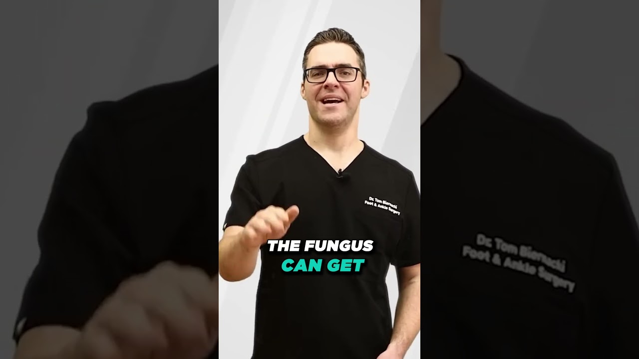
If you’ve ever looked down at a thick, yellow, crumbling toenail and wondered whether it’s just “old age” or something you can actually fix — you’re not alone. Toenail fungus is one of the most common conditions I see in my Michigan podiatry clinic, and it’s also one of the most undertreated. People live with it for years because they assume it’s cosmetic. It isn’t. Untreated fungal infections spread, thicken further, and — for people with diabetes — can become a serious health risk.
This guide covers everything you need to know: how toenail fungus looks, what causes it, how we diagnose it, and every treatment option from home remedies to prescription medications to laser therapy.
What Is Toenail Fungus?
Toenail fungus, medically called onychomycosis, is a fungal infection of the nail plate, nail bed, or both. It’s caused by dermatophyte fungi in about 90% of cases — the same family of fungi responsible for athlete’s foot and ringworm. In the remaining cases, Candida yeast or non-dermatophyte molds are the culprit, and treatment may differ.
Onychomycosis affects approximately 10% of the general population — rising to 20% of people over 60, and up to 50% of people over 70. In our clinic, we see it frequently in athletes, people with diabetes, anyone who uses public pools or locker rooms regularly, and patients who’ve had long-term nail trauma from ill-fitting shoes. Toenails are far more commonly affected than fingernails because fungi thrive in the warm, dark, moist environment inside shoes.
Key takeaway: Toenail fungus is a genuine infection, not a cosmetic issue. It’s caused by fungi that live in your nail tissue and won’t resolve on their own without treatment.
Symptoms: What Toenail Fungus Looks Like
Toenail fungus rarely causes pain in the early stages — which is exactly why people ignore it. The primary changes are visual and structural. Here’s what to look for, in rough order of how the infection typically progresses:
- White or yellow spots under the nail tip — often the first sign. The nail looks slightly discolored near the free edge.
- Yellow, brown, or white discoloration spreading toward the base of the nail. The nail may look dull rather than shiny.
- Thickened nail — one of the hallmarks. A healthy toenail is about 0.5–1mm thick; fungal nails can reach 2–3mm and become difficult to trim.
- Brittle, crumbly, or ragged edges — the nail breaks or crumbles when cut rather than trimming cleanly.
- Distorted nail shape — the nail may curve inward, outward, or lose its normal contour.
- Debris under the nail — white or yellowish chalky material (nail keratin debris) builds up between the nail and nail bed.
- Separation from the nail bed (onycholysis) — the nail lifts away from the skin underneath, creating a gap that can trap dirt and worsen infection.
- Foul odor — more common in advanced cases with significant nail destruction.
In my clinic, patients often tell me they assumed the discoloration was from nail polish or a bruise from a stubbed toe. The key difference: nail polish stains wash off eventually, and bruised nails grow out (you’ll see the discoloration move toward the tip as the nail grows). Fungal discoloration doesn’t move — it starts distally and progresses toward the cuticle as the infection deepens.
Key takeaway: Fungal discoloration progresses from the nail tip toward the cuticle and doesn’t grow out like a bruise. Thickening and brittleness are the most reliable signs that separate fungal infection from cosmetic staining.
What Causes Toenail Fungus?
Dermatophyte fungi — primarily Trichophyton rubrum (responsible for roughly 70% of cases) — are the main culprits. These fungi feed on keratin, the protein that makes up your nails and skin. They’re contagious and spread through direct contact with infected surfaces. Common sources of exposure include:
- Public floors — pool decks, gym locker rooms, yoga studios, hotel bathrooms. Walking barefoot dramatically increases risk.
- Shared nail tools — clippers, files, and nail buffers used at nail salons or shared within a household. A medical pedicure uses sterile, single-use instruments — a safer alternative to salon nail care.
- Athlete’s foot — untreated tinea pedis easily spreads from the skin to the nail.
- Tight or occlusive footwear — shoes that create a warm, moist microenvironment accelerate fungal growth.
- Nail trauma — a damaged nail provides a portal of entry for fungi. This is why runners and athletes have higher rates.
Several risk factors make people more susceptible regardless of exposure:
- Age — nail growth slows with age, giving fungi more time to establish
- Diabetes — impaired circulation and immune response increase susceptibility significantly
- Peripheral vascular disease — reduced blood flow limits the nail’s ability to fight infection
- Immunosuppression — from medications (steroids, biologics) or conditions like HIV
- Hyperhidrosis — excessive sweating of the feet
- Prior fungal infections — onychomycosis tends to recur
How Is Toenail Fungus Diagnosed?
This is where a clinical visit makes a meaningful difference. In our office, we don’t just look at the nail — we confirm the infection before prescribing systemic medication. Visual diagnosis alone is accurate only about 50% of the time. Studies show that up to 30% of nails that look fungal actually have another cause — psoriasis, lichen planus, traumatic dystrophy, or simple keratin buildup.
Our diagnostic workup typically includes:
- Nail clipping and subungual debris collection — a small sample of the discolored nail or debris under the nail is sent to a lab.
- KOH (potassium hydroxide) preparation — a rapid in-office test that dissolves nail tissue to reveal fungal hyphae under a microscope. Results in minutes.
- Fungal culture — grows the organism to confirm species and guide antibiotic selection if resistance is a concern. Takes 4–6 weeks.
- PCR testing — the most sensitive test available; identifies the specific fungal species within 24–48 hours. We use this for recurrent or treatment-resistant cases.
If you’ve been treated twice before without success, we strongly recommend PCR testing before starting a third round of antifungals — the problem may not be Trichophyton at all, and different species respond to different medications.
Toenail Fungus Treatment Options
Treatment selection depends on the severity of infection, the number of nails involved, your overall health, and which fungal species is causing the problem. Here’s the full treatment ladder we use at Balance Foot & Ankle:
Topical Antifungals
Topical medications (prescription or OTC) are the lowest-risk option but have the lowest cure rates for established infections. Efinaconazole (Jublia) and tavaborole (Kerydin) are FDA-approved prescription topicals with cure rates around 17–18% — much lower than oral medications, but appropriate for mild distal infection, elderly patients who can’t take systemic medications, or as maintenance after oral treatment. OTC options (undecylenic acid, tolnaftate) are useful for very early or superficial infections, but rarely cure established onychomycosis.
Oral Antifungals (Most Effective Option)
Terbinafine (Lamisil) is the gold standard — 70–80% mycological cure rate, taken daily for 6 weeks (fingernails) or 12 weeks (toenails). It works by inhibiting an enzyme essential to fungal cell membrane synthesis. We always check liver function tests before starting, and we rarely see significant side effects at the recommended dose and duration. Itraconazole is an alternative for patients who can’t take terbinafine or who have Candida-type infections — given in pulse doses (1 week on, 3 weeks off for 2–3 cycles).
Important caveat: even after a confirmed mycological cure (lab tests show no living fungus), the nail may take 12–18 months to look normal because it must physically grow out and replace the damaged portion. Clear nail at the base growing toward the tip is the sign that treatment is working.
Laser Therapy (PACT / Nd:YAG)
We offer laser treatment for patients who prefer to avoid oral medications or for whom systemic antifungals are contraindicated. PACT (photodynamic antimicrobial therapy) and Nd:YAG laser use targeted light energy to destroy fungal organisms within the nail. Studies show cure rates of 30–60% depending on infection severity — lower than oral terbinafine, but without systemic drug exposure. Typically requires 3–4 sessions. We use laser as a standalone option for mild-to-moderate infections and as a combination add-on to oral treatment for severe cases.
Nail Debridement
In-office nail debridement — thinning and cleaning the infected nail — is performed at nearly every visit. It serves two purposes: it reduces the fungal load physically, and it allows topical medications to penetrate more effectively. Severely thickened nails are often painful in shoes; debridement provides immediate relief while antifungal treatment takes effect over months.
Restoring the nail’s appearance during treatment: because clearing fungus takes months, many patients pair their treatment with KeryFlex nail restoration — a flexible, medical-grade resin nail applied in office that looks natural while the healthy nail grows out underneath.
Nail Removal (Avulsion)
Chemical or surgical nail avulsion (permanent or temporary removal of the nail) is reserved for cases with severe nail dystrophy, pain, or repeated treatment failure. Temporary avulsion allows topical antifungals direct access to the nail bed. Permanent avulsion (destroying the nail matrix) is considered only when the nail has become structurally non-viable and is causing recurrent ingrown nails or soft tissue damage.
Key takeaway: Oral terbinafine (12 weeks) achieves the highest cure rates. Expect 12–18 months for the nail to look normal even after successful treatment. Topical medications alone rarely cure established infections.
🧦 Reduce Fungal Recurrence: Anti-Fungal Socks
Toenail fungus thrives in warm, moist environments. Standard cotton socks retain moisture against the skin for hours — creating ideal conditions for fungal regrowth after treatment. DASS copper-infused moisture-wicking socks significantly reduce the damp environment that fuels recurrence. I recommend these during and after antifungal treatment for any patient with hyperhidrosis or a history of repeat infections.
Shop DASS Antimicrobial Compression Socks → | ~$25–35
Affiliate disclosure: As an Amazon Associate and Foundation Wellness partner, we earn from qualifying purchases at no extra cost to you.
Home Remedies: What Actually Works (and What Doesn’t)
Patients ask me about home remedies constantly — and I give them an honest answer rather than dismissing all of them. Here’s the evidence-based breakdown:
Vicks VapoRub
Sounds odd, but there’s actual research behind it. A 2011 study in Journal of the American Board of Family Medicine found that 83% of patients using Vicks daily for 48 weeks showed partial improvement and 27.8% achieved mycological cure. The active ingredients — thymol, camphor, and eucalyptus oil — have antifungal properties. It won’t work as fast or as reliably as prescription medication, but for mild early infections in patients who decline oral medication, it’s a reasonable option. Apply a small amount to the infected nail daily after bathing.
Tea Tree Oil
A small randomized trial found tea tree oil comparable to clotrimazole cream for nail improvement (though not mycological cure). Its antifungal properties are real but modest. Useful for maintenance or very mild distal infections. Apply with a cotton swab to the nail surface twice daily.
Apple Cider Vinegar Soaks
No high-quality clinical evidence for direct antifungal efficacy. The acidic pH may slow fungal growth, and soaking does help soften thickened nails for easier trimming. Not harmful, but not a reliable treatment for established infection.
Bleach Foot Soaks
Dilute bleach (1 teaspoon per gallon of water) does have antifungal activity and is occasionally used for resistant tinea pedis. For nail infections, penetration is too poor to achieve meaningful reduction in fungal load. Not recommended due to skin irritation risk with little proven benefit.
The most common mistake I see: spending 12+ months trying home remedies on a moderate-to-severe infection while the fungus spreads to adjacent nails and the nail becomes more structurally damaged. Home remedies are appropriate for very mild early infections only. If you’re on your third year of trying Vicks and the nail looks worse — come in.
Warning Signs: When to See a Podiatrist
⚠️ See a podiatrist promptly if you have:
- Diabetes, peripheral neuropathy, or peripheral vascular disease — fungal nails can progress to serious secondary infections
- Pain when wearing shoes or walking — thickened nails pressing on adjacent toes cause ulcers in diabetic feet
- More than 3 nails affected, or infection spreading to fingernails
- Previous treatment failure (2+ courses without improvement)
- Signs of secondary bacterial infection: redness, warmth, swelling, or discharge around the nail
- Nail lifting completely from the nail bed (full onycholysis)
- Skin cracks between toes — athlete’s foot left untreated becomes a portal for bacterial cellulitis
Prevention: How to Stop Toenail Fungus From Coming Back
Recurrence rates for toenail fungus are significant — up to 50% within 3 years even after successful treatment. Prevention is an active process, not just “being careful.” Here’s what actually reduces recurrence risk:
- Treat athlete’s foot aggressively — don’t let tinea pedis persist on the skin. It will reinfect the nails.
- Wear moisture-wicking socks — synthetic or merino wool, not cotton. Change socks after workouts.
- Wear shower shoes in public — in gyms, pools, hotel rooms, and nail salons.
- Disinfect your nail tools — clippers and files should be cleaned with 70% isopropyl alcohol after use, or replaced periodically.
- Rotate shoes — allow at least 24 hours between wearings to let shoes dry out. Use UV shoe sanitizers or antifungal powders in frequently worn shoes.
- Trim nails properly — straight across, not too short. Short nails with rounded corners are more prone to trauma that creates fungal entry points.
- Choose breathable footwear — leather, mesh, or canvas uppers over synthetic materials.
- Maintenance topical antifungals — for high-risk patients (diabetics, prior recurrences), ongoing use of a prescription topical or quality OTC antifungal after completing oral treatment significantly reduces recurrence.
Frequently Asked Questions
Is toenail fungus contagious to other people?
Yes — toenail fungus is moderately contagious through shared surfaces and direct contact. Sharing nail tools, walking barefoot on contaminated floors, or having prolonged skin-to-skin contact with an infected person can transmit the fungus. Household transmission is common; studies show roughly 30–40% of household contacts of people with onychomycosis carry the same fungal species. This is one reason we recommend treating promptly rather than waiting.
Can toenail fungus go away on its own?
Spontaneous resolution without treatment is rare — estimated at less than 5% of cases, and primarily in very early superficial infections. In the vast majority of cases, toenail fungus progresses without treatment, spreading to more nails and worsening the structural damage. “Waiting to see” for more than 2–3 months after noticing changes is not recommended.
Is toenail fungus the same as athlete’s foot?
They’re caused by the same family of fungi (dermatophytes), but they’re distinct infections in different tissue. Athlete’s foot (tinea pedis) infects the superficial skin of the foot — particularly between the toes. Toenail fungus (onychomycosis) infects the nail plate and nail bed. Athlete’s foot is easier to treat; onychomycosis is harder to eliminate because the nail acts as a physical barrier to medications. Athlete’s foot frequently precedes or accompanies toenail fungus.
Can I paint my toenails if I have fungus?
We recommend against nail polish during active treatment. Nail polish seals moisture under the nail, creating conditions that promote fungal growth, and prevents topical antifungal medications from penetrating. Once treatment is complete and the nail has regrown, normal nail polish use is fine. Some patients use antifungal nail polish as maintenance.
How do I know if toenail fungus treatment is working?
The earliest sign that treatment is working is new clear nail growth at the base (cuticle end) of the nail. This appears within 4–8 weeks of starting oral antifungals. The clear zone slowly advances toward the tip as the nail grows out over 12–18 months. Lab tests (repeat KOH or culture) at 3 months can confirm mycological cure before the visual improvement is obvious. If you see no new clear nail growth at 3 months, the treatment may not be working and a species change should be investigated.
Sources
- Gupta AK, et al. “Onychomycosis: a review.” JAAD. 2023.
- Derby R, et al. “Novel treatment of onychomycosis using over-the-counter mentholated ointment.” J Am Board Fam Med. 2011.
- Elewski BE, et al. “Efinaconazole topical solution, 10%: phase III trials for toenail onychomycosis.” JAAD. 2013.
- Scher RK, et al. “Onychomycosis: diagnosis and definition of cure.” JAAD. 2007.
- Hay RJ. “Onychomycosis.” NEJM. 2023;388(13):1204–1213.
See also: Best Socks for Toenail Fungus 2026 | Vicks VapoRub for Toenail Fungus: Does It Work? | Toenail Psoriasis vs Fungus: How to Tell the Difference
Ready for Expert Care?
Same-week toenail fungus evaluation in Howell & Bloomfield Township, MI. We confirm your diagnosis with lab testing before prescribing treatment.
4.9★ | 1,193 Reviews | 3,000+ Surgeries
Or call: (810) 206-1402
More Toenail Fungus Resources
- When to See a Foot Doctor for Toenail Fungus
- Best Over-the-Counter Toenail Fungus Treatments
- Best Socks for Toenail Fungus
- How to Fix Severe Toenail Fungus
- Keratin Granulations vs. Toenail Fungus
Toenail Fungus & Nail Care Resources
Expert guides on toenail fungus, nail conditions, and care:
- Bruised Toenail
- Laser Treatment for Toenail Fungus 2026
- Dark Streak Toenail Subungual Melanoma 2026
- Laser Toenail Fungus Treatment in Michigan
- Black Toenail Causes Care 2026
- How to Treat an Ingrown Toenail at Home
- Toenail Psoriasis vs Fungus
- Medical Nail Care Bloomfield Township MI 2026
- Toenail Discoloration Causes & Treatment 2026
- Ingrown Toenail Auburn Hills MI 2026
- Subungual Hematoma Treatment 2026
- Ingrown Toenail Treatment Howell MI
- Toenail Fungus Laser Treatment 2026
- How to Treat Toenail Fungus at Home
- Toenail Fungus Laser Oral Treatment 2026
- How to Prevent Toenail Fungus
- Senior Toenail Care
- Pediatric Ingrown Toenail Children 2026
- Ingrown Toenail Surgery Nail Avulsion 2026
- Ingrown Toenail Permanent Removal 2026
- Home Remedies for Toenail Fungus 2026
- Nail Fungus Onychomycosis Treatment 2026
- Ingrown Toenail Treatment Bloomfield Township
- Ingrown Toenail Treatment Auburn Hills MI 2026
- Ingrown Toenail Surgery 2026
- Thick Toenails
- Prevent Toenail Fungus Spreading 2026
- Hydrogen Peroxide for Toenail Fungus
- What Causes Toenail Fungus? Risk Factors & Prevention
- Ingrown Toenail Home Remedies vs Podiatrist 2026
- What Causes Ingrown Toenails? Prevention & Treatment
Related: does Vicks VapoRub actually work on toenail fungus and thick toenails? A podiatrist explains.
Related foot & ankle guides
- Toenail ridges explained
- Nail fungus home remedies
- Apple cider vinegar for foot fungus
- Urea cream for feet
- Common foot skin disorders
- Sweaty feet: causes & fixes
Searching for toenail fungus laser treatment near you? Balance Foot & Ankle offers in-office laser toenail fungus treatment plus prescription and topical antifungal care at our Bloomfield Township office (Oakland County) and Howell office (Livingston County). We give you honest, realistic timelines — no gimmicks. Book online or call (810) 206-1402.
Prefer we call you?
Leave your info and our team calls back within 2 hours, the same business day — to get you scheduled with a board-certified podiatrist at our Howell or Bloomfield Township office. Same-week appointments; most insurance accepted, and we verify your benefits up front.
Nail & skin condition guides
Toenail conditions
- Fungal Toenail Removal
- Laser Toenail Fungus Treatment
- Toenail Infection Treatment
- Nail Discoloration Causes
- Toenail Discoloration Causes
- Paronychia (Toe Infection) Treatment
Foot skin conditions
Toenail fungus that won’t clear?
When creams fail, prescription and laser options work. In Michigan? our podiatrists confirm it’s fungus and treat it properly. Same-week visits in Howell & Bloomfield Township, most insurance accepted.
📅 Book an Appointment 📞 (810) 206-1402★ 4.9 from 1,193+ Google reviews · Same-week visits · We verify your insurance free — most visits are just a copay.
Clinical sources
- A Systematic Review of Onychomycosis Clinical Trials. Journal of Fungi (MDPI). Reference
- Terbinafine for onychomycosis — treatment-duration evidence. American Family Physician, 2023. AFP
- Treatment Options for Onychomycosis: Efficacy, Side Effects & Adherence. Journal of Clinical and Aesthetic Dermatology. JCAD
To lower reinfection risk while treating a fungal nail, wear moisture-wicking, best antifungal socks that keep the skin dry between treatments.
Toenail fungus treatment — towns we treat
Fungal nails clear slowly, so we keep follow-up visits short and close to home. Pick your city for directions and appointment options.
Treated at our Howell office
Brighton · Hartland · Pinckney · Hamburg · Fenton · Whitmore Lake · Ann Arbor · South Lyon · New Hudson · Milford · Highland · White Lake
Treated at our Bloomfield Township office
Birmingham · Troy · Rochester Hills · Royal Oak · Madison Heights · Southfield · Novi · Farmington Hills · West Bloomfield · Keego Harbor · Wolverine Lake · Walled Lake · Commerce Township · Wixom · Waterford · Pontiac · Clarkston · Auburn Hills · Lake Orion · Livonia
Howell office · Bloomfield Township office · See both offices
Related
Nail changes that are often mistaken for fungus:
- Onycholysis — when the nail lifts rather than thickens
- Black toenail — fungal, bruise, or something else
Almost everyone has tried something at home before booking an appointment, and soaking in Listerine is the one that comes up most. It is worth knowing what a topical soak can and cannot reach through the nail plate before spending another season on it.