

Medically reviewed by Dr. Tom Biernacki, DPM, FACFAS
Board-certified podiatric surgeon | Balance Foot & Ankle | Last reviewed: May 2026
Quick answer: Gout is caused by monosodium urate crystal deposition in joints — most commonly the first metatarsophalangeal joint (podagra). Acute attacks are treated with colchicine, NSAIDs, or corticosteroids. Long-term control requires urate-lowering therapy (allopurinol or febuxostat) targeting a serum uric acid below 6 mg/dL. Without urate-lowering therapy, attacks become more frequent, joints are permanently damaged, and tophi (urate deposits) develop in soft tissue.
Watch Dr. Tom Biernacki DPM explain gout treatment — causes, diagnosis, and home remedies at Balance Foot & Ankle. MichiganFootDoctors YouTube
What Is Gout? (Pathophysiology)
Gout is a crystal deposition arthropathy caused by the precipitation of monosodium urate (MSU) crystals in joint spaces, periarticular tissues, and tendons. It is not simply “high uric acid” — many people with elevated serum uric acid (hyperuricemia) never develop gout, while some gout patients have normal serum levels during an acute attack. The triggering event is crystal nucleation and shedding into the joint space, which activates the NLRP3 inflammasome, triggering a cascade of IL-1β release, neutrophil recruitment, and intense inflammatory synovitis.
The result is one of medicine’s most dramatic pain presentations: sudden, severe, often nocturnal joint pain that reaches maximum intensity within 12–24 hours, accompanied by warmth, swelling, erythema, and tenderness so detailed that the weight of a bedsheet is intolerable.
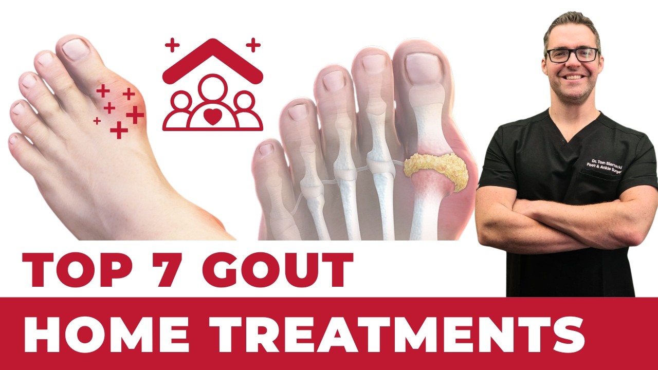
Why the big toe? The first metatarsophalangeal (MTP) joint — the base of the big toe — is affected in approximately 70% of initial gout attacks (called podagra). Urate crystals precipitate preferentially in cooler peripheral tissues (the first MTP is one of the coolest joints in the body) and in areas of prior trauma or lower synovial fluid volume. Urate solubility decreases significantly as temperature falls — at 37°C (core body temperature) the solubility threshold is ~6.8 mg/dL; at 30°C (peripheral joint temperature) it falls to approximately 4.5 mg/dL.
The Gout Disease Spectrum: Acute to Tophaceous
Gout is not a single event — it is a progressive disease with four recognizable stages if uric acid is not controlled:
- Asymptomatic hyperuricemia: Elevated uric acid without crystal deposition or symptoms. Does not require pharmacologic treatment but warrants dietary and lifestyle counseling.
- Acute intermittent gout: Discrete, intensely painful attacks separated by completely symptom-free intervals (intercritical periods). Early in the disease, attacks self-resolve in 7–10 days even without treatment. Over time, attacks become longer, more frequent, and involve additional joints (ankle, knee, wrist).
- Intercritical gout: The period between attacks. The joint is not actively inflamed, but crystal deposits remain and subclinical inflammation persists. This is the optimal window for starting urate-lowering therapy.
- Chronic tophaceous gout: After years of uncontrolled hyperuricemia, MSU crystals accumulate as visible, palpable nodular deposits (tophi) in soft tissues — commonly the helix of the ear, Achilles tendon, olecranon bursa, and periarticular tissues of the hands and feet. Tophaceous disease causes chronic dull joint pain and progressive joint destruction. Tophi can ulcerate through skin, creating chalky white drainage and infection risk.
Critical point: Most patients seek care only during acute attacks and discontinue treatment once the pain resolves. This is the most common management failure in gout. The disease continues to progress silently — urate crystals accumulate, attacks become more frequent, and permanent joint damage accrues — until tophaceous disease is present. Urate-lowering therapy must be continued long-term, not just during attacks.
Who Gets Gout?
- Prevalence: Affects approximately 9 million Americans (4% of adults); the most common inflammatory arthritis in men over 40
- Sex: Men predominate 3–4:1; women gain risk rapidly after menopause (estrogen is uricosuric)
- Primary gout: Genetic underexcretion of uric acid by the kidney (90% of cases) or, less commonly, overproduction
- Secondary gout: Caused by identifiable conditions — chronic kidney disease (reduced urate excretion), diuretic use (thiazide and loop diuretics concentrate uric acid), cyclosporine use, myeloproliferative disorders with high cell turnover, psoriasis, and lead nephropathy
- Dietary triggers: Red meat, organ meats, shellfish (high purine load), alcohol (especially beer and spirits — fructose from alcohol metabolism increases urate production and impairs renal excretion), high-fructose corn syrup beverages
- Protective factors: Dairy consumption (casein and lactalbumin promote uricosuria), vitamin C, and coffee consumption have modest protective associations in epidemiologic studies
Symptoms of Gout
The acute gout attack presents with:
- Sudden onset severe joint pain — typically maximal within 12–24 hours, often beginning at night
- Intense warmth, redness (erythema), and swelling over the affected joint — can mimic cellulitis
- Allodynia: pain with even light touch or the pressure of bedsheets
- Low-grade fever is common; high fever suggests concurrent infection
- In classic podagra: the first MTP joint is markedly swollen and warm, the patient cannot bear weight or wear a shoe
- Self-resolution in 7–10 days even without treatment (in early disease)
Common trigger events for an acute attack: dietary purine excess, alcohol binge, dehydration, prolonged fasting (surgery, illness), rapid uric acid change (starting or stopping allopurinol), trauma to the joint, and acute intercurrent illness.
Diagnosing Gout
Gold Standard: Joint Aspiration
Definitive diagnosis is by synovial fluid analysis under polarized light microscopy. Monosodium urate crystals appear as needle-shaped, negatively birefringent crystals (bright yellow when parallel to the polarizer, blue when perpendicular). This also rules out septic arthritis, which can present identically and is a medical emergency. In a classic first-MTP podagra presentation in an otherwise healthy patient with high uric acid, most clinicians treat empirically — but aspiration is essential when the diagnosis is uncertain or when joint infection cannot be excluded.
Serum Uric Acid
A useful but imperfect test. Normal is typically cited as under 6.8 mg/dL (the physiologic saturation point). Important caveat: during an acute attack, serum uric acid can be falsely normal or low because crystals are being actively deposited in joints, pulling uric acid from the serum. Do not use a normal acute-phase uric acid to rule out gout. Levels should be rechecked 2–4 weeks after an attack resolves to establish a true baseline.
Imaging
X-ray: Early gout shows only soft-tissue swelling. Chronic tophaceous disease produces characteristic “punched-out” erosions with sclerotic margins and overhanging edges (the “rat-bite” appearance) at joint margins, distinctly different from the marginal erosions of rheumatoid arthritis.
Ultrasound: Can visualize the “double contour sign” — a hyperechoic line over the articular cartilage surface caused by urate crystal deposition — and tophaceous deposits within tendons and soft tissue. Useful for early diagnosis before X-ray changes develop.
Dual-energy CT (DECT): The most sensitive imaging modality, color-coding urate deposits throughout the body. Valuable for confirming diagnosis in atypical presentations and quantifying tophaceous burden before initiating urate-lowering therapy.
Gout vs. Similar Conditions
- Septic arthritis (joint infection): The most dangerous mimicker of gout — both present with acute red, hot, swollen joint and fever. Cannot be reliably distinguished clinically. Joint aspiration is essential: septic joint shows >50,000 WBC/mm³ with >90% neutrophils, organisms on Gram stain, and positive culture. Gout shows crystals but no organisms. When in doubt, treat both until cultures finalize — a septic joint requires urgent surgical washout.
- Pseudogout (calcium pyrophosphate deposition, CPPD): Presents with nearly identical acute attacks but most commonly affects the knee, wrist, and ankle rather than the first MTP. Crystals are rhomboid-shaped and positively birefringent under polarized light. X-ray shows chondrocalcinosis (calcification within cartilage). Risk factors include hyperparathyroidism, hemochromatosis, and hypothyroidism — workup differs accordingly.
- Cellulitis: Skin infection over the dorsal first MTP can be difficult to distinguish from podagra — both cause erythema, warmth, and swelling over the same area. Key differentiator: cellulitis produces no joint crepitus or decreased range of motion (the skin is inflamed, not the joint); gout is maximally painful with joint palpation and passive motion. Cellulitis should not improve rapidly with colchicine or NSAIDs.
- Psoriatic arthritis: Can cause inflammatory arthritis at the first MTP and other foot joints, often with dactylitis (sausage digit), nail changes (pitting, onycholysis), and skin plaques. Seronegative (negative RF, normal uric acid). Enthesitis (Achilles, plantar fascia) is characteristic and absent in gout.
Treatment Options for Gout
Acute Attack Treatment (Reduce Inflammation)
The goal during an acute attack is to suppress the inflammatory response as quickly as possible. Three proven options:
Colchicine (first-line for most patients): The evidence-based approach is low-dose colchicine — 1.2mg at first symptom, then 0.6mg one hour later — rather than the older high-dose regimen, which achieves equivalent efficacy with dramatically fewer GI side effects (diarrhea, nausea). Colchicine inhibits microtubule polymerization and neutrophil migration, blocking the inflammatory cascade. Most effective when started within 24 hours of attack onset; less effective once the attack is fully established. Dose must be adjusted in renal impairment and with CYP3A4 inhibitors (statins, clarithromycin).
NSAIDs (indomethacin, naproxen, ibuprofen): Highly effective for acute gout when started early. Full prescription-dose NSAIDs (indomethacin 50mg TID, naproxen 500mg BID) are used for 3–5 days. Contraindicated in CKD (stages 3–5), anticoagulated patients, and those with peptic ulcer disease. Add a proton pump inhibitor when using NSAIDs in higher-risk patients.
Corticosteroids: Used when colchicine and NSAIDs are contraindicated — renal impairment, anticoagulation, or GI intolerance. Oral prednisone (30–40mg/day, tapered over 10–14 days) or intramuscular triamcinolone are effective. In-office intra-articular corticosteroid injection into the first MTP joint provides rapid, targeted relief without systemic exposure — particularly useful in our clinic for isolated podagra.
What NOT to do during an acute attack: Do not start or adjust urate-lowering therapy during an acute attack — this can precipitate crystal shedding and prolong or worsen the episode. Continue existing allopurinol at the current dose; do not initiate new ULT until the attack fully resolves.
Urate-Lowering Therapy (Prevent Future Attacks)
ULT is indicated after a second confirmed gout attack, after a first attack in any patient with CKD stage 2+, uric acid >9 mg/dL, or presence of tophi on exam or imaging. The target is serum uric acid below 6 mg/dL (below 5 mg/dL for tophaceous disease) — well below the 6.8 mg/dL saturation threshold to drive crystal dissolution over time.
Allopurinol (xanthine oxidase inhibitor — first-line ULT): Blocks urate synthesis. Start at 100mg/day (50mg in CKD), uptitrate by 100mg every 2–4 weeks to achieve target uric acid. Maximum dose 800mg/day. HLA-B*5801 allele testing before starting is recommended in patients of Asian descent (risk of severe allopurinol hypersensitivity reaction). Prophylactic colchicine 0.6mg daily for 3–6 months when initiating to prevent mobilization flares.
Febuxostat (alternative XO inhibitor): More potent urate reduction, useful in allopurinol intolerance or in patients needing uric acid control without dose titration. A cardiovascular safety signal (increased CV events vs. allopurinol) in the CARES trial means it is now reserved for allopurinol-intolerant patients. Dose: 40–80mg daily.
Probenecid (uricosuric): Promotes renal urate excretion; useful in underexcretors with adequate kidney function. Requires adequate fluid intake (kidney stone risk). Less commonly used than XO inhibitors.
Dietary and Lifestyle Modifications
Diet alone rarely normalizes uric acid sufficiently to prevent attacks (typical dietary change reduces serum urate by 1–2 mg/dL), but it is an important adjunct and reduces attack frequency:
- Eliminate or significantly reduce: red meat, organ meats, shellfish, beer, spirits, high-fructose corn syrup beverages (sodas, fruit juices)
- Limit (not eliminate): wine in moderation, fructose-containing fruits
- Increase: dairy (low-fat), water intake (target 2L/day to promote uricosuria), vitamin C 500mg/day (modest uricosuric effect), coffee
- Weight loss: obesity is strongly associated with hyperuricemia; even modest weight reduction improves urate control. Avoid crash dieting — rapid weight loss increases uric acid through ketoacidosis and tissue breakdown
- Review all medications for urate-elevating agents: thiazide diuretics (hydrochlorothiazide), loop diuretics (furosemide), low-dose aspirin, cyclosporine, niacin. When clinically feasible, substituting losartan (mildly uricosuric) for a thiazide or switching to amlodipine improves gout control
Joint Aspiration and Injection
In-office joint aspiration of the first MTP joint serves two purposes: it confirms the diagnosis (crystal identification under polarized microscopy), and it reduces joint pressure immediately, providing pain relief. Following aspiration, intra-articular corticosteroid injection delivers anti-inflammatory medication directly to the pathologic site. We perform this routinely in our clinic for acute podagra that does not tolerate oral therapy or requires faster relief.
Management of Tophi
Tophi resolve slowly with adequate urate-lowering therapy — most studies show complete resolution at 1–3 years of sustained uric acid below 5 mg/dL. Surgical removal of tophi is reserved for specific indications: ulceration with secondary infection, mechanical impingement on tendons causing functional limitation, or severe cosmetic deformity affecting patient quality of life. Surgical debridement alone without ULT invariably leads to recurrence.
Most Common Mistakes
- Stopping urate-lowering therapy after symptoms improve: Allopurinol is a lifetime medication for most gout patients. Stopping it after attacks resolve allows uric acid to rebound, crystals to re-accumulate, and the disease to progress — often with a severe rebound attack. Many patients are never told this by prescribers who treat gout as an acute-only condition.
- Initiating allopurinol during an acute attack: Starting ULT during active inflammation causes mobilization flares — the sudden change in uric acid levels triggers additional crystal shedding. The correct approach is to treat the acute attack fully, then start allopurinol 2–4 weeks after complete resolution, with colchicine prophylaxis coverage for the first 3–6 months.
Red Flags — When to See a Podiatrist Immediately
- Fever above 38.5°C (101.3°F) with an acutely inflamed joint — mandates joint aspiration to rule out septic arthritis, which is a surgical emergency
- Open, draining tophi — ulcerated tophi through the skin create a direct portal for infection; require urgent wound care and assessment for underlying bone involvement (osteomyelitis)
- Rapidly enlarging swelling with skin color change (purple, dusky, gray) — may indicate septic joint, necrotizing fasciitis, or vascular compromise; requires urgent evaluation
- Inability to bear any weight — significant injury must be excluded (fracture, complete tendon rupture) before attributing inability to weight-bear to gout alone
- Gout attacks in multiple joints simultaneously (polyarticular gout) — most common in undertreated chronic disease, but can also represent a septic joint in one location and gout in others; requires careful evaluation and often aspiration of each affected joint
- Any diabetic patient with acute foot joint inflammation — diabetics with neuropathy may have impaired pain perception; infection and gout coexist; same-day evaluation is essential
In-Office Gout Treatment at Balance Foot & Ankle
Our podiatric team manages the full gout spectrum: acute attack treatment with in-office joint aspiration and corticosteroid injection, coordination with primary care physicians for urate-lowering therapy initiation, dietary counseling, and long-term monitoring. We have on-site digital X-ray and musculoskeletal ultrasound for same-day imaging. Most major Michigan insurance plans cover gout treatment. Same-week appointments available.
Howell: 4330 E Grand River Ave, Howell MI 48843
Bloomfield Hills: 43494 Woodward Ave #208, Bloomfield Hills MI 48302
Phone: (810) 206-1402 | Book online →
Sources
- FitzGerald JD, et al. 2020 American College of Rheumatology Guideline for the Management of Gout. Arthritis Rheumatol. 2020;72(6):879–895.
- Dalbeth N, Merriman TR, Stamp LK. Gout. Lancet. 2016;388(10055):2039–2052.
- Richette P, Bardin T. Gout. Lancet. 2010;375(9711):318–328.
- Choi HK, et al. Purine-rich foods, dairy and protein intake, and the risk of gout in men. N Engl J Med. 2004;350(11):1093–1103.
- White WB, et al. Cardiovascular safety of febuxostat or allopurinol in patients with gout (CARES). N Engl J Med. 2018;378(13):1200–1210.
- Neogi T. Gout. N Engl J Med. 2011;364(5):443–452.
Acute Gout Attack? Get Fast In-Office Relief.
Joint aspiration & same-day treatment — Howell & Bloomfield Hills, MI
4.9★ | 1,123 Reviews | 3,000+ Surgeries
Or call: (810) 206-1402
Related Treatments at Balance Foot & Ankle
Common conditions we treat in our Howell and Bloomfield Hills offices.