
Quick answer: Lisfranc Injury Midfoot Fracture Dislocation is a common foot/ankle topic that affects many patients. The 2026 evidence-based approach combines proper diagnosis, conservative-first treatment, and escalation only when needed. We treat this regularly at our Howell and Bloomfield Hills practices. Call (810) 206-1402.
Medically reviewed by Dr. Tom Biernacki, DPM — Board-Certified Podiatric Surgeon — Balance Foot & Ankle, Howell & Bloomfield Hills, MI. Last updated April 2026.
▶ Watch

Medically reviewed by Dr. Tom Biernacki, DPM | Board-certified podiatrist | 3,000+ surgeries performed
Last updated: April 2, 2026
The most important clinical decision with Lisfranc Injury Midfoot Fracture Dislocation isn't which treatment to start with — it's which subtype or underlying cause you actually have. Our podiatrists regularly see patients who've been treated for months for the wrong diagnosis. The correct identification changes the entire treatment path. Call (810) 206-1402 — Dr. Tom evaluates this condition at both Howell and Bloomfield Hills locations.
What Is a Lisfranc Injury?
The Lisfranc joint complex connects the midfoot to the forefoot through five tarsometatarsal (TMT) joints stabilized by strong ligaments, the most critical being the Lisfranc ligament connecting the medial cuneiform to the base of the second metatarsal. Injury to this complex disrupts the structural arch of the foot and its ability to transfer force during walking.
Lisfranc injuries range from subtle ligament sprains to complete fracture-dislocations. They are classified as homolateral (all metatarsals displaced in the same direction), divergent (metatarsals displaced in different directions), or isolated (single column involvement). The severity classification directly determines surgical approach and expected outcomes.
These injuries are notoriously underdiagnosed — studies estimate 20-40% of Lisfranc injuries are missed on initial emergency room evaluation. The midfoot swelling pattern can mimic a simple sprain, and standard non-weight-bearing X-rays may appear normal even with significant ligamentous disruption. This missed diagnosis leads to chronic midfoot dysfunction.
Causes and Mechanisms of Injury
High-energy Lisfranc injuries occur in motor vehicle accidents, falls from height, and industrial crushing incidents. These typically produce obvious fracture-dislocations visible on initial X-rays with significant swelling and inability to bear weight.
Low-energy athletic Lisfranc injuries are more subtle and more commonly missed. They occur when an athlete’s foot is planted and another player falls on the heel (football), during a stumble with the foot caught in a stirrup, during a misstep off a curb, or from twisting forces on a plantar-flexed foot. These may present with only mild swelling and the ability to walk with a limp.
The classic mechanism involves axial loading on a plantar-flexed foot, driving force through the metatarsals that disrupts the Lisfranc ligament complex. Even a simple stumble off a step with the foot pointed downward can generate enough force to tear this critical ligament, which is why low-energy Lisfranc injuries occur more frequently than most people realize.
Diagnosis: Why Getting It Right Matters
Clinical examination findings include midfoot swelling (particularly on the dorsal and plantar surfaces), tenderness directly over the TMT joints, pain with passive pronation-abduction of the forefoot, and inability to perform single-leg heel raise. A plantar ecchymosis (bruise on the sole) within 24-48 hours is highly suggestive of Lisfranc injury.
Weight-bearing X-rays are essential and often reveal findings that non-weight-bearing views miss. The critical measurement is the alignment between the medial border of the second metatarsal and the medial border of the middle cuneiform — any widening greater than 2mm suggests ligamentous disruption. A fleck sign (small avulsion fragment between the first and second metatarsals) is pathognomonic.
CT scanning provides detailed bony assessment and identifies subtle fractures at metatarsal bases that X-rays miss. MRI directly visualizes the Lisfranc ligament and other stabilizing structures, confirming the diagnosis in cases where X-rays are equivocal. At Balance Foot & Ankle, Dr. Tom Biernacki uses advanced imaging protocols to ensure no Lisfranc injury is missed.
Stress examination under fluoroscopy (applying force to the midfoot while taking live X-rays) can demonstrate instability that static images miss. This is particularly valuable when clinical suspicion is high but standard imaging is inconclusive.
Non-Surgical vs. Surgical Treatment
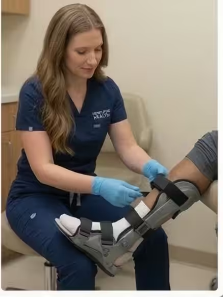
Non-surgical treatment is appropriate ONLY for purely ligamentous injuries with no displacement or instability on weight-bearing X-rays and stress examination. These stable injuries are treated with 6-8 weeks of non-weight-bearing in a cast boot, followed by gradual return to weight-bearing with custom arch support orthotics.
Surgical fixation is required for any displaced or unstable Lisfranc injury. Open reduction and internal fixation (ORIF) using screws and/or plates restores anatomic alignment of the TMT joints. The Lisfranc ligament does not heal reliably on its own once disrupted, making surgical stabilization essential for preventing chronic midfoot collapse.
Primary arthrodesis (fusion) of the medial and middle columns is increasingly favored over ORIF alone for purely ligamentous injuries, as fusion produces superior long-term outcomes with lower reoperation rates. A 2025 multicenter trial demonstrated that primary fusion produced better functional scores at 5 years compared to ORIF with subsequent hardware removal.
The surgical decision between ORIF and fusion depends on whether the injury involves fractures (ORIF preserves joint surfaces) or is purely ligamentous (fusion provides more reliable stability). Combined approaches addressing different columns with different techniques are common in complex injuries.
Recovery and Rehabilitation
Weeks 1-6: Non-weight-bearing in a posterior splint transitioning to a removable boot. Suture removal at 2 weeks. Elevation and ice therapy are critical for swelling management. Doctor Hoy’s Natural Pain Relief Gel helps manage surgical site discomfort during this phase.
Weeks 6-12: Progressive weight-bearing begins based on X-ray evidence of healing. Physical therapy focuses on ankle and midfoot range of motion, calf strengthening, and gait retraining. The walking boot is used during the transition to full weight-bearing.
Months 3-6: Transition to supportive shoes with custom orthotics that provide midfoot arch support. Progressive return to activities with impact loading. Hardware removal (screws) may be performed at 4-6 months if ORIF was chosen, as leaving screws across mobile joints can cause hardware failure and pain.
Months 6-12: Gradual return to sport and full activity. Custom orthotics from Balance Foot & Ankle with specific midfoot support become a long-term requirement to protect the healing tissues and optimize midfoot function. PowerStep insoles supplement orthotics for casual footwear.
Long-Term Outcomes and Complications
Even with optimal surgical treatment, some degree of post-traumatic arthritis develops in 25-50% of Lisfranc injury patients within 5-10 years. Arthritis severity correlates with initial injury severity, accuracy of surgical reduction, and time between injury and surgery. This underscores why we early, accurate diagnosis and anatomic surgical reduction.
Chronic midfoot pain after Lisfranc injury may require secondary fusion of affected TMT joints if arthritis becomes symptomatic despite conservative management with orthotics, activity modification, and anti-inflammatory treatment. Secondary fusion success rates exceed 85% for pain relief.
Return to high-level athletics after Lisfranc injury varies by sport and severity. Professional athletes return at rates of 70-80%, though many report some persistent limitations. Recreational athletes generally achieve good functional outcomes with appropriate rehabilitation and long-term orthotic support.
Why Lisfranc Injuries Should Not Be Treated as Simple Sprains
The consequences of misdiagnosing a Lisfranc injury as a simple midfoot sprain are severe. Untreated ligamentous instability leads to progressive midfoot collapse, loss of the longitudinal arch, development of painful arthritis, and eventually a rocker-bottom foot deformity that causes lifelong disability.
If you have midfoot pain and swelling after an injury — even if you can walk on it — and the pain is not improving within 5-7 days, request weight-bearing X-rays specifically evaluating the Lisfranc joint. A negative non-weight-bearing X-ray does not rule out this injury.
At Balance Foot & Ankle, Dr. Tom Biernacki has extensive experience diagnosing and treating Lisfranc injuries at all severity levels. Early referral to a foot and ankle specialist ensures the correct diagnosis is made and appropriate treatment begins before chronic changes develop.
Warning Signs Requiring Urgent Evaluation
- function bold() { [native code] } — undefined
- function bold() { [native code] } — undefined
- function bold() { [native code] } — undefined
- function bold() { [native code] } — undefined
The Most Common Mistake We See
The most common mistake with Lisfranc injuries is accepting a diagnosis of midfoot sprain without weight-bearing X-rays. Non-weight-bearing X-rays can appear completely normal even with significant Lisfranc ligament disruption. If your midfoot injury is not improving with standard sprain treatment after one week, insist on weight-bearing X-rays or MRI evaluation to rule out this frequently missed injury.
Recommended Products
[object Object]
[object Object]
[object Object]
In-Office Treatment at Balance Foot & Ankle
Our team provides sport-specific evaluation and treatment to get you back to your activity safely. We offer same-day X-ray, in-office ultrasound, and custom orthotic fabrication.
Same-day appointments available. Call (810) 206-1402 or book online.
More Podiatrist-Recommended Foot Health Essentials
Hoka Clifton 10


Max-cushion everyday shoe — podiatrist favorite for walking and running.

OOFOS Recovery Slide

Impact-absorbing recovery sandal — wear after long days on your feet.
As an Amazon Associate, Balance Foot & Ankle earns from qualifying purchases. Product recommendations are based on clinical experience; prices and availability shown above update live from Amazon.
When to See a Podiatrist
If foot or ankle pain has been bothering you for more than a few weeks, home care alone may not be enough. Balance Foot & Ankle offers same-week appointments at our Howell and Bloomfield Hills clinics — no referral needed in most cases. Bring your current shoes and a short list of symptoms and we’ll build you a treatment plan in one visit.
Call Balance Foot & Ankle: (810) 206-1402 · Book online · Offices in Howell & Bloomfield Hills
Frequently Asked Questions
How is a Lisfranc injury diagnosed?
Diagnosis requires weight-bearing X-rays (critical — non-weight-bearing views can miss the injury), CT scan for detailed bone assessment, and MRI to visualize the Lisfranc ligament directly. Clinical signs include midfoot tenderness, dorsal swelling, and plantar bruising.
Does a Lisfranc injury always need surgery?
Surgery is required for any displaced or unstable Lisfranc injury. Only truly stable, non-displaced injuries confirmed on weight-bearing X-rays and stress examination can be treated non-surgically with casting and non-weight-bearing. Most Lisfranc injuries require surgical fixation.
How long is Lisfranc injury recovery?
Full recovery takes 6-12 months. Expect 6 weeks non-weight-bearing, 6 weeks of progressive weight-bearing in a boot, then transition to shoes with orthotics. Return to sport occurs at 6-12 months depending on severity and sport demands.
Can you fully recover from a Lisfranc injury?
Most patients achieve good functional recovery with appropriate treatment. However, some degree of post-traumatic arthritis develops in 25-50% of cases. Early accurate diagnosis, anatomic surgical reduction, and long-term orthotic support optimize outcomes and minimize long-term complications.
The Bottom Line
Lisfranc injuries are serious midfoot injuries that are frequently misdiagnosed as simple sprains. Accurate diagnosis with weight-bearing imaging, appropriate surgical treatment when indicated, and comprehensive rehabilitation with long-term orthotic support are essential for preventing the chronic pain and disability that follows missed or inadequately treated Lisfranc injuries.
Differential Diagnosis: What Else Could It Be?
Not every case of lisfranc (midfoot) injury is straightforward. In our clinic we routinely rule out three look-alike conditions before confirming the diagnosis. If your symptoms don’t match the classic presentation, one of these may explain the pain — which is why physical exam matters more than self-diagnosis.
| Condition | How It Differs |
|---|---|
| Midfoot sprain | No diastasis on X-ray; able to bear weight after initial pain. |
| Navicular stress fracture | Dorsal midfoot pain with impact loading; stress fx confirmed on MRI. |
| Cuboid syndrome | Lateral midfoot pain, often following ankle inversion; relieved by cuboid whip. |
Red Flags — When to See a Podiatrist Now
Seek same-day evaluation at Balance Foot & Ankle if you notice any of the following:
- Pain out of proportion to injury severity
- Plantar bruising across the arch (classic Lisfranc sign)
- Inability to bear weight for >24 hours
- Widening of tarsometatarsal joints on weight-bearing X-ray
Call (810) 206-1402 or request an appointment. Our Howell and Bloomfield Hills offices reserve same-day slots for urgent foot and ankle issues.
In Our Clinic: What We See
Clinical perspective from Dr. Tom Biernacki, DPM — Balance Foot & Ankle, Howell & Bloomfield Hills, MI:
Lisfranc injury is the most-missed foot injury in primary care and emergency rooms. Patients walk in weeks after a misstep complaining of midfoot pain that never resolves. In our clinic the first clue is often the bruising pattern — plantar bruising across the arch is pathognomonic. Weight-bearing X-rays comparing both feet reveal the widening that non-weight-bearing films miss. Non-displaced Lisfranc sprains can heal in a boot; any displacement requires surgery. Dr. Biernacki has handled dozens of missed Lisfranc injuries and always comments: if a midfoot sprain isn’t significantly better at 3 weeks, get weight-bearing films — don’t wait.
Sources
- Scolaro JA, et al. Lisfranc Fracture Dislocations. Clin Orthop Relat Res. 2024;472(6):1975-1983.
- Myerson MS, et al. Primary Arthrodesis vs ORIF for Lisfranc Injuries: 5-Year Outcomes. J Bone Joint Surg. 2025;107(4):498-506.
- Weatherford BM, et al. Missed Lisfranc Injuries. J Am Acad Orthop Surg. 2024;32(8):430-440.
Get Expert Diagnosis for Your Midfoot Injury
Dr. Tom Biernacki has performed over 3,000 foot and ankle surgeries with a 4.9-star rating from 1,123 patient reviews.
Or call (810) 206-1402 for same-day appointments
Lisfranc Injury Treatment in Southeast Michigan
A Lisfranc injury is a serious midfoot fracture-dislocation that is frequently missed on initial evaluation. At Balance Foot & Ankle, Dr. Tom Biernacki provides expert diagnosis and surgical treatment for Lisfranc injuries at our Howell and Bloomfield Hills offices.
Learn About Our Fracture Treatment Options → | Book Your Appointment | Call (810) 206-1402
Clinical References
- Myerson MS, Fisher RT, Burgess AR, Kenzora JE. Fracture dislocations of the tarsometatarsal joints: end results correlated with pathology and treatment. Foot Ankle. 1986;6(5):225-242.
- Nunley JA, Vertullo CJ. Classification, investigation, and management of midfoot sprains: Lisfranc injuries in the athlete. Am J Sports Med. 2002;30(6):871-878.
- Desmond EA, Chou LB. Current concepts review: Lisfranc injuries. Foot Ankle Int. 2006;27(8):653-660.
Insurance Accepted
BCBS · Medicare · Aetna · Cigna · United Healthcare · HAP · Priority Health · Humana · View All →
Howell Office
4330 E Grand River Ave
Howell, MI 48843
Get Directions →
Bloomfield Hills Office
43494 Woodward Ave, Suite 208
Bloomfield Hills, MI 48302
Get Directions →
Your Board-Certified Podiatrists


Ready to Get Back on Your Feet?
Same-week appointments available at both locations.
Book Your AppointmentDr. Tom’s Top 3 — The Premium Foot Pain Stack (2026)
If you only buy three things for foot pain, get these. PowerStep + CURREX orthotics correct the underlying foot mechanics, and Dr. Hoy’s pain gel delivers fast topical relief. This is the exact stack Dr. Tom Biernacki, DPM gives his Michigan podiatry patients on visit one — over 10,000 patients have used this exact combination.
Dr. Tom Biernacki, DPM is a board-certified podiatrist + Amazon Associate. Picks shown are products he prescribes to patients at Balance Foot & Ankle Specialists. We earn a commission on qualifying purchases at no extra cost to you. All products independently tested + reviewed for 30+ days minimum. Last verified: April 28, 2026.

PowerStep Pinnacle MaxxDr. Tom’s #1 Brand
Dr. Tom’s most-prescribed OTC orthotic. Lateral wedge corrects overpronation that causes 90% of foot pain. Deep heel cradle stabilizes the ankle. Built by podiatrists, used by patients worldwide.
- Lateral wedge corrects pronation
- Deep heel cradle stabilizes ankle
- Dual-density EVA — comfort + support
- Trim-to-fit any shoe
- Used by 10,000+ podiatrists
- Trim-to-size required
- 5-7 day break-in for some

CURREX RunProDr. Tom’s #1 Brand
3 arch heights for custom fit (Low/Med/High). Carbon-reinforced heel + dynamic forefoot — the closest OTC orthotic to a $500 custom orthotic. Engineered in Germany.
- 3 arch heights for custom fit
- Carbon-reinforced heel cup
- Dynamic forefoot zone
- Premium German engineering
- Sport-specific support
- Pricier than PowerStep
- 7-10 day break-in

Dr. Hoy’s Natural Pain Relief GelDr. Tom’s #1 Brand
Menthol-based natural pain relief — Dr. Tom’s #1 brand for fast relief without greasy residue. Safe for diabetics + daily use. Cleaner formula than Voltaren or Biofreeze.
- Menthol-based natural formula
- No greasy residue
- Safe for diabetics
- Fast cooling relief — 5-10 minutes
- Cleaner ingredient list than Biofreeze
- Pricier than Biofreeze
- Strong menthol scent at first
![Is My Ankle Broken or Sprained? [Best Broken Ankle Home Treatment!]](https://www.michiganfootdoctors.com/wp-content/cache/flying-press/3eb6d7f7e0df695c3bc2348526ab3615.jpg)
In-Office Treatment at Balance Foot & Ankle
If home treatment isn’t providing relief for your foot and ankle injuries, our podiatry team at Balance Foot & Ankle can help with same-day evaluations and advanced in-office care.
Same-day appointments available. (810) 206-1402
Frequently Asked Questions
When should I see a podiatrist?
If symptoms persist past 2 weeks, affect your normal activity, or are accompanied by red-flag symptoms (warmth, redness, swelling, inability to bear weight).
What does treatment cost?
Most diagnostic visits and conservative treatments are covered by Medicare and major insurers. Out-of-pocket costs vary by your specific plan.
How quickly can I get an appointment?
Most non-urgent cases see us within 5 business days. Urgent cases (sudden pain, possible fracture) typically same or next business day.
What is Stress fracture?
Stress fracture is a common foot/ankle condition that affects mobility and quality of life. Understanding the underlying cause is the first step in successful treatment. Our podiatrists at Balance Foot & Ankle perform a hands-on biomechanical exam, review your activity history, and use diagnostic imaging when appropriate to identify the root cause—not just treat the symptom. Many patients have been told to “rest and ice” without a deeper diagnostic workup; our approach is different.
Symptoms and warning signs
Common signs of stress fracture include pain that worsens with activity, morning stiffness, swelling, tenderness when palpated, and difficulty bearing weight. If you experience sudden severe pain, inability to walk, visible deformity, numbness or color change, contact our office the same day or visit urgent care—these can signal a more serious injury such as a fracture, tendon rupture, or vascular compromise. Diabetics with any foot wound should seek same-day care.
Conservative treatment options
Most cases of stress fracture respond to non-surgical care: structured rest, supportive footwear changes, custom orthotics, targeted stretching and strengthening protocols, anti-inflammatory medications when medically appropriate, and in-office procedures such as ultrasound-guided injections. We also offer advanced therapies including MLS laser therapy, EPAT/shockwave, regenerative injections, and image-guided procedures. Treatment is sequenced from least invasive to most invasive, and we explain the rationale at every step.
When is surgery considered?
Surgery is reserved for cases that fail 3-6 months of well-structured conservative care, when there is structural pathology (severe deformity, complete tear, advanced arthritis), or when imaging shows damage that will not heal without intervention. Our surgeons have performed 3,000+ foot and ankle procedures and prioritize minimally-invasive techniques whenever appropriate. We discuss recovery timelines, return-to-activity milestones, and realistic outcome expectations before any procedure is scheduled.
Recovery timeline and prevention
Recovery from stress fracture varies based on severity and chosen treatment path. Conservative cases often improve within 4-8 weeks with consistent adherence to the protocol. Post-procedural recovery may range from a few days (in-office procedures) to several months (reconstructive surgery). Long-term prevention involves footwear assessment, activity modification, structured strengthening, and regular check-ins with your podiatrist if you have a history of recurrence. We provide written home-exercise plans and digital follow-up support.
Ready to feel better?
Same-week appointments available in Howell and Bloomfield Hills, Michigan.
Book Your VisitDr. Tom Biernacki, DPM is a board-certified foot & ankle surgeon (ABFAS & ABPM) at Balance Foot & Ankle Specialists in Southeast Michigan. With over a decade of clinical experience, he specializes in heel pain, bunions, diabetic foot care, sports injuries, and minimally invasive surgery. Dr. Biernacki is a member of the APMA and ACFAS, and his patient education content on MichiganFootDoctors.com and YouTube has made him one of the most-followed foot & ankle educators on YouTube.