


Posterior tibial tendon tear or rupture causes the medial arch to collapse — and unlike most tendon injuries, the diagnosis is often delayed until the flatfoot is rigid and difficult to fix without surgery.
You’re in the right place. Dr. Tom Biernacki, DPM, FACFAS — board-certified foot & ankle surgeon with 3,000+ surgeries — explains exactly what posterior tibial tendon tear means and what works. Call (810) 206-1402 for same-day appointment at Howell or Bloomfield Hills.
Quick answer: Treatment for posterior tibial tendon tear rupture symptoms treatment follows a stepwise approach: 1) conservative care first (rest, ice, supportive footwear, OTC anti-inflammatories), 2) physical therapy and targeted exercises, 3) in-office treatments (injections, custom orthotics) if conservative fails at 4-6 weeks, 4) surgery for refractory cases. Most patients resolve at step 1 or 2. Call (810) 206-1402.
![Heel Bursitis & Achilles Tendon Bursitis [Best HOME Treatment!]](https://www.michiganfootdoctors.com/wp-content/cache/flying-press/f2850da852ba9539b6bf4f72ae5062fe.jpg)
Watch: Heel Bursitis & Achilles Tendon Bursitis [Best HOME Treatment!] — MichiganFootDoctors YouTube
Medically reviewed by Dr. Tom Biernacki, DPM — Board-Certified Podiatric Surgeon — Balance Foot & Ankle, Howell & Bloomfield Hills, MI. Last updated April 2026.
▶ Watch
Medically reviewed by Dr. Tom Biernacki, DPM | Board-certified podiatrist | 3,000+ surgeries performed
Last updated: April 2, 2026
The most important clinical decision with Posterior Tibial Tendon Tear Rupture Symptoms Treatment isn’t which treatment to start with — it’s which subtype or underlying cause you actually have. That distinction changes everything. Call us: (810) 206-1402
What Is the Posterior Tibial Tendon and Why It Matters
The posterior tibial tendon runs from the posterior tibial muscle in the deep calf compartment, courses behind the medial malleolus (inside ankle bone), and inserts broadly on the navicular and cuneiforms of the midfoot. It is the primary dynamic support for the medial longitudinal arch, contracting during every step to lock the midfoot joints and convert the foot into a rigid lever for push-off.
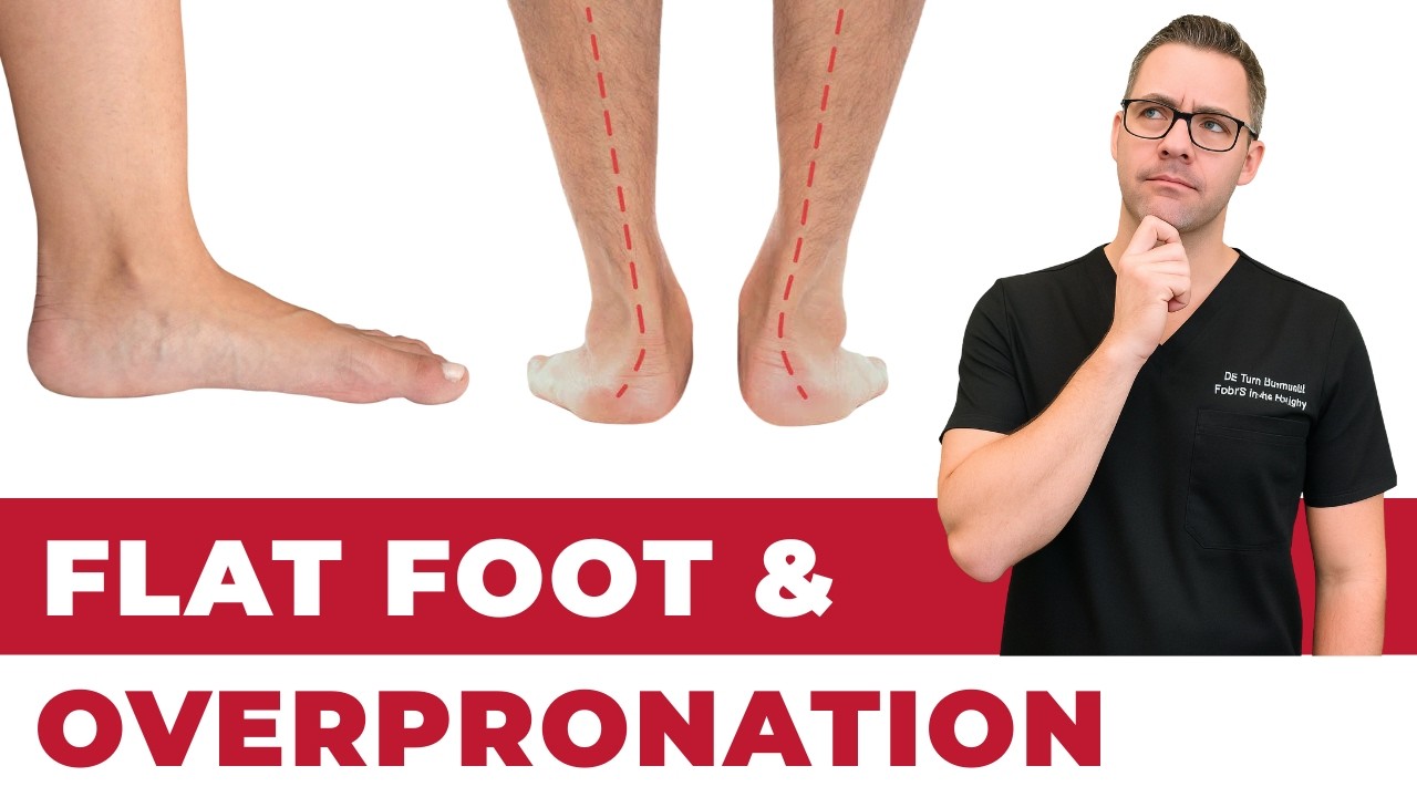
When the posterior tibial tendon (PTT) weakens from degeneration, tear, or rupture, the arch loses its dynamic support and progressively collapses under body weight. The heel tilts outward (valgus), the midfoot sags, and the forefoot abducts—a deformity pattern called adult acquired flatfoot deformity (AAFD) that worsens without treatment.
PTT dysfunction is the most common cause of acquired flatfoot in adults, affecting an estimated 3-10% of the population over age 40. A 2024 Journal of Bone and Joint Surgery study confirmed that untreated PTT dysfunction progresses through predictable stages, emphasizing why we early diagnosis before irreversible joint changes develop.
Stages of Posterior Tibial Tendon Dysfunction
Stage I involves tendon inflammation (tenosynovitis) without deformity. Patients have medial ankle pain and swelling along the tendon course but maintain normal foot alignment and can perform a single-leg heel raise. This stage is fully reversible with appropriate treatment.
Stage II is defined by flexible flatfoot deformity—the arch collapses during weight-bearing but can be manually corrected. Patients cannot perform a single-leg heel raise on the affected side, and the too-many-toes sign (visible toes lateral to the heel when viewed from behind) becomes positive. This is the most common stage at presentation and the critical window for intervention.
Stage III involves rigid flatfoot deformity that cannot be manually corrected due to joint contracture and arthritic changes in the subtalar and talonavicular joints. Stage IV adds ankle joint involvement with valgus tilting of the talus within the ankle mortise. Stages III-IV require surgical fusion for correction because the joints are no longer flexible enough for realignment osteotomies.
Recognizing Symptoms at Each Stage
Early symptoms include pain and swelling along the inside of the ankle and arch, particularly after prolonged standing or walking. The pain worsens during push-off activities like stair climbing, running, and hiking. Morning stiffness and tenderness directly behind and below the medial malleolus are characteristic. These early symptoms are often dismissed as overuse or mild tendinitis.
As the tendon weakens and the arch begins to collapse, patients notice their foot shape changing—the arch flattening, the ankle rolling inward, and the forefoot pointing outward. Shoes wear unevenly on the medial side. Walking on uneven terrain becomes difficult, and activities that previously caused no problems now produce significant pain and fatigue.
Late-stage symptoms include constant aching in the ankle and sinus tarsi (the space on the outer ankle that becomes impinged as the heel tilts outward), stiffness in the midfoot joints, difficulty fitting into shoes, and progressive limitation of walking distance. Some patients develop lateral ankle pain from subfibular impingement as the tilted calcaneus compresses against the fibula.
Conservative Treatment for Early-Stage PTT Dysfunction
Stage I treatment focuses on reducing tendon inflammation and preventing progression. Immobilization in a CAM boot for 4-6 weeks allows the inflamed tendon to rest and heal. After the acute phase, custom orthotics with medial arch support and rearfoot posting control pronation and reduce tendon strain during daily activities.
Physical therapy targets posterior tibial tendon strengthening through progressive resistance exercises: seated heel raises progressing to standing heel raises, resisted inversion with resistance bands, toe curls, and single-leg balance activities. Eccentric exercises (lowering slowly from a heel raise) stimulate tendon remodeling and improve load tolerance.
Stage II management adds more aggressive bracing with an ankle-foot orthosis (AFO) or Arizona brace when custom orthotics alone don’t adequately control the deformity. These devices provide external arch support and hindfoot alignment control that compensates for the weakened tendon. Weight management is emphasized because each additional pound increases tendon loading proportionally.
Surgical Options When Conservative Treatment Fails
Stage II surgical reconstruction typically combines flexor digitorum longus (FDL) tendon transfer to replace the failed PTT, medial displacement calcaneal osteotomy to realign the heel, and spring ligament repair to restore medial arch stability. Additional procedures like Cotton osteotomy or lateral column lengthening address residual forefoot deformity as needed.
Stage III-IV reconstruction requires hindfoot fusion (triple or double arthrodesis) because the joints are rigid and arthritic. Fusion corrects the deformity by permanently positioning the joints in corrected alignment. Stage IV adds deltoid ligament reconstruction or ankle fusion depending on the degree of ankle joint damage.
Dr. Biernacki tailors the surgical plan to each patient’s specific deformity pattern using weight-bearing radiographs and CT imaging. The goal is to address all components of the deformity in a single surgical session to avoid the need for revision surgery. Modern fixation techniques and rehabilitation protocols have significantly improved outcomes for all stages of PTT reconstruction.
Prevention and Long-Term Management
Preventing PTT dysfunction progression requires maintaining strong posterior tibial muscle function through regular exercises, wearing supportive footwear with adequate arch support, maintaining a healthy body weight, and addressing biomechanical risk factors with custom orthotics. Individuals with naturally flat feet or excessive pronation benefit from prophylactic orthotic support.
After surgical reconstruction, lifelong commitment to custom orthotic use, regular exercise, and weight management protects the surgical correction and adjacent joints. Annual follow-up evaluations monitor for any recurrence of deformity or development of adjacent joint problems.
Early recognition of PTT dysfunction symptoms—medial ankle pain, arch collapse, inability to heel raise—and prompt evaluation prevents the condition from progressing to stages that require complex reconstruction. Most patients who are diagnosed in Stage I-early Stage II can be managed successfully with conservative treatment alone.
Warning Signs Requiring Urgent Evaluation
- function bold() { [native code] } — undefined
- function bold() { [native code] } — undefined
- function bold() { [native code] } — undefined
- function bold() { [native code] } — undefined
Frequently Asked Questions
How long does treatment take to work?
Most patients see improvement in 4-8 weeks with consistent conservative care. Persistent symptoms after 8 weeks need imaging and escalation.
When is surgery needed?
Surgery is reserved for cases that fail 3-6 months of conservative care, structural deformities, or fractures requiring stabilization.
Is this covered by insurance?
Most diagnostic visits and conservative treatments are covered by Medicare and major insurers. Custom orthotics often require diabetic or post-surgical justification.
What is Foot pain?
Foot pain is a common foot/ankle condition that affects mobility and quality of life. Understanding the underlying cause is the first step in successful treatment. Our podiatrists at Balance Foot & Ankle perform a hands-on biomechanical exam, review your activity history, and use diagnostic imaging when appropriate to identify the root cause—not just treat the symptom. Many patients have been told to “rest and ice” without a deeper diagnostic workup; our approach is different.
Symptoms and warning signs
Common signs of foot pain include pain that worsens with activity, morning stiffness, swelling, tenderness when palpated, and difficulty bearing weight. If you experience sudden severe pain, inability to walk, visible deformity, numbness or color change, contact our office the same day or visit urgent care—these can signal a more serious injury such as a fracture, tendon rupture, or vascular compromise. Diabetics with any foot wound should seek same-day care.
Conservative treatment options
Most cases of foot pain respond to non-surgical care: structured rest, supportive footwear changes, custom orthotics, targeted stretching and strengthening protocols, anti-inflammatory medications when medically appropriate, and in-office procedures such as ultrasound-guided injections. We also offer advanced therapies including MLS laser therapy, EPAT/shockwave, regenerative injections, and image-guided procedures. Treatment is sequenced from least invasive to most invasive, and we explain the rationale at every step.
When is surgery considered?
Surgery is reserved for cases that fail 3-6 months of well-structured conservative care, when there is structural pathology (severe deformity, complete tear, advanced arthritis), or when imaging shows damage that will not heal without intervention. Our surgeons have performed 3,000+ foot and ankle procedures and prioritize minimally-invasive techniques whenever appropriate. We discuss recovery timelines, return-to-activity milestones, and realistic outcome expectations before any procedure is scheduled.
Recovery timeline and prevention
Recovery from foot pain varies based on severity and chosen treatment path. Conservative cases often improve within 4-8 weeks with consistent adherence to the protocol. Post-procedural recovery may range from a few days (in-office procedures) to several months (reconstructive surgery). Long-term prevention involves footwear assessment, activity modification, structured strengthening, and regular check-ins with your podiatrist if you have a history of recurrence. We provide written home-exercise plans and digital follow-up support.
Dr. Tom’s PTT Tear Recovery Protocol
- PowerStep Maxx — Posterior tibial tendon tear with arch collapse: PowerStep Maxx is the highest OTC medial column support available — primary insole for Stage 1-2 PTTD.
- Doctor Hoy’s Natural Pain Relief Gel — PTT tendon pain and peritendinous inflammation: arnica + camphor gel applied to the medial ankle and arch 3-4x daily during conservative management.
- DASS Medical Compression Socks — PTT tear with ankle edema: graduated compression reduces peritendinous swelling that worsens with prolonged standing during Stage 1-2 recovery.
Arch visibly collapsing or PTT pain not improving after 6 weeks of insoles + bracing? Stage 3-4 PTT dysfunction requires surgical evaluation. Balance Foot & Ankle → (810) 206-1402
Ready to feel better?
Same-week appointments available in Howell and Bloomfield Hills, Michigan.
Book Your VisitIn-Office Treatment at Balance Foot & Ankle
If home treatment isn’t providing relief for your Achilles tendon conditions, our podiatry team at Balance Foot & Ankle can help with same-day evaluations and advanced in-office care.
Same-day appointments available. (810) 206-1402
Get Expert Care at Balance Foot & Ankle
Same-week appointments at our Howell and Bloomfield Hills offices. Board-certified podiatric surgeons. Most insurance accepted.
Same-Week Appointments in Howell & Bloomfield Hills
Three board-certified podiatric surgeons. 1,123+ five-star reviews. Most insurance accepted.
Dr. Tom Biernacki, DPM is a board-certified foot & ankle surgeon (ABFAS & ABPM) at Balance Foot & Ankle Specialists in Southeast Michigan. With over a decade of clinical experience, he specializes in heel pain, bunions, diabetic foot care, sports injuries, and minimally invasive surgery. Dr. Biernacki is a member of the APMA and ACFAS, and his patient education content on MichiganFootDoctors.com and YouTube has made him one of the most-followed foot & ankle educators on YouTube.