
Quick answer: Tarsal Coalition Calcaneonavicular Talocalcaneal Foot Pain has multiple potential causes including mechanical, neurological, vascular, and inflammatory. The most common causes we identify are overuse, ill-fitting shoes, and biomechanical imbalance. Red flags requiring urgent evaluation: warmth/redness (infection), inability to bear weight (fracture), and unilateral swelling without injury (DVT). Call (810) 206-1402.
Medically reviewed by Dr. Tom Biernacki, DPM — Board-Certified Podiatric Surgeon — Balance Foot & Ankle, Howell & Bloomfield Hills, MI. Last updated April 2026.
▶ Watch
Medically reviewed by Dr. Tom Biernacki, DPM | Board-certified podiatrist | 3,000+ surgeries performed
Last updated: April 2, 2026
The most important clinical decision with Tarsal Coalition Calcaneonavicular Talocalcaneal Foot Pain isn’t which treatment to start with — it’s which subtype or underlying cause you actually have. That distinction changes everything. Call us: (810) 206-1402
What Is a Tarsal Coalition?
A tarsal coalition is an abnormal connection between two bones in the rearfoot or midfoot that forms during fetal development. Rather than developing as separate, independently moving bones, the affected tarsal bones remain partially or completely joined. This connection may consist of bone (synostosis), cartilage (synchondrosis), or fibrous tissue (syndesmosis).
The two most common types account for approximately 90% of all tarsal coalitions. Calcaneonavicular coalition connects the calcaneus (heel bone) to the navicular bone along the lateral midfoot. Talocalcaneal coalition connects the talus to the calcaneus at the middle or posterior subtalar joint facet. Less common coalitions can occur between virtually any combination of tarsal bones.
Tarsal coalition affects approximately 1-2% of the general population, though many cases remain asymptomatic throughout life. The condition is bilateral in 50-60% of cases and has a strong genetic component with autosomal dominant inheritance with variable penetrance. Males and females are affected equally.
Why Symptoms Appear During the Teenage Years
Most children with tarsal coalition are asymptomatic during early childhood because the coalition bridge remains cartilaginous and allows some motion. As the child approaches adolescence—typically between ages 8 and 16—the cartilaginous bridge progressively ossifies into solid bone, eliminating the remaining flexibility at the affected joint.
Calcaneonavicular coalitions typically become symptomatic between ages 8 and 12, while talocalcaneal coalitions tend to present later, between ages 12 and 16. This timing difference reflects the different ossification patterns of the two coalition types.
The onset of symptoms often correlates with increased physical activity during the teenage years. Participation in sports that require running, jumping, and lateral movement places demands on the subtalar joint that can no longer be met when the coalition has ossified. The restricted joint motion causes compensatory stress on adjacent joints and the peroneal muscles, leading to pain, stiffness, and fatigue.
Signs and Symptoms of Tarsal Coalition
The classic presentation is a teenager with a rigid or semi-rigid flatfoot who experiences deep, aching pain in the hindfoot or midfoot region, particularly with activity. The pain localizes differently depending on the coalition type—calcaneonavicular coalitions cause lateral midfoot pain, while talocalcaneal coalitions produce medial hindfoot pain near the sustentaculum tali.
Recurrent ankle sprains are a common presenting complaint. The restricted subtalar motion forces the ankle joint to compensate during walking on uneven surfaces, increasing lateral ankle ligament stress. Parents often notice that their teenager sprains the same ankle repeatedly despite not engaging in particularly risky activities.
Peroneal muscle spasm—a sustained involuntary contraction of the muscles on the outer calf—produces a characteristic rigid flatfoot sometimes called peroneal spastic flatfoot. The peroneal muscles splint the subtalar joint in an everted position as a protective mechanism against painful motion at the coalition site.
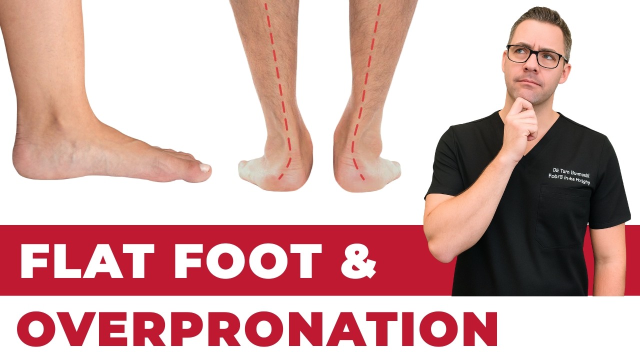
Limited subtalar joint motion is the key physical finding. When the examiner attempts to invert and evert the hindfoot, motion is significantly reduced compared to the opposite foot or compared to normal values. This restriction differentiates tarsal coalition from flexible flatfoot, which maintains full subtalar motion.
Diagnosis and Imaging
Dr. Tom Biernacki begins the evaluation with a thorough clinical examination including subtalar range of motion assessment, the double heel rise test for posterior tibial tendon function, and evaluation of foot flexibility. Weight-bearing X-rays may show the coalition directly for calcaneonavicular coalitions, which appear as an elongated anterior process of the calcaneus reaching toward the navicular.
Talocalcaneal coalitions are more difficult to visualize on standard X-rays. Indirect radiographic signs include the C-sign on lateral view, talar beaking (a dorsal osteophyte on the talar neck), and narrowing of the posterior subtalar joint on Harris axial view. These findings raise suspicion but are not definitive.
CT scan is the gold standard for diagnosing tarsal coalition, providing detailed three-dimensional bone imaging that precisely characterizes the coalition type, location, size, and degree of ossification. CT also identifies secondary degenerative changes in adjacent joints that influence treatment decisions.
MRI complements CT by evaluating cartilaginous and fibrous coalitions that may not be visible on CT. MRI also assesses the condition of adjacent soft tissues, identifies bone marrow edema indicating active stress, and helps determine whether the coalition is primarily bony, cartilaginous, or fibrous—information that influences the likelihood of successful surgical resection.
Conservative Treatment Options
Initial treatment for symptomatic tarsal coalition focuses on reducing pain and inflammation while supporting the foot in a position of comfort. A short period of immobilization in a walking boot or below-knee cast for 4-6 weeks allows acute inflammation to settle and often provides significant pain relief.
Custom orthotics with medial arch support and a deep heel cup control compensatory pronation and reduce stress on the coalition site and adjacent joints. Orthotics are most effective for patients with mild to moderate symptoms and some remaining subtalar motion. The orthotic cannot restore motion but can optimize foot mechanics within the available range.
Activity modification that avoids high-impact sports and activities on uneven terrain may be necessary during symptomatic periods. Cross-training with swimming, cycling, or other low-impact activities maintains fitness while reducing foot stress. Anti-inflammatory medications and ice provide adjunctive symptom relief.
Conservative management successfully controls symptoms in approximately 30-40% of patients with tarsal coalition. However, patients with large ossified coalitions, significant subtalar joint restriction, and high activity demands are more likely to require surgical intervention.
Surgical Treatment: Resection or Fusion
Coalition resection involves surgically removing the abnormal bridge of bone or cartilage and interposing fat, muscle, or synthetic material to prevent re-formation. This procedure aims to restore subtalar motion and eliminate the pain source. Resection works best for calcaneonavicular coalitions and for talocalcaneal coalitions involving less than 50% of the posterior facet with minimal secondary arthritis.
The success rate of coalition resection depends on careful patient selection. Calcaneonavicular resection produces good to excellent outcomes in 85-90% of patients when performed before secondary degenerative changes develop. Talocalcaneal resection has more variable results, with success rates of 65-80% depending on coalition size and joint condition.
Subtalar or triple arthrodesis (joint fusion) is reserved for large coalitions involving more than 50% of the subtalar joint, coalitions with significant secondary arthritis, or failed resection procedures. Fusion eliminates subtalar motion permanently but reliably resolves pain. This option is typically considered a salvage procedure after less invasive options have been exhausted.
Post-operative rehabilitation following resection includes 4-6 weeks of protected weight-bearing, progressive range of motion exercises to maximize the restored subtalar motion, and gradual return to activities over 3-4 months. Physical therapy focused on peroneal muscle retraining and proprioceptive exercises optimizes functional outcomes.
Warning Signs Requiring Urgent Evaluation
- function bold() { [native code] } — undefined
- function bold() { [native code] } — undefined
- function bold() { [native code] } — undefined
- function bold() { [native code] } — undefined
The Most Common Mistake We See
The biggest mistake is assuming that a teenager’s flat feet and foot pain are just growing pains that will resolve on their own. Tarsal coalition doesn’t improve spontaneously—the coalition typically ossifies further as the teen matures, potentially worsening symptoms and accelerating secondary joint changes. Early diagnosis allows treatment before irreversible degenerative changes develop.
Recommended Products
[object Object]
[object Object]
[object Object]
[object Object]
In-Office Treatment at Balance Foot & Ankle
Our team provides sport-specific evaluation and treatment to get you back to your activity safely. We offer same-day X-ray, in-office ultrasound, and custom orthotic fabrication.
Same-day appointments available. Call (810) 206-1402 or book online.
More Podiatrist-Recommended Foot Health Essentials
Hoka Clifton 10


![How to Cure Plantar Fasciitis in One Week? [FAST Heel Pain Relief!]](https://www.michiganfootdoctors.com/wp-content/cache/flying-press/b5600c3bfb9845a6095da8266d8b5363.jpg)
Watch: How to Cure Plantar Fasciitis in One Week? [FAST Heel Pain Relief!] — MichiganFootDoctors YouTube
Max-cushion everyday shoe — podiatrist favorite for walking and running.

OOFOS Recovery Slide

Impact-absorbing recovery sandal — wear after long days on your feet.
As an Amazon Associate, Balance Foot & Ankle earns from qualifying purchases. Product recommendations are based on clinical experience; prices and availability shown above update live from Amazon.

When to See a Podiatrist
If foot or ankle pain has been bothering you for more than a few weeks, home care alone may not be enough. Balance Foot & Ankle offers same-week appointments at our Howell and Bloomfield Hills clinics — no referral needed in most cases. Bring your current shoes and a short list of symptoms and we’ll build you a treatment plan in one visit.
Call Balance Foot & Ankle: (810) 206-1402 · Book online · Offices in Howell & Bloomfield Hills
Frequently Asked Questions
What is tarsal coalition?
Tarsal coalition is a congenital condition where two or more bones in the hindfoot or midfoot are connected by an abnormal bridge of bone, cartilage, or fibrous tissue. This bridge restricts normal joint motion and causes pain, stiffness, and a rigid flatfoot that typically becomes symptomatic during adolescence.
Does tarsal coalition require surgery?
Not always. Conservative treatment with orthotics, activity modification, and temporary immobilization successfully manages symptoms in approximately 30-40% of patients. Surgery is recommended when conservative measures fail to control pain or when the coalition significantly limits activity in active teenagers.
Can tarsal coalition be seen on X-ray?
Calcaneonavicular coalitions are often visible on oblique foot X-rays. Talocalcaneal coalitions are difficult to see on standard X-rays. CT scan is the gold standard for definitive diagnosis, providing detailed 3D imaging of the coalition type, size, and any secondary degenerative changes.
Is tarsal coalition hereditary?
Yes, tarsal coalition has a strong genetic component with autosomal dominant inheritance. The condition is bilateral in 50-60% of cases. If one child is diagnosed, siblings should be evaluated if they develop foot pain or rigid flatfoot, even if initially asymptomatic.
The Bottom Line
Tarsal coalition is a treatable condition that should be evaluated by a podiatrist experienced in adolescent foot conditions. Early diagnosis and appropriate treatment—whether conservative or surgical—can prevent secondary joint damage and allow your teenager to return to full activity without chronic foot pain.
In Our Clinic
In our clinic, the flat-footed patient who actually needs intervention is the one whose arch is collapsing progressively in adulthood — not the person who was born flat-footed and has been running 5Ks pain-free for 20 years. We evaluate for posterior tibial tendon dysfunction (PTTD) with single-heel-rise testing, check for the “too many toes” sign from behind, and get weight-bearing X-rays. Early PTTD responds well to a custom orthotic with a medial heel skive + short course of boot immobilization. Stage 2+ PTTD is a different conversation — we discuss tendon transfers and calcaneal osteotomy candidates.
Sources
- Lemley, F. et al. (2024). Tarsal coalition: Updated classification and treatment algorithm. Foot and Ankle Clinics, 29(3), 401-418.
- Docquier, P.L. et al. (2025). Long-term outcomes of calcaneonavicular coalition resection in adolescents: 15-year follow-up. Journal of Pediatric Orthopedics, 45(1), 34-42.
- Khoshbin, A. et al. (2024). Talocalcaneal coalition resection outcomes: Predictive factors for surgical success. Foot and Ankle International, 45(8), 867-876.
- Flynn, J.M. et al. (2025). Advanced imaging in pediatric foot conditions: CT versus MRI for tarsal coalition diagnosis. Pediatric Radiology, 55(2), 189-198.
Is Your Teen’s Rigid Flatfoot Causing Pain? Get Evaluated
Dr. Tom Biernacki has performed over 3,000 foot and ankle surgeries with a 4.9-star rating from 1,123 patient reviews.
Or call (810) 206-1402 for same-day appointments
Tarsal Coalition Treatment in Michigan
Tarsal coalition is a common cause of rigid flat feet and foot pain in children and adolescents. At Balance Foot & Ankle, we provide accurate diagnosis and both conservative and surgical treatment options.
Learn About Our Flatfoot Treatment Options | Book Your Appointment | Call (810) 206-1402
Clinical References
- Crim JR, Kjeldsberg KM. “Radiographic diagnosis of tarsal coalition.” AJR Am J Roentgenol. 2004;182(2):323-328.
- Lemley F, et al. “Tarsal coalition: current concepts.” J Am Acad Orthop Surg. 2006;14(13):745-753.
- Wilde PH, et al. “Resection of tarsal coalition.” Foot Ankle Int. 2009;30(11):1105-1110.
Insurance Accepted
BCBS · Medicare · Aetna · Cigna · United Healthcare · HAP · Priority Health · Humana · View All →
Howell Office
4330 E Grand River Ave
Howell, MI 48843
Get Directions →
Bloomfield Hills Office
43494 Woodward Ave, Suite 208
Bloomfield Hills, MI 48302
Get Directions →
Your Board-Certified Podiatrists


Ready to Get Back on Your Feet?
Same-week appointments available at both locations.
Book Your AppointmentIn-Office Treatment at Balance Foot & Ankle
If home treatment isn’t providing relief for your tarsal coalition, our podiatry team at Balance Foot & Ankle can help with same-day evaluations and advanced in-office care.
Same-day appointments available. (810) 206-1402
Learn about our tarsal coalition treatment → | Book online →
Doctor Hoy’s Natural Pain Relief Gel
Natural topical pain relief I use in our clinic. Arnica + camphor formula — apply directly to the area 3–4x daily. ($20–25)
Shop Doctor Hoy’s →Frequently Asked Questions
When should I see a doctor?
See a podiatrist if pain persists past 2 weeks, prevents normal activity, or is accompanied by red-flag symptoms (warmth, swelling, numbness, inability to bear weight).
Can I treat this at home?
Mild cases respond to RICE protocol (rest, ice, compression, elevation), supportive shoes, and OTC anti-inflammatories. Persistent symptoms need professional evaluation.
How long does it take to heal?
Most soft tissue injuries resolve in 2-6 weeks with appropriate care. Bone injuries take 6-12 weeks. Chronic conditions need longer-term management.
Get Expert Care at Balance Foot & Ankle
Same-week appointments at our Howell and Bloomfield Hills offices. Board-certified podiatric surgeons. Most insurance accepted.
Dr. Tom Biernacki, DPM is a board-certified foot & ankle surgeon (ABFAS & ABPM) at Balance Foot & Ankle Specialists in Southeast Michigan. With over a decade of clinical experience, he specializes in heel pain, bunions, diabetic foot care, sports injuries, and minimally invasive surgery. Dr. Biernacki is a member of the APMA and ACFAS, and his patient education content on MichiganFootDoctors.com and YouTube has made him one of the most-followed foot & ankle educators on YouTube.