
You are in the right place. Dr. Tom Biernacki, DPM, FACFAS — board-certified foot & ankle surgeon with 3,000+ surgeries — explains exactly what calcaneal osteotomy types evans dwyer koutsogiannis means and what works. Call (810) 206-1402 for same-day appointment at Howell or Bloomfield Hills.
Medically reviewed by Dr. Tom Biernacki, DPM · Board-Certified Podiatric Surgeon · Last reviewed: April 2026 · Editorial Policy
The most important clinical decision with Calcaneal Osteotomy Types Evans Dwyer Koutsogiannis isn’t which treatment to start with — it’s identifying the correct subtype. That changes everything. Call (810) 206-1402.
Quick Answer
Calcaneal Osteotomy Types 2026: Evans, Dwyer & Koutsogi relates to foot pain — typically caused by overuse, footwear, or biomechanics. Most patients improve in 6-12 weeks with conservative care. Same-week appointments in Howell + Bloomfield Hills: (810) 206-1402.

Medically reviewed by Dr. Tom Biernacki, DPM — Board-certified foot & ankle surgeon, 3,000+ surgeries performed. Updated April 2026 with current clinical evidence. This article reflects real practice experience from Balance Foot & Ankle Specialists in Howell and Bloomfield Hills, Michigan.
Quick Answer
Most foot and ankle problems respond to conservative care — proper footwear, supportive inserts, activity modification, and targeted stretching — within 4-8 weeks. Persistent pain beyond that window, or any symptom that prevents walking, warrants a podiatric evaluation to rule out fracture, tendon tear, or systemic cause.
Watch: Dr. Tom Biernacki, DPM

Medically reviewed by Dr. Tom Biernacki, DPM — Board-Certified Podiatric Surgeon — Balance Foot & Ankle, Howell & Bloomfield Hills, MI. Last updated April 2026.
▶ Watch

Medically Reviewed by Dr. Tom Biernacki, DPM — Board-Certified Podiatrist, Balance Foot & Ankle Specialists, Michigan. Last updated April 2026.
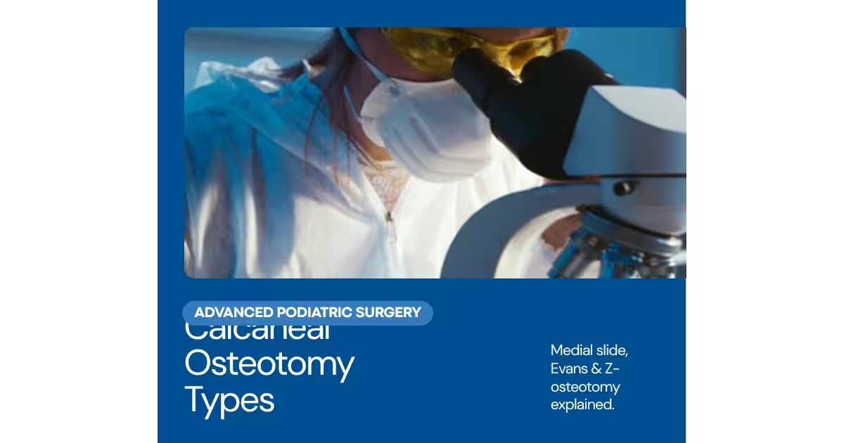
Calcaneal osteotomies are among the most powerful tools in reconstructive foot surgery — they allow the surgeon to fundamentally alter hindfoot alignment by repositioning the calcaneal body relative to the talus and the rest of the foot. The four most commonly performed calcaneal osteotomies — Evans, Dwyer, Koutsogiannis (medial displacement calcaneal osteotomy, MDCO), and the Z-cut (also called the combined osteotomy) — each address different deformity planes and are selected based on the specific biomechanical problem being corrected.
Evans Osteotomy (Lateral Column Lengthening)
The Evans osteotomy is a lateral column lengthening procedure performed through an oblique cut in the anterior calcaneus, typically 1.5 cm posterior to the calcaneocuboid joint. A structural bone graft (typically tricortical iliac crest or allograft) is impacted into the osteotomy site to lengthen the lateral column. The biomechanical effect is to reduce forefoot abduction and restore the talar head coverage that is lost in flexible flatfoot deformity — essentially by pushing the forefoot laterally relative to the hindfoot, recreating the lateral arch and reducing the valgus hindfoot moment. The Evans osteotomy is the primary procedure for correcting significant forefoot abduction in symptomatic flexible flatfoot reconstruction. Potential complications include calcaneocuboid joint impingement if placed too far anterior, and lateral column overload producing lateral midfoot pain if overcorrected.
Dwyer Osteotomy (Lateral Closing Wedge)
The Dwyer osteotomy is a lateral closing wedge calcaneal osteotomy that removes a laterally-based wedge from the calcaneal body, allowing the posterior fragment to shift medially and correct varus hindfoot alignment. It is the primary procedure for correcting hindfoot varus in cavovarus foot reconstruction — often combined with plantar fascia release and peroneal tendon procedures for comprehensive cavovarus correction. The degree of wedge removal directly determines the degree of varus correction; careful pre-operative planning with weight-bearing hindfoot alignment radiographs guides wedge sizing. The Dwyer osteotomy is the opposite of the MDCO — it corrects varus rather than valgus.
Koutsogiannis / Medial Displacement Calcaneal Osteotomy (MDCO)
The MDCO — also called the Koutsogiannis or Gleich osteotomy — is a transverse oblique cut through the calcaneal body that allows the entire posterior tuberosity to slide medially, typically 8–12mm. This medial shift reduces the valgus moment on the hindfoot, decreasing subtalar joint stress and offloading the failing posterior tibial tendon in Stage II PTTD (posterior tibial tendon dysfunction / adult acquired flatfoot deformity). The MDCO is the most commonly performed hindfoot osteotomy in adult flatfoot reconstruction — it is reliable, technically reproducible, and provides consistent reduction of hindfoot valgus when combined with soft tissue procedures (FDL tendon transfer, spring ligament repair).
Z-Cut / Combined Osteotomy
The Z-cut osteotomy combines elements of lateral column lengthening and medial displacement into a single calcaneal cut shaped like a Z or step-cut configuration. This allows simultaneous correction of forefoot abduction (through lateral column lengthening) and hindfoot valgus (through medial displacement of the tuberosity) without requiring two separate osteotomy sites. The Z-cut is particularly valuable in moderate-to-severe flexible flatfoot with both significant forefoot abduction and hindfoot valgus requiring correction in both planes. Dr. Biernacki at Balance Foot & Ankle performs comprehensive hindfoot reconstruction including calcaneal osteotomy selection and surgical planning. Call (810) 206-1402 for a surgical consultation at our Bloomfield Hills or Howell office.
📧 Get Dr. Tom’s Free Lab Test Guide
Discover the 5 lab tests every person over 35 should ask their doctor about — explained in plain English by a board-certified physician.
📍 Located in Michigan?
Our board-certified podiatrists treat this condition at two convenient locations. Same-day appointments often available.
Insurance Accepted
BCBS · Medicare · Aetna · Cigna · United Healthcare · HAP · Priority Health · Humana · View All →
Howell Office
4330 E Grand River Ave
Howell, MI 48843
Get Directions →
Bloomfield Hills Office
43494 Woodward Ave, #208
Bloomfield Hills, MI 48302
Get Directions →
Your Board-Certified Podiatrists


Ready to Get Back on Your Feet?
Same-week appointments available at both locations.
Book Your AppointmentIn-Office Treatment at Balance Foot & Ankle
If home care isn’t resolving your your foot or ankle concern, a visit with a board-certified podiatrist is the fastest path to accurate diagnosis and a personalized plan. At Balance Foot & Ankle Specialists, Dr. Tom Biernacki, Dr. Carl Jay, and Dr. Daria Gutkin offer same-day and next-day appointments at both our Howell and Bloomfield Hills offices. We perform on-site diagnostic ultrasound, digital X-ray, conservative care, advanced regenerative treatments, and minimally invasive surgery when indicated.
Call (810) 206-1402 or request an appointment online. Most insurance plans accepted, including Medicare, Blue Cross Blue Shield, Aetna, Cigna, and United Healthcare.
Most Common Mistake We See
The most common mistake we see is: Waiting too long before seeking care. Fix: any foot pain lasting more than 4 weeks, or any sudden severe symptom, deserves a professional evaluation rather than more rest.
Warning Signs That Need Same-Day Care
Seek immediate evaluation at Balance Foot & Ankle if you experience any of the following:
- Unable to bear weight
- Severe swelling with skin colour change
- Fever with foot pain (possible infection)
- Diabetes plus any new foot symptom
Call (810) 206-1402 — same-day and next-day appointments at our Howell and Bloomfield Hills offices.
More Podiatrist-Recommended Surgery Essentials
OOFOS Recovery Slide


Post-op approved — impact-absorbing slide for early recovery.
HOKA Ora 3 Recovery Slide
Max-cushion recovery sandal — comfort for post-surgical swelling.
Hoka Bondi 9
Max-cushion walking shoe — ease into return-to-walking post-surgery.
As an Amazon Associate, Balance Foot & Ankle earns from qualifying purchases. Product recommendations are based on clinical experience; prices and availability shown above update live from Amazon.

When to See a Podiatrist
Foot and ankle surgery in 2026 is dramatically different than a decade ago — most procedures are now minimally-invasive, outpatient, and allow weight-bearing within days. Balance Foot & Ankle surgeons have performed 3,000+ foot/ankle surgeries with modern techniques. If another surgeon has recommended a traditional open procedure, a second opinion may reveal a faster, less-invasive option.
Call Balance Foot & Ankle: (810) 206-1402 · Book online · Offices in Howell & Bloomfield Hills
Watch: Dr. Tom explains
Podiatrist-recommended products
As an Amazon Associate, Dr. Tom earns from qualifying purchases.
Controlled healing.
View on Amazon →Arch support post-osteotomy.
View on Amazon →Post-op swelling.
View on Amazon →Post-osteotomy rehab.
View on Amazon →Related resources
Ready to solve this? Book today.
Same-week appointments · Howell & Bloomfield Hills · 4.9★ (1,123+ reviews)
☎ (810) 206-1402Book Online →Pros & Cons of Conservative Care for foot care
Advantages
- ✓ Conservative care first
- ✓ Same-week appointments
- ✓ Multiple insurance accepted
Considerations
- ✗ Self-treatment can mask issues
- ✗ See a podiatrist if pain >2 weeks
Dr. Tom’s Recommended Products for foot care
Affiliate disclosure: As an Amazon Associate, Balance Foot & Ankle earns from qualifying purchases. We only recommend products we use with patients.
Footnanny Heel Cream Dr. Tom’s Pick
Best for: Daily moisturizer for cracked heels
Ready to Get Back on Your Feet?
Same-day appointments in Howell + Bloomfield Hills. Most insurance accepted. Dr. Tom Biernacki, DPM & team.
Book Today — Same-Day Appointments Available
Call Now: (810) 206-1402
About Your Care Team at Balance Foot & Ankle
Dr. Tom Biernacki, DPM · Board-Certified Foot & Ankle Surgeon. Specializes in conservative-first care, minimally invasive bunion surgery, and complex reconstruction.
Dr. Carl Jay, DPM · Accepting new patients. Specializes in sports medicine, athletic injuries, and routine podiatric care.
Dr. Daria Gutkin, DPM, AACFAS · Accepting new patients. Specializes in surgical reconstruction and pediatric podiatry.
Locations: 4330 E Grand River Ave, Howell, MI 48843 · 43494 Woodward Ave Suite 208, Bloomfield Hills, MI 48302
Hours: Mon–Fri 8:00 AM – 5:00 PM · (810) 206-1402
Doctor Hoy’s Natural Pain Relief Gel
Natural topical pain relief I use in our clinic. Arnica + camphor formula — apply directly to the area 3–4x daily. ($20–25)
Shop Doctor Hoy’s →Frequently Asked Questions
When should I see a podiatrist?
If symptoms persist past 2 weeks, affect your normal activity, or are accompanied by red-flag symptoms (warmth, redness, swelling, inability to bear weight).
What does treatment cost?
Most diagnostic visits and conservative treatments are covered by Medicare and major insurers. Out-of-pocket costs vary by your specific plan.
How quickly can I get an appointment?
Most non-urgent cases see us within 5 business days. Urgent cases (sudden pain, possible fracture) typically same or next business day.
What is Foot pain?
Foot pain is a common foot/ankle condition that affects mobility and quality of life. Understanding the underlying cause is the first step in successful treatment. Our podiatrists at Balance Foot & Ankle perform a hands-on biomechanical exam, review your activity history, and use diagnostic imaging when appropriate to identify the root cause—not just treat the symptom. Many patients have been told to “rest and ice” without a deeper diagnostic workup; our approach is different.
Symptoms and warning signs
Common signs of foot pain include pain that worsens with activity, morning stiffness, swelling, tenderness when palpated, and difficulty bearing weight. If you experience sudden severe pain, inability to walk, visible deformity, numbness or color change, contact our office the same day or visit urgent care—these can signal a more serious injury such as a fracture, tendon rupture, or vascular compromise. Diabetics with any foot wound should seek same-day care.
Conservative treatment options
Most cases of foot pain respond to non-surgical care: structured rest, supportive footwear changes, custom orthotics, targeted stretching and strengthening protocols, anti-inflammatory medications when medically appropriate, and in-office procedures such as ultrasound-guided injections. We also offer advanced therapies including MLS laser therapy, EPAT/shockwave, regenerative injections, and image-guided procedures. Treatment is sequenced from least invasive to most invasive, and we explain the rationale at every step.
When is surgery considered?
Surgery is reserved for cases that fail 3-6 months of well-structured conservative care, when there is structural pathology (severe deformity, complete tear, advanced arthritis), or when imaging shows damage that will not heal without intervention. Our surgeons have performed 3,000+ foot and ankle procedures and prioritize minimally-invasive techniques whenever appropriate. We discuss recovery timelines, return-to-activity milestones, and realistic outcome expectations before any procedure is scheduled.
Recovery timeline and prevention
Recovery from foot pain varies based on severity and chosen treatment path. Conservative cases often improve within 4-8 weeks with consistent adherence to the protocol. Post-procedural recovery may range from a few days (in-office procedures) to several months (reconstructive surgery). Long-term prevention involves footwear assessment, activity modification, structured strengthening, and regular check-ins with your podiatrist if you have a history of recurrence. We provide written home-exercise plans and digital follow-up support.
Ready to feel better?
Same-week appointments available in Howell and Bloomfield Hills, Michigan.
Book Your VisitReady to fix this for good?
Reading goes so far. The fastest path is a 30-minute office visit. Same-day Howell or Bloomfield Hills. Call (810) 206-1402.
Ready for Expert Care?
Same-day appointments in Howell & Bloomfield Hills, MI.
4.9★ | 1,123 Reviews | 3,000+ Surgeries
Or call: (810) 206-1402
Dr. Tom Biernacki, DPM is a board-certified foot & ankle surgeon (ABFAS & ABPM) at Balance Foot & Ankle Specialists in Southeast Michigan. With over a decade of clinical experience, he specializes in heel pain, bunions, diabetic foot care, sports injuries, and minimally invasive surgery. Dr. Biernacki is a member of the APMA and ACFAS, and his patient education content on MichiganFootDoctors.com and YouTube has made him one of the most-followed foot & ankle educators on YouTube.