

Medically reviewed by Dr. Tom Biernacki, DPM
Board-certified podiatric surgeon | Balance Foot & Ankle
Last reviewed: May 2026
Swelling inside Kager’s triangle is one of the earliest detectable signs of Achilles pathology — but there’s one specific MRI finding within this space that determines whether conservative treatment will succeed or whether the patient is on a path toward surgery. Call (810) 206-1402 — expert podiatric care across Michigan.
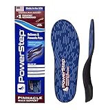
- Slightly Angled Heel Post: PowerStep Pinnacle Maxx insoles for men & women keep the feet from rolling inward, improve stability, and prevent problems caused by overpronation including flat feet and plantar fasciitis.
- Firm But Flexible Design: Our orthotics Pinnacle PowerStep insoles for overpronation are equipped with a deep heel insert cradle for increased comfort, stability, and motion control for standard arch support and immediate heel pain relief.
- Premium Dual Layer Cushioning: For enhanced comfort from heel to toe. As the thickest of our Pinnacle PowerStep orthotics, these need to be worn in shoes where the factory insole can be removed. HSA & FSA Eligible. Made in the USA
- The Perfect Balance of Comfort and Support: These PowerStep Pinnacle arch support inserts for men & women are unlike most other orthotics as they help to correct over-pronation which can cause ankle, knee, and hip pain in your daily routine
- No Trimming: Our arch support PowerStep insoles men & women can use can be worn in a variety of shoe styles & fit in any type with no trimming required! Unlike most insoles for flat feet and other heel pain relief products, these come ready to wear.


- COMFORTABLE SUPPORT: 1/4 inch thick felt metatarsal pads provide gentle cushioning under the ball of the foot for daily wear
- VALUE PACK: Includes 6 pairs (12 pieces) to ensure long-lasting comfort and convenience
- ADHESIVE BACKING: Strong adhesive keeps the pad in place inside shoes or directly on the foot
- MADE IN THE USA: Proudly designed, cut, and manufactured in the United States
- EASY TO CUSTOMIZE: Can be trimmed with scissors for a better fit inside a variety of footwear
As an Amazon Associate, Balance Foot and Ankle earns from qualifying purchases. Product links are affiliate links and we may earn a commission at no extra cost to you. This does not influence our clinical recommendations. Last update on 2026-09-07 / Images from Amazon Product Advertising API
What Is Kager’s Triangle?
Kager’s triangle is an anatomical space at the posterior ankle first described by Hans Kager in 1939. On a lateral radiograph of the ankle, it appears as a radiolucent (dark) triangular region — hence the term “triangle.” On MRI and ultrasound, it is visualized as a bright, fatty signal mass filling this space.
The boundaries of Kager’s triangle are:
- Posterior boundary: The Achilles tendon (anterior surface)
- Anterior boundary: The flexor hallucis longus (FHL) muscle belly
- Inferior boundary: The superior surface of the calcaneus (heel bone)
The fat pad filling this space is known as the pre-Achilles fat pad or Kager’s fat pad. It serves several critical biomechanical functions: cushioning the Achilles tendon during ankle plantarflexion and dorsiflexion, facilitating tendon gliding by reducing friction, and transmitting compressive and tensile forces between the tendon and bone. It has its own neurovascular supply — it is not passive filler tissue.
Key takeaway: Kager’s triangle is a real anatomical structure with a functional role in Achilles tendon biomechanics. Pathology within this fat pad is a distinct clinical entity, not just ‘Achilles pain.’
What Is Kager’s Fat Pad Impingement?
Kager’s fat pad impingement occurs when the fat pad is abnormally compressed, inflamed, or fibrotic, causing posterior heel pain and limited ankle range of motion. In our clinic, we identify this condition in three distinct patterns:
1. Primary Impingement (Mechanical)
Forced dorsiflexion — common in squatting athletes, rock climbers, and dancers — drives the Achilles tendon anteriorly and compresses the fat pad against the superior calcaneus. Repeated compression leads to fat pad edema, micro-hemorrhage, and eventually fibrous metaplasia. The fat pad loses its normal soft compliance and becomes a rigid, painful impingement mass.
2. Secondary Impingement (Adjacent Pathology)
Insertional Achilles tendinopathy, retrocalcaneal bursitis, or a calcaneal exostosis (Haglund’s deformity) can each invade the Kager’s triangle space, compressing the fat pad secondarily. MRI studies show that fat pad signal changes are present in over 60% of patients with insertional Achilles tendinopathy — meaning the fat pad is almost always involved even when it is not the primary diagnosis.
3. Post-Surgical or Post-Traumatic Fibrosis
Achilles tendon repair, calcaneal fracture, or even aggressive physical therapy can cause scar tissue to form within the Kager’s triangle, replacing the normally mobile fat pad with fibrous adhesions. This is a particularly challenging form to treat because the biomechanical scaffold is permanently altered.
Key takeaway: Fat pad impingement can be primary (mechanical compression), secondary (adjacent tendon or bone pathology), or post-traumatic (fibrosis). The treatment approach differs for each.
Symptoms of Kager’s Fat Pad Impingement
The symptoms of Kager’s fat pad impingement overlap with several more commonly diagnosed conditions, which is why it is frequently missed on the first clinical encounter. The hallmark features that distinguish it are:
- Posterior heel pain that is specifically anterior to the Achilles tendon, not within the tendon itself. Patients often point to the space “in front of” the Achilles rather than the tendon body.
- Pain with dorsiflexion (pulling the foot up). This is a key differentiator from mid-portion Achilles tendinopathy, which is typically more painful with plantarflexion loading.
- Restricted ankle dorsiflexion due to fat pad bulk occupying the posterior ankle space. Patients often cannot perform a heel-to-wall test at less than 4–5 cm, even without calf tightness.
- Palpable fullness in the hollow alongside the Achilles. Normally the skin on either side of the Achilles tendon sinks slightly inward. In Kager’s fat pad impingement, this hollow feels full and occasionally tender.
- Pain with deep squatting or positions of maximum ankle dorsiflexion such as kneeling, yoga positions, or hill running.
- Absence of classic plantar fasciitis morning pain — the pain is posterior, not plantar, and does not follow the classic first-step pattern.
How Is Kager’s Fat Pad Impingement Diagnosed?
Kager’s fat pad impingement is a diagnosis that requires imaging — clinical examination alone is insufficient because the fat pad sits too deep for reliable palpation assessment. Here is how we approach diagnosis at Balance Foot & Ankle:
Clinical Examination
I perform a structured posterior ankle examination including: resisted plantarflexion testing (to rule out Achilles rupture or tendon pathology), two-finger squeeze test of the retrocalcaneal space (for bursitis), weight-bearing dorsiflexion range of motion measurement, and direct palpation of the pre-Achilles space with the ankle in dorsiflexion. A positive “Kager’s impingement sign” involves pain provoked when the examiner compresses the fat pad anteriorly while the ankle is brought into maximal dorsiflexion.
Diagnostic Ultrasound
Ultrasound is the first-line imaging tool in our clinic. It allows dynamic assessment — we can visualize how the fat pad moves (or fails to move) during ankle dorsiflexion in real time. Abnormal findings include: increased echogenicity (brighter signal indicating fibrosis), loss of the normal wedge-shaped outline, fat pad protrusion into the retrocalcaneal recess, and tethering to the adjacent Achilles tendon. Doppler ultrasound can detect increased vascularity indicating active inflammation.
MRI
MRI is the definitive imaging study for Kager’s triangle pathology. Normal Kager’s fat pad appears as uniformly bright (high signal) on T1-weighted sequences. Pathological findings include: loss of the normal bright T1 signal replaced by intermediate signal (fibrosis), T2-weighted edema or fluid signal within the fat pad, fat pad herniation into adjacent spaces, and associated Achilles tendon or calcaneal findings that explain secondary involvement. MRI also rules out other posterior heel pathology including stress fractures, which can present similarly.
Differential Diagnosis
Conditions I routinely consider in the differential for posterior heel pain that may involve or mimic Kager’s pathology:
- Retrocalcaneal bursitis: Bursa sits between the Achilles insertion and calcaneus; imaging distinguishes it from fat pad involvement
- Insertional Achilles tendinopathy: Often coexists with fat pad changes; MRI separates the two
- Haglund’s deformity (calcaneal exostosis): Bony prominence that compresses fat pad secondarily; visible on X-ray
- Posterior ankle impingement syndrome: Involves the posterior talar process or os trigonum; different pattern on imaging
- FHL tenosynovitis: Inflammation of the flexor hallucis longus, which forms the anterior boundary of Kager’s triangle
- Calcaneal stress fracture: Must be ruled out; bone scan or MRI is diagnostic
⚠️ When to seek urgent evaluation for posterior heel pain:
- Pain severe enough to prevent weight-bearing
- Sudden pop or snap followed by weakness pushing off (Achilles rupture)
- Swelling, bruising, or warmth spreading beyond the heel region
- Night pain or pain at rest unrelated to recent activity
- History of inflammatory arthritis, gout, or recent steroid injection in the area
- Pain developing after calcaneal fracture or Achilles surgery
Treatment Options for Kager’s Fat Pad Impingement
Treatment is staged based on whether the fat pad changes are primarily inflammatory (reversible) or fibrotic (structural). In our clinic, we approach this in a stepwise progression:
Stage 1: Load Management and Biomechanical Correction (Weeks 1–4)
The first priority is removing the compressive force. This means: activity modification (eliminating deep squats, kneeling, and hill running), heel lift orthotics (a modest 6–8mm heel elevation reduces the amount of dorsiflexion required for normal gait, immediately decompressing the fat pad), and footwear with structured heel counters that limit excessive posterior compression. This is not rest — it is substituting tolerable loading for harmful loading.
Stage 2: Physical Therapy (Weeks 3–8)
Physical therapy for Kager’s fat pad impingement is different from standard Achilles tendinopathy rehab. The emphasis is on posterior ankle mobilization rather than heavy tendon loading. Talocrural joint posterior glides (Maitland grade III–IV mobilizations) restore dorsiflexion range of motion that has been lost due to fat pad bulk and associated soft tissue restriction. FHL stretching addresses the anterior boundary tightness. Eccentric calf work is introduced late, once dorsiflexion has been restored, to prevent recurrence through tendon and muscle balance.
Stage 3: Image-Guided Injection (Weeks 4–12 if conservative fails)
For inflammatory fat pad impingement that has not responded to 4–6 weeks of conservative management, I perform ultrasound-guided corticosteroid injection directly into the Kager’s fat pad under real-time imaging. This is critically different from a blind retrocalcaneal or peri-tendinous injection — the target is the fat pad specifically. A 2019 study in Skeletal Radiology reported significant VAS pain score reduction in 78% of patients following US-guided fat pad injection. We typically use a small volume (1–2 mL) of betamethasone with a local anesthetic.
For fibrotic fat pad cases, ultrasound-guided hydrodissection (injecting sterile saline to mechanically free scar tissue adhesions) has emerging evidence. Platelet-rich plasma (PRP) injection into the fat pad is used in select cases to stimulate tissue remodeling, though evidence specific to fat pad (as opposed to tendon) is still developing.
Stage 4: Surgical Intervention
Surgical treatment of isolated Kager’s fat pad impingement is uncommon but effective when conservative and injection-based approaches fail. The procedure of choice is endoscopic debridement of the Kager’s fat pad via posterior ankle arthroscopy. Using two portals at the posterior ankle, the hypertrophied or fibrotic fat pad tissue is resected under direct visualization until full, pain-free dorsiflexion is restored. Recovery is typically 6–8 weeks to return to full activity. A 2023 systematic review found excellent functional outcomes in over 85% of surgical cases at minimum 2-year follow-up.
When Kager’s impingement is secondary to Haglund’s deformity or insertional Achilles disease, the primary pathology is addressed simultaneously (calcaneal osteotomy, Achilles debridement), and the fat pad is assessed and decompressed as part of the same surgical procedure.
Key takeaway: Most Kager’s fat pad impingement cases resolve with heel lifts, targeted PT, and one image-guided injection. Surgery is effective but rarely needed — most patients never reach Stage 4.
How Long Does Recovery Take?
Recovery timelines for Kager’s fat pad impingement vary based on how long the condition has been present and whether fibrosis has set in:
- Acute inflammatory impingement (<6 weeks): 4–8 weeks with load management, PT, and possible injection. Most patients return to full activity without surgery.
- Subacute (<6 months): 8–16 weeks. Injection is typically needed alongside PT. Activity modification must be consistent.
- Chronic fibrotic impingement (>6 months): 4–6 months of conservative and injection treatment before surgical candidacy is assessed. Surgical cases recover in 6–10 weeks post-operatively.
The most common mistake I see is patients who were told they had “Achilles tendinitis,” treated with aggressive eccentric calf raises, and worsened — because the compressive load of calf raises is exactly what the impinged fat pad cannot tolerate. If your “Achilles tendinitis” worsened with eccentric loading, Kager’s fat pad impingement should be high on the differential.
Posterior Heel Pain That Won’t Resolve?
Kager’s fat pad impingement and Achilles pathology require accurate diagnosis. Same-day appointments available in Howell & Bloomfield Township, MI.
4.9★ | 1,123 Reviews | 3,000+ Surgeries
Or call: (810) 206-1402
Frequently Asked Questions
What is Kager’s triangle on an MRI?
Kager’s triangle on MRI refers to the pre-Achilles fat pad space at the back of the ankle. On T1-weighted sequences, normal fat pad appears uniformly bright (hyperintense). When your radiologist reports “abnormal signal in Kager’s triangle,” it typically means fat pad edema (T2 bright signal), fibrosis (T1 signal loss), or hemorrhage — each indicating different stages of fat pad impingement pathology.
Is Kager’s fat pad impingement the same as retrocalcaneal bursitis?
No — though they co-occur frequently. The retrocalcaneal bursa sits between the Achilles tendon insertion and the posterior calcaneus. Kager’s fat pad fills the space anterior to the Achilles tendon above the bursa. They are adjacent structures that can both become inflamed with insertional Achilles disease, but they require separate diagnosis on imaging and have different injection targets when treated.
Can Kager’s fat pad impingement be seen on X-ray?
The Kager’s triangle space is visible on lateral ankle X-ray as a radiolucent wedge — this is normal anatomy. However, soft tissue pathology within the fat pad (edema, fibrosis, inflammation) cannot be seen on X-ray. X-ray is useful for identifying associated bony changes like Haglund’s exostosis, calcaneal spurs, or os trigonum, but MRI or ultrasound is required to diagnose fat pad impingement itself.
What exercises should I avoid with Kager’s fat pad impingement?
Avoid any exercise that forces the ankle into maximal dorsiflexion under load: deep squats, kneeling stretches, downhill running, stair descent (especially with a forefoot strike), box jumps landing in deep knee bend, and heavy eccentric heel drop exercises. These movements compress the fat pad against the calcaneus and perpetuate the impingement cycle. Swimming (with a relaxed ankle, not a forced dolphin kick) and cycling on a flat road are generally well tolerated.
Will I need surgery for Kager’s fat pad impingement?
The majority of patients — in my practice, roughly 80–85% — resolve with conservative management (heel lifts, PT, one to two ultrasound-guided injections) without surgery. Surgery is reserved for chronic fibrotic cases unresponsive to 4–6 months of comprehensive conservative treatment, or when posterior ankle arthroscopy is already planned for a concurrent pathology like os trigonum removal or Achilles debridement.
The Bottom Line
Kager’s triangle is not an obscure radiological curiosity — it is a functionally important anatomical space that, when pathological, causes significant posterior heel pain that is frequently misdiagnosed as Achilles tendinopathy. The fat pad inside this triangle cushions and lubricates the Achilles tendon; when it becomes inflamed or fibrotic through compressive overload, treating it as a tendon problem leads patients down the wrong treatment path.
Accurate imaging, precise injection targeting, and biomechanical modification (heel lifts, dorsiflexion restriction) produce excellent outcomes in most cases. If you have posterior heel pain that has not responded to standard Achilles tendinopathy treatment, Kager’s fat pad impingement is worth a dedicated imaging evaluation.
Sources
- Theobald P, et al. The functional anatomy of Kager’s fat pad in relation to retrocalcaneal problems and other hindfoot conditions. J Anat. 2006.
- Lohrer H, et al. Kager’s fat pad impingement — a review of the current evidence. Foot Ankle Int. 2019.
- Lyman J, et al. Endoscopic debridement for posterior heel impingement: systematic review of outcomes. Arthrosc Tech. 2023.
- van Dijk CN. Hindfoot endoscopy for posterior ankle impingement. J Bone Joint Surg Am. 2006.
- Hess GW. Achilles tendon rupture: a review of etiology, population, anatomy, risk factors, and fluids. Foot Ankle Spec. 2010.
Frequently Asked Questions: Kager’s Triangle Fat Pad
What is Kager’s triangle and why does it matter?
Kager’s triangle is the radiolucent fat-filled space at the back of the ankle on lateral X-ray, bounded by the Achilles tendon, the flexor hallucis longus, and the calcaneus. Loss or blurring of this triangle on imaging is an early indicator of Achilles tendinopathy, retrocalcaneal bursitis, or posterior ankle impingement. Podiatrists and radiologists specifically look for Kager’s fat pad obliteration as a sign of posterior ankle inflammation.
Can Kager’s fat pad impingement be treated without surgery?
In most cases, yes. Conservative treatment includes heel lifts to reduce Achilles tendon compression, physical therapy targeting posterior chain tightness, and ultrasound-guided corticosteroid injection into the retrocalcaneal bursa (not the fat pad itself). If conservative care fails after 3-6 months, surgical debridement of impinging fat pad tissue and retrocalcaneal bursectomy provides reliable pain relief with low recurrence rates.
When should I see a podiatrist for Achilles or posterior ankle pain?
See a podiatrist if posterior ankle pain persists beyond 3 weeks, worsens with activity, or you notice morning stiffness that lasts more than 30 minutes. Early evaluation with ultrasound or MRI identifies whether the problem is tendinopathy, bursitis, or fat pad impingement — each requiring a different treatment approach. Same-day appointments at Balance Foot & Ankle — (810) 206-1402.
For a complete clinical overview: Our Complete Ankle Pain & Conditions Guide — explains all common ankle pain conditions, diagnostic approach & evidence-based treatments from a DPM.
According to the heel and Achilles pain overview (APMA), conditions affecting the Kager’s fat pad are closely linked to Achilles tendinopathy and heel pain syndromes.
When Kager’s triangle changes matter clinically
Loss of the normal lucency in Kager’s fat pad on a lateral ankle radiograph is a radiological sign, not a diagnosis, and it is easy to over-read. It becomes clinically meaningful when it accompanies a matching examination: a palpable gap or thickening in the tendon, a positive Thompson test, weakness on single-leg heel raise, or pain that is reproduced two to six centimetres above the insertion. In those settings the sign supports Achilles tendon pathology or a retrocalcaneal bursitis and warrants ultrasound or MRI. In isolation, on an incidental film in someone with no posterior symptoms, it usually needs nothing at all. Anyone with posterior heel pain who cannot perform a single-leg heel raise should be assessed promptly rather than managed as a simple strain.
Balance Foot & Ankle sees patients at two Michigan offices: our Howell podiatry office, serving Livingston County, and our Bloomfield Township podiatry office, serving Bloomfield Hills, Birmingham, Pontiac and the rest of Oakland County. Dr. Tom Biernacki sees patients at both. Call (810) 206-1402 to book an appointment.

Dr. Tom Biernacki, DPM is a board-certified foot & ankle surgeon (ABFAS & ABPM) at Balance Foot & Ankle Specialists in Southeast Michigan. With over a decade of clinical experience, he specializes in heel pain, bunions, diabetic foot care, sports injuries, and minimally invasive surgery. Dr. Biernacki is a member of the APMA and ACFAS, and his patient education content on MichiganFootDoctors.com and YouTube has made him one of the most-followed foot & ankle educators on YouTube.
More questions patients ask
What is Kager's triangle fat pad?
Kager's triangle is a wedge-shaped fatty space at the back of the ankle visible on MRI and ultrasound. It contains Kager's fat pad, which sits in front of the Achilles tendon and helps it glide. Impingement or inflammation of this fat pad causes posterior heel pain.
What causes Kager's fat pad pain?
Kager's fat pad syndrome results from repetitive compression of the fat pad between the Achilles tendon and calcaneus. It often accompanies Achilles tendinopathy, posterior ankle impingement, or Haglund's deformity and is common in runners and jumping athletes.
How is Kager's fat pad impingement treated?
Treatment includes rest, anti-inflammatory medication, heel lifts to unload the fat pad, physical therapy, and ultrasound-guided corticosteroid injection. In resistant cases, surgical fat pad resection or debridement may be necessary.
What is Kager triangle fat pad?
Kager fat pad (Kager triangle) is a triangular fat-filled space anterior to the Achilles tendon on lateral ankle X-ray, bordered by the Achilles posteriorly, deep flexors anteriorly, and calcaneus inferiorly. It cushions the Achilles and contains valuable diagnostic information — abnormalities suggest Achilles tendinopathy or retrocalcaneal bursitis.
What does Kager triangle look like on imaging?
Normal Kager triangle on lateral X-ray: triangular fat density (radiolucent) anterior to Achilles. Abnormal findings: blurring or obliteration suggests inflammation, edema, or hemorrhage. MRI shows greater detail: fat signal abnormalities, edema, retrocalcaneal bursitis. Ultrasound also useful for dynamic assessment. Loss of fat pad clarity is a sensitive sign of posterior heel pathology.
Why is Kager triangle important?
Kager triangle is diagnostically important because it cushions the Achilles tendon, hosts the deep flexor tendons, and shows the earliest signs of posterior heel pathology before bony or tendon changes appear on imaging. Abnormalities in Kager fat pad guide diagnosis of Achilles tendinopathy, retrocalcaneal bursitis, and Haglund deformity.
What is Kager's triangle and why does it matter for Achilles problems?
Kager's triangle is a wedge-shaped fat-filled space visible on lateral ankle X-rays and MRI, sitting behind the ankle between the Achilles tendon and the heel bone. It matters because obliteration or distortion of this fat pad is an early indicator of Achilles tendon pathology — often detectable before the tendon itself shows structural damage on imaging.
What does obliteration of Kager's triangle fat pad mean?
Obliteration of the Kager's triangle fat pad on MRI indicates significant inflammation around the Achilles tendon, typically from retrocalcaneal bursitis or advanced insertional Achilles tendinopathy. This finding guides podiatrists toward more aggressive conservative treatment including load management and potentially corticosteroid injection in the bursa.
Can Kager's triangle inflammation heal without surgery?
In the majority of cases, yes. Conservative treatment combining load reduction, heel lifts to offload the insertion, and physical therapy for the Achilles tendon resolves Kager's triangle fat pad inflammation in 4-12 weeks. Surgical debridement is reserved for chronic insertional Achilles cases failing 6 or more months of conservative care.
Still have a question about coverage or cost? Book online and we will check your benefits before your visit: Book in Howell · Book in Bloomfield Township. Prefer to talk it through first? Call (810) 206-1402.