


Wondering what a normal foot X-ray looks like? Here is a side-by-side guide for patients to compare with their own.
You are in the right place. Dr. Tom Biernacki, DPM, FACFAS — board-certified foot & ankle surgeon with 3,000+ surgeries — explains exactly what normal foot and ankle X-ray images means and what works. Call (810) 206-1402 for same-day appointment at Howell or Bloomfield Hills.
Quick answer: Normal Foot And Ankle X Ray Images is a common concern we evaluate and treat regularly. Effective treatment starts with a targeted diagnosis, conservative-first treatment, and escalation only when needed. We treat this regularly at our Howell and Bloomfield Hills practices. Call (810) 206-1402.
Normal Foot and Ankle X Ray Images
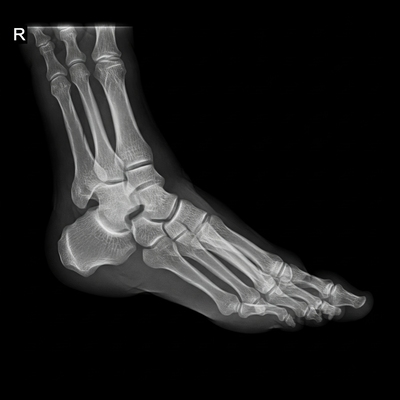 Normal Foot and Ankle X Rays: The human foot has 26 bones and 33 joints. There are more than a hundred muscles, tendons and ligaments. This is a very complex structure due to the need to support your entire body weight. The foot is a very stable composition of bones supported by strong ligaments.
Normal Foot and Ankle X Ray Anatomy:
The Phalanges (14): Each of the lesser toes are composed of 3 bones each. The big toe is made up of only 2 bones. The great toe is called the hallux. The Metatarsal bones (5): first, second, third, fourth, and fifth metatarsal bone. These are the longest bones in the foot. The tarsal bones (7): the talus, calcaneus, cuneiformes (3), cuboid, and navicular. The tibia and fibula make up just above the ankle.Normal Foot and Ankle X Ray Anatomy:
The foot and ankle can be subdivided into 4 different parts: the rear foot, the mid foot, and the forefoot: The Ankle is comprised of the talus, the tibia and the fibula. The rearfoot is is composed of the talus and the calcaneus (heel bone). The two long bones of the lower leg, the tibia and fibula, are connected to the top of the talus to form the ankle. The midfoot is composed of 5 bones: These bones are the cuboid, the navicular, and three cuneiform bones. These 5 bones compose the arch of the foot. These 5 bones connect the rear foot to the forefoot. The forefoot is composed of five toes and 5 metatarsals. The bones of the toes are called phalanges and the big toe has two phalanges while the other four toes have three phalanges each. The joints between the phalanges are called interphalangeal and those between the metatarsus and phalanges are called metatarsophalangeal joints. Pain in the Feet has many causes and can be localized to different areas (toenails, heels, big toes). In addition, pain in each location can be caused by many conditions and health problems. Scan the list below, and let us help you find the cause of your chronic pain in the feet.Michigan Podiatric Care at Balance Foot & Ankle: Comprehensive Foot and Ankle Services
At Balance Foot & Ankle, Michigan patients receive expert podiatric care for the full range of foot and ankle conditions, delivered by fellowship-trained podiatric surgeons at our Howell (4330 E Grand River) and Bloomfield Hills (43494 Woodward Ave #208) offices. Same-week new patient appointments are available; we accept Blue Cross Blue Shield of Michigan, Aetna, UnitedHealthcare, Cigna, Medicare, and most Medicare Advantage plans. In-office digital X-ray and musculoskeletal ultrasound allow same-visit diagnostic imaging. Our services include custom orthotics, diabetic foot care, toenail care, wound care, injection therapy, EPAT for tendinopathy, and the full spectrum of foot and ankle surgical procedures. Michigan patients with any foot or ankle concern can call (810) 206-1402 to schedule with Balance Foot & Ankle.
Foot Pain Evaluation and Treatment in Michigan: Balance Foot & Ankle
Michigan patients with foot pain deserve a thorough clinical evaluation that identifies the specific cause and directs effective treatment. At Balance Foot & Ankle, our fellowship-trained podiatric surgeons use weight-bearing digital X-rays, musculoskeletal ultrasound, and detailed clinical examination to establish accurate diagnoses for foot pain conditions — distinguishing plantar fasciitis from Baxter’s nerve entrapment, metatarsalgia from Morton’s neuroma, posterior tibial tendon dysfunction from tarsal tunnel syndrome, and stress fractures from soft tissue injuries. Treatment follows the diagnosis: custom orthotics, injection therapy, EPAT, bracing, physical therapy, or surgical correction depending on the condition and its severity. Michigan foot pain patients can call Balance Foot & Ankle at (810) 206-1402 for a diagnostic evaluation at our Howell or Bloomfield Hills location.
Michigan Foot Doctor: Why Patients Choose Balance Foot & Ankle
Related Treatment Guides
Michigan patients choose Balance Foot & Ankle for podiatric care because our fellowship-trained surgeons combine clinical depth with a patient-centered approach — thorough evaluations, clear explanations, and treatment plans designed for the individual patient’s goals and circumstances. We provide same-week new patient appointments at our Howell (4330 E Grand River) and Bloomfield Hills (43494 Woodward Ave #208) offices, with in-office digital X-ray for same-visit imaging. We accept Blue Cross Blue Shield of Michigan, Aetna, UnitedHealthcare, Cigna, Medicare, and most Medicare Advantage plans. From conservative care — custom orthotics, injection therapy, EPAT — to surgical correction of foot and ankle conditions, our Michigan patients receive evidence-based care that addresses the root cause of their foot problems. Call Balance Foot & Ankle at (810) 206-1402 to schedule your appointment.
Medical References & Sources
- American Podiatric Medical Association — Patient Education
- American Orthopaedic Foot & Ankle Society — Foot Conditions
Need Foot or Ankle X-Rays?
Our clinic provides on-site digital X-ray imaging for immediate diagnosis of fractures, arthritis, bone spurs, and structural deformities. Weight-bearing X-rays are essential for accurate assessment of foot and ankle alignment.
References
- Barg A, et al. Weightbearing computed tomography of the foot and ankle: emerging technology. Foot Ankle Int. 2018;39(3):376-386.
- Coughlin MJ, et al. Radiographic analysis of hallux valgus deformity. Foot Ankle Int. 2006;27(10):804-811.
- Saltzman CL, et al. Measurement of the alignment of the hindfoot using weight-bearing radiographs. J Bone Joint Surg Am. 1995;77(12):1791-1802.

Insurance Accepted
BCBS · Medicare · Aetna · Cigna · United Healthcare · HAP · Priority Health · Humana · View All →
Howell Office
4330 E Grand River Ave
Howell, MI 48843
Get Directions →
Bloomfield Hills Office
43494 Woodward Ave, #208
Bloomfield Hills, MI 48302
Get Directions →
Your Board-Certified Podiatrists


Ready to Get Back on Your Feet?
Same-week appointments available at both locations.
Book Your AppointmentPodiatrist-Recommended Products
🏆 Doctor Hoy’s Natural Pain Relief Gel — Top recommendation for reducing foot pain and inflammation naturally.
PowerStep Pinnacle Orthotic Insoles — Physician-grade arch support in an OTC package.
CURREX Support Insoles — Dynamic arch support in multiple profiles.
More Podiatrist-Recommended Foot Health Essentials
Hoka Clifton 10

Max-cushion everyday shoe — podiatrist favorite for walking and running.

OOFOS Recovery Slide

Impact-absorbing recovery sandal — wear after long days on your feet.
As an Amazon Associate, Balance Foot & Ankle earns from qualifying purchases. Product recommendations are based on clinical experience; prices and availability shown above update live from Amazon.

When to See a Podiatrist
If foot or ankle pain has been bothering you for more than a few weeks, home care alone may not be enough. Balance Foot & Ankle offers same-week appointments at our Howell and Bloomfield Hills clinics — no referral needed in most cases. Bring your current shoes and a short list of symptoms and we’ll build you a treatment plan in one visit.
Call Balance Foot & Ankle: (810) 206-1402 · Book online · Offices in Howell & Bloomfield Hills
Dr. Tom’s Foot & Ankle Health Maintenance Stack
- Doctor Hoy’s Natural Pain Relief Gel — Foot or ankle pain that prompted X-ray evaluation: arnica + camphor gel applied to the symptomatic area provides targeted anti-inflammatory support while awaiting imaging interpretation.
- PowerStep Pinnacle — Arch and alignment correction to prevent pathologic X-ray findings: PowerStep Pinnacle reduces the progressive arch collapse and stress fracture risk that develops from years of inadequate support — maintaining the normal alignment you see on healthy X-rays.
- Plantar Fasciitis Compression Socks — Lower extremity swelling that motivated imaging: graduated compression reduces the venous pooling that causes soft tissue changes around bones and joints visible on X-ray.
X-ray findings showing stress fracture, arthritis, or alignment concerns? Clinical interpretation and treatment at Balance Foot & Ankle. Balance Foot & Ankle → (810) 206-1402
In-Office Treatment at Balance Foot & Ankle
If home treatment isn’t providing relief for your foot and ankle conditions, our podiatry team at Balance Foot & Ankle can help with same-day evaluations and advanced in-office care.
Frequently Asked Questions
When should I see a podiatrist?
If symptoms persist past 2 weeks, affect your normal activity, or are accompanied by red-flag symptoms (warmth, redness, swelling, inability to bear weight).
What does treatment cost?
Most diagnostic visits and conservative treatments are covered by Medicare and major insurers. Out-of-pocket costs vary by your specific plan.
How quickly can I get an appointment?
Most non-urgent cases see us within 5 business days. Urgent cases (sudden pain, possible fracture) typically same or next business day.
Ready to feel better?
Same-week appointments available in Howell and Bloomfield Hills, Michigan.
Book Your VisitGet Expert Care at Balance Foot & Ankle
Same-week appointments at our Howell and Bloomfield Hills offices. Board-certified podiatric surgeons. Most insurance accepted.
Dr. Tom Biernacki, DPM is a board-certified foot & ankle surgeon (ABFAS & ABPM) at Balance Foot & Ankle Specialists in Southeast Michigan. With over a decade of clinical experience, he specializes in heel pain, bunions, diabetic foot care, sports injuries, and minimally invasive surgery. Dr. Biernacki is a member of the APMA and ACFAS, and his patient education content on MichiganFootDoctors.com and YouTube has made him one of the most-followed foot & ankle educators on YouTube.