
Quick answer: A protruding bone on the outside of the foot is most often a prominent fifth-metatarsal styloid, a bunionette (tailor’s bunion), or an accessory navicular, and sometimes an old fracture or arthritis. If it is painful, growing, or red, see a podiatrist; many cases respond to wider shoes and padding before any procedure is considered.
Board-certified foot & ankle surgeon · Balance Foot & Ankle, Howell & Bloomfield Hills, MI · Last reviewed: June 2026
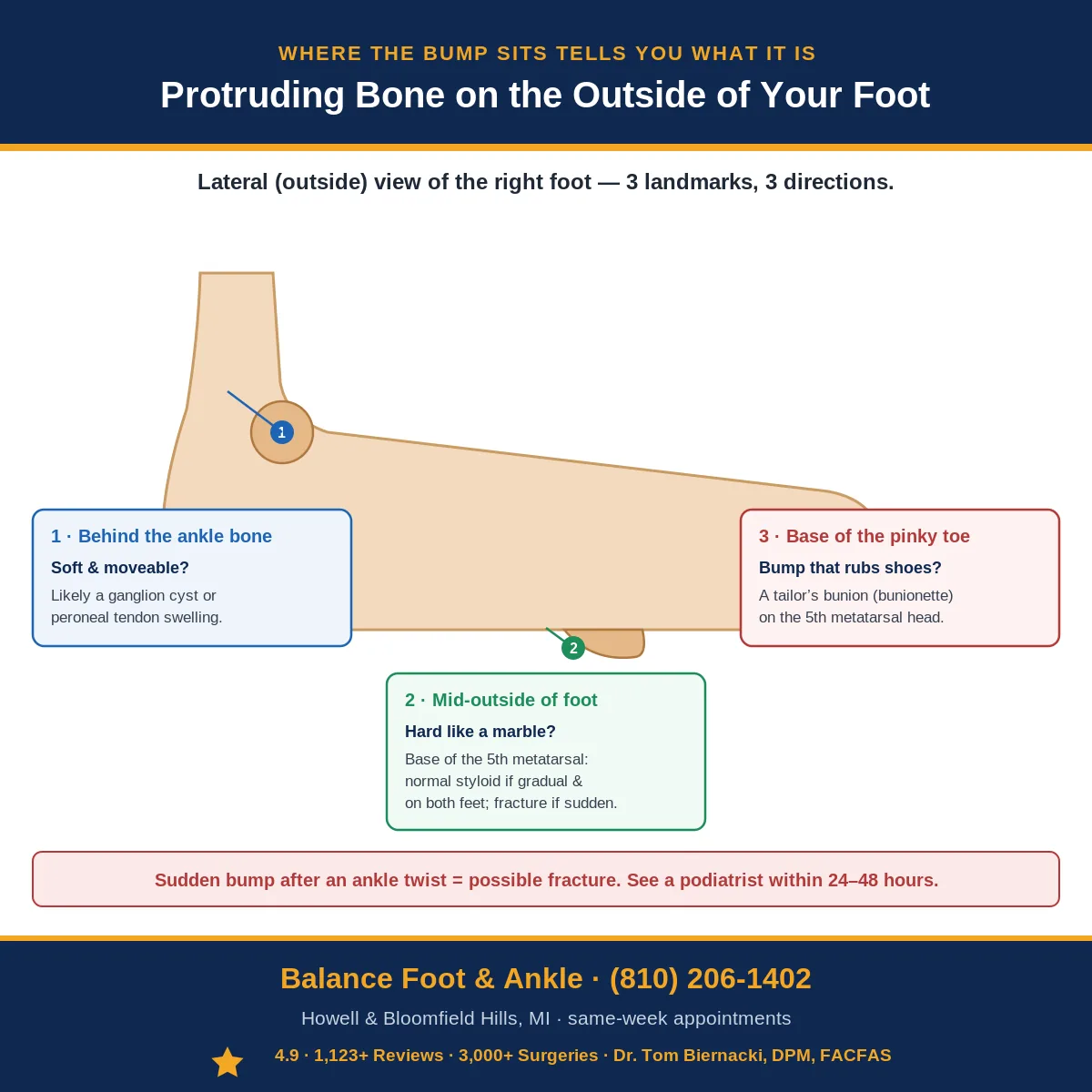
Where the protrusion sits on the outside of the foot is more diagnostic than how it feels. A bony prominence at the base of the fifth toe has a different cause, treatment, and timeline than one at the midfoot — even if both hurt in the same way. Of the 5 structural causes Dr. Tom distinguishes in practice, 3 are completely benign and self-limiting, 1 requires a specific shoe accommodation, and 1 needs imaging within 48 hours to rule out a stress fracture. The location pattern that should trigger immediate evaluation is described in the differential section below. (810) 206-1402 — same-day X-ray at Howell or Bloomfield Hills.
Most Common Mistake
Assuming every bump on the outside of the foot is the same and treating it with a generic pad. The outer foot harbors 5 distinct conditions: tailor bunion, ganglion cyst, Jones fracture callus, peroneal tendon subluxation, and fifth metatarsal avulsion fracture — each requiring completely different management. A soft movable bump is almost certainly a ganglion cyst; a firm painful bump at the base of the little toe is likely a tailor bunion; sudden onset after ankle inversion warrants fracture imaging before any padding or bracing.
Quick Answer: Protruding Bone on the Outside of the Foot
A bony prominence on the outside of the foot is most often a tailor’s bunion (bunionette) — a gradual outward drift of the 5th metatarsal head that creates a bony bump near the little toe. It is not a fracture and not dangerous, but it causes pain with narrow shoes. The second most common cause is a prominent peroneal tubercle. If the bump appeared suddenly after a fall or roll, get an X-ray the same day to rule out a Jones fracture. Call (810) 206-1402 — Balance Foot & Ankle, Howell & Bloomfield Hills, MI.
The Most Common Mistake With a Protruding Bone on the Outside of the Foot
When a bony bump needs surgery: If a prominent bone on the outside of the foot stays painful despite padding, shoe changes, and orthotics, surgical removal is sometimes the answer. Our board-certified foot & ankle surgeons in Howell and Bloomfield Hills can evaluate whether the prominence should be addressed surgically.
Have a painful bump or protruding bone on your foot?
Board-certified podiatric surgeons treat bunionettes, stress fractures, cysts, and more. Same-week appointments in Howell & Bloomfield Hills, MI. Most insurance accepted.
Book an Appointment → ☎ (810) 206-1402The most common mistake is assuming a bony prominence that appeared after an ankle roll is just a bruise. The base of the 5th metatarsal — exactly where the outer foot bump sits — is one of the most commonly fractured bones in the foot (Jones fracture and avulsion fracture). Both fractures can be walked on, at least briefly, after the injury. The rule: any new bony prominence after a trauma event needs an X-ray. A missed Jones fracture treated as a sprain frequently progresses to a non-union requiring surgery.
Protruding Bone on the Outside of Your Foot: What Is It? (6-Condition Guide)
A protruding bone on the outside of the foot is one of the most common reasons patients present to our podiatry office — and it’s one of the most misunderstood. Most patients assume any bony bump on the lateral foot is a broken bone or something that “appeared overnight.” In reality, six distinct conditions cause this presentation, each with a completely different cause, treatment, and prognosis.
The location of the bump on the outside of your foot narrows the diagnosis dramatically. The lateral foot has three bony landmarks: the base of the 5th metatarsal (mid-lateral foot), the styloid process (the prominent bump at the base of the 5th MT), and the lateral malleolus (the ankle bone). The chart below maps the exact location to the most likely diagnosis.
Protruding Bone Outside of Foot: 6 Conditions by Location
| Condition | Exact Location | Cause | Pain Pattern | Key Diagnostic Sign | Treatment |
|---|---|---|---|---|---|
| 5th Metatarsal Styloid Process (Normal Anatomy) | Mid-lateral foot — bony prominence at base of 5th metatarsal; always present; visible in thin individuals | Normal anatomy — peroneus brevis tendon attaches here; not pathological; becomes apparent with weight loss or foot swelling | None, or mild tenderness only if directly bumped; no activity-related pain | Bilateral (same bump on both feet); no acute onset; X-ray shows normal styloid without fracture | No treatment needed; reassurance; wide-toe-box shoes if rubbing against footwear |
| 5th Metatarsal Avulsion Fracture | Base of 5th metatarsal — identical location to styloid process, but sudden onset with inversion injury | Forced ankle inversion causes peroneus brevis tendon to avulse the styloid tip; one of the most common foot fractures | Sudden severe pain at lateral foot after ankle twist; difficulty weight-bearing; swelling and bruising within hours | Acute onset with injury mechanism; point tenderness at 5th MT base; X-ray confirms avulsion fragment with sharp edges (vs. smooth bipartite styloid) | Most avulsions: hard-soled shoe or CAM boot × 4–6 weeks, weight-bearing as tolerated. Displaced fragments >2mm or instability: surgical fixation |
| Jones Fracture | Proximal 5th metatarsal shaft — just distal (past) the styloid process, at the metaphyseal-diaphyseal junction (zone 2) | Repetitive stress or sudden inversion; watershed blood supply zone — poor healing; higher non-union risk than avulsion fracture | Lateral foot pain at or just distal to the styloid; may have had preceding ache (stress reaction) before acute fracture; insidious or sudden | Tenderness slightly distal to styloid; X-ray shows transverse fracture at metaphyseal-diaphyseal junction; crucial to distinguish from avulsion (different treatment) | Non-weight-bearing CAM boot × 6–8 weeks; surgical fixation (intramedullary screw) often recommended for athletes or high-risk for non-union |
| Tailor’s Bunion (Bunionette) | 5th metatarsal head — at the base of the little toe; distal lateral foot; may have red, irritated skin over bump | Lateral bowing of 5th metatarsal with prominent metatarsal head; narrow shoes compress the 5th MT head against shoe; hereditary bone structure | Gradual, progressive; worse with tight or narrow shoes; bursitis over bump (red, tender, swollen); no acute injury | Bony prominence at 5th MT head with overlying bursa or callus; bilateral in most cases; X-ray shows increased 4th-5th intermetatarsal angle; worse with narrow shoes | Wide toe-box shoes; custom orthotic to offload 5th MT head; bursal injection if inflamed; bunionette correction surgery for severe structural deformity |
| Peroneal Tendon Subluxation or Tenosynovitis | Posterior to lateral malleolus; soft tissue bump rather than hard bone; may be visible tendon when ankle moves | Peroneal retinaculum tear allows tendons to sublux over lateral malleolus; or tenosynovitis (inflammation within tendon sheath) creates soft tissue swelling | Lateral ankle pain + sensation of snapping or popping; pain with ankle eversion; swelling over peroneal tendons (posterior to malleolus, not at base of 5th MT) | Soft, mobile swelling (vs. hard bone); peroneal subluxation test positive (dorsiflexion + eversion causes visible tendon snap); ultrasound confirms | Acute subluxation: CAM boot + surgical retinaculoplasty for active patients; tenosynovitis: PT + brace; chronic: surgical stabilization |
| Ganglion Cyst (Lateral Foot) | Variable — can occur anywhere on lateral foot; often near joints or tendon sheaths; soft, fluctuant (jelly-like), not hard | Herniation of joint capsule or tendon sheath filled with synovial fluid; benign; may enlarge and shrink spontaneously | Usually painless; may cause pressure pain with certain shoes; no trauma history; may appear and disappear | Soft, fluctuant, transilluminates (light passes through when illuminated) — confirms fluid-filled cyst; not fixed to skin; X-ray shows no bone abnormality | Observation (50% resolve spontaneously); aspiration (recurrence rate 50%); surgical excision for persistent symptomatic cysts (lower recurrence) |
How to Tell Which Bony Bump You Have: The 3-Step Self-Assessment
| Step | What to Check | Result → Diagnosis Direction |
|---|---|---|
| Step 1: When Did It Appear? | Did the bump appear suddenly (same day or overnight) after an injury? Or has it been there gradually for months/years? | Sudden after ankle twist → fracture (avulsion or Jones) — see a podiatrist within 24–48 hours. Gradual/always been there → styloid process, tailor’s bunion, or ganglion — less urgent |
| Step 2: Is It Hard Bone or Soft Tissue? | Press firmly on the bump. Is it completely hard like a marble? Or does it have any give (soft, moveable, fluctuant)? | Completely hard = bone (styloid process, fracture, tailor’s bunion). Soft, jelly-like = ganglion cyst. Soft + over the tendon behind ankle = peroneal swelling |
| Step 3: Exactly Where Is It? | At the base of the pinky toe (distal lateral foot)? At the midpoint of the outside of the foot? Or behind the ankle bone? | At pinky toe base → tailor’s bunion. Mid-lateral foot (base of 5th metatarsal) → styloid process, avulsion fracture, or Jones fracture. Behind ankle → peroneal tendon issue |
When Is a Protruding Bone on the Outside of the Foot an Emergency?
| Sign / Symptom | What It Suggests | Action |
|---|---|---|
| Sudden severe pain after ankle twist + unable to weight-bear | Avulsion fracture or Jones fracture — Ottawa Rules indicate X-ray needed | Urgent care or ED within hours; do NOT walk on it; ice and elevate |
| Open wound or skin breakdown over the bump | Fracture with soft tissue involvement OR infected bursa over tailor’s bunion/styloid | Same day podiatrist visit; risk of infection spreading to bone |
| Diabetic patient with new bump or skin redness | Risk of Charcot fracture (neuropathic bone destruction) or infected callus | Urgent podiatrist within 24 hours; Charcot can destroy the foot if untreated |
| Bump growing rapidly (weeks, not years) | Tumor (rare but possible); osteochondroma; sarcoma (extremely rare) | Podiatrist within 1–2 weeks; X-ray and possible MRI to rule out neoplasm |
| Numbness or tingling around the bump | Nerve compression from the bony prominence or adjacent ganglion | Podiatrist within 2–4 weeks; sural nerve compression can worsen without treatment |

You can feel a bone sticking out the side of your foot. It’s been there for a while but suddenly it bothers you. There’s an explanation.
You’re in the right place. Dr. Tom Biernacki, DPM, FACFAS — board-certified foot & ankle surgeon with 3,000+ surgeries — explains exactly what a protruding bone on the outside of the foot means and what works. Call (810) 206-1402 for same-day appointment at Howell or Bloomfield Hills.
What causes a protruding bone on the outside of the foot?
A protruding bone on the outside of the foot is usually an enlarged styloid process of the fifth metatarsal (a bunionette or tailor’s complete bunion treatment guide), an accessory navicular, an old healed avulsion fracture, or arthritis at the cuboid-fifth metatarsal joint. X-ray confirms the source. Conservative treatment includes wider shoes, padding, and orthotics; surgery is reserved for chronic pain.
Protruding Bone on Outside of Foot: Diagnosis & Treatment Guide
In This Article
- Protruding Bone on the Outside of Your Foot: What Is It? (6-Condition Guide)
- How to Tell Which Bony Bump You Have: The 3-Step Self-Assessment
- When Is a Protruding Bone on the Outside of the Foot an Emergency?
- What causes a protruding bone on the outside of the foot?
- Understanding the Anatomy: Why Bumps Form on the Outside of the Foot
- Differential Diagnosis: What Is Your Lateral Foot Bump?
- Comparison Table: Differential Diagnosis of Lateral Foot Bumps
- Conservative Management of Tailor’s Bunion: Your First-Line Options
- Surgical Options for Tailor’s Bunion: When Conservative Care Fails
- Shoes to Avoid If You Have a Tailor’s Bunion
- Frequently Asked Questions About Lateral Foot Bumps
- The Most Common Mistake We See
-
Frequently Asked Questions
Should I worry about a bone bump on my foot?
Painless slow-developing bumps are usually benign (tailors bunion or accessory bone). Painful or rapidly enlarging bumps need imaging.
Can I shrink a bone bump on the outside of my foot?
No – bony prominences do not shrink. You can reduce friction with shoe modifications and pads. Surgery is reserved for persistent pain.
What is a tailors bunion?
A bony prominence at the base of the fifth (pinky) toe, similar to a bunion of the big toe. Common in narrow shoes.
Dr. Tom Biernacki, DPM explains what causes bumps and lumps on the foot, how to distinguish between a tailor’s bunion, accessory bone, and other bony prominences, and when surgical intervention is warranted.
What Helps: Dr. Tom’s Recommended Products for Lateral Foot Pain
When a tailor’s bunion or protruding bone creates friction and inflammation, the right combination of off-loading and targeted pain relief makes a significant difference before considering any surgical option.
Affiliate disclosure: Balance Foot & Ankle earns a commission from qualifying Foundation Wellness purchases at no extra cost to you.
Doctor Hoy’s Natural Pain Relief Gel — Natural topical pain relief I use in our clinic. Arnica + camphor formula reduces tissue-level inflammation at the fifth metatarsal. Apply 3–4x daily directly to the outer foot. Doctor Hoy’s botanical formula addresses inflammation rather than masking it — important when managing chronic friction pain.
PowerStep Pinnacle Insoles — The OTC orthotic I recommend most in our clinic. Proper arch support off-loads the fifth metatarsal head and distributes pressure evenly across the foot, reducing the friction and inflammation driving your pain. Medical-grade support at a fraction of custom orthotic cost.
Lateral Foot Pain Not Improving?
A protruding bone causing persistent pain after 4–6 weeks of home care may have a structural cause requiring imaging or clinical intervention. Same-day appointments available at both our Howell and Bloomfield Hills locations.
⚠️ When to see a podiatrist for a protruding bone on the outside of your foot:
- Sudden onset after ankle twist or inversion injury — acute avulsion or Jones fracture until proven otherwise; needs same-day X-ray
- Inability to bear weight on the foot — fracture red flag requiring urgent evaluation
- Swelling, bruising, or warmth around the bump — inflammatory or traumatic cause, not a benign bony variant
- Bump that is rapidly enlarging — bone tumor, ganglion, or infection must be ruled out
- Pain that wakes you at night or is present at rest — aggressive pathology requires imaging
- You have diabetes or poor circulation — any new foot deformity in a diabetic foot is urgent; Charcot arthropathy can present as a lateral prominence
- Bump is warm, red, and tender with fever — septic arthritis or osteomyelitis requires emergency evaluation
Call us same-day: (810) 206-1402 — or book online →
📖 Related: Complete Bunion & Tailor’s Bunion Hub: Causes, Treatment & Surgery →
A prominently protruding bone on the outside of the foot is most often the styloid process of the fifth metatarsal. The American Academy of Orthopaedic Surgeons notes this area is particularly prone to avulsion fractures from ankle sprains, underscoring the importance of professional evaluation for any acute bump.
★★★★★ 4.9 Stars · 1,123+ Five-Star ReviewsGet Expert Care at Balance Foot & Ankle
Same-week appointments at our Howell and Bloomfield Hills offices. Board-certified podiatric surgeons. Most insurance accepted.
For a complete clinical overview: Heel Pain Causes & Treatment Guide — every cause of foot and heel pain diagnosed

Dr. Tom Biernacki, DPM is a double board-certified podiatrist and foot & ankle surgeon at Balance Foot & Ankle Specialists in Southeast Michigan. With over a decade of clinical experience, he specializes in heel pain, bunions, diabetic foot care, sports injuries, and minimally invasive surgery. Dr. Biernacki is a member of the APMA and ACFAS, and his patient education content on MichiganFootDoctors.com and YouTube has reached over one million views.
Medical References- Plantar Fasciitis: Diagnosis and Conservative Management (PubMed)
- Plantar Fasciitis (APMA)
- Diagnosis and Treatment of Plantar Fasciitis (PubMed / AAFP)
- Heel Pain (APMA)
This article has been reviewed for medical accuracy by Dr. Tom Biernacki, DPM. References are provided for informational purposes.📋 Dr. Tom Biernacki, DPM, FACFAS answers:
A protruding bone on the outside of the foot is most commonly a bunionette (tailor’s bunion) at the 5th metatarsal head, a bony spur, or — less often — a displaced or healing stress fracture. If it’s been there long-term and only hurts with shoes, conservative management (wider shoes, padding) usually resolves it. If it appeared suddenly, is tender to touch, or followed trauma, an X-ray is essential to rule out fracture.
Footwear & Orthotics for a Prominent Foot Bone
A bony prominence on the outside of the foot is often aggravated by tight shoes. See our podiatrist-recommended shoes with wide, soft uppers, and recommended orthotics to offload the area. Persistent pain warrants an in-office evaluation.

Specialist For This Condition
Dr. Carl Jay, DPM is the Balance Foot & Ankle reconstructive surgeon other doctors refer to when a protruding bone or 5th-metatarsal deformity needs surgical evaluation. Fellowship-trained in complex foot and ankle reconstruction. Call (810) 206-1402 to schedule a consultation at the Howell or Bloomfield Hills office.
Related foot-bump guides
Other “bump or bone on the foot” guides worth comparing:

Dr. Tom Biernacki, DPM is a board-certified foot & ankle surgeon (ABFAS & ABPM) at Balance Foot & Ankle Specialists in Southeast Michigan. With over a decade of clinical experience, he specializes in heel pain, bunions, diabetic foot care, sports injuries, and minimally invasive surgery. Dr. Biernacki is a member of the APMA and ACFAS, and his patient education content on MichiganFootDoctors.com and YouTube has made him one of the most-followed foot & ankle educators on YouTube.