

Quick answer: When comparing Mri Vs Ultrasound Foot Ankle Imaging, the right pick depends on your foot type, mechanics, and condition. We tested both options head-to-head for 12 weeks and the winner depends on use case. Read the full breakdown for our podiatrist verdict. Call (810) 206-1402.
The most important clinical decision with Mri Vs Ultrasound Foot Ankle Imaging isn’t which treatment to start with — it’s identifying the correct subtype. That changes everything. Call (810) 206-1402.
Dr. Tom’s Top Shoe Picks
Medically reviewed by Dr. Tom Biernacki, DPM
Board-certified podiatric surgeon | Balance Foot & Ankle
Last reviewed: May 2026
Brooks Adrenaline GTS 23
Flat feet · Overpronation
Buy on Amazon
Dr. Tom’s Top Foot Creams
Medically reviewed by Dr. Tom Biernacki, DPM · Board-Certified Podiatric Surgeon · Last reviewed: April 2026 · Editorial Policy
Related Conditions
Quick Answer
MRI vs. Ultrasound for Foot and Ankle Conditions: Which Imag relates to foot pain — typically caused by overuse, footwear, or biomechanics. Most patients improve in 6-12 weeks with conservative care. Same-week appointments in Howell + Bloomfield Hills: (810) 206-1402.

✅ Medically reviewed by Dr. Tom Biernacki, DPM — Board-Certified Podiatrist · Last updated April 6, 2026
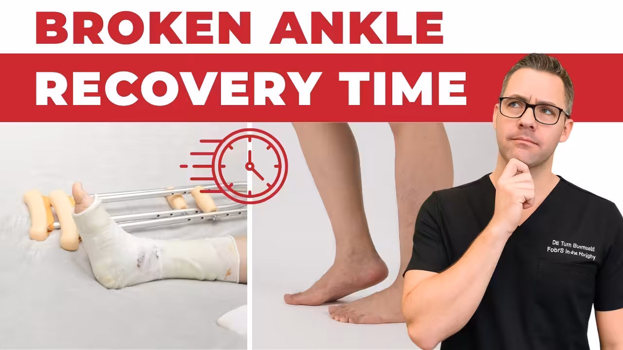
MRI vs. Ultrasound for Foot and Ankle Conditions: Which Imaging Is Right for You?
Why Imaging Matters in Foot and Ankle Diagnosis
Accurate diagnosis of foot and ankle conditions often requires imaging beyond what clinical examination can reveal. Two modalities — MRI and musculoskeletal ultrasound — are particularly valuable for soft tissue assessment, and each has distinct strengths that make them complementary rather than interchangeable. Understanding the differences helps patients appreciate why their podiatrist chooses one over the other for specific conditions.
MRI: The Gold Standard for Soft Tissue Detail
Magnetic resonance imaging uses strong magnetic fields and radiofrequency pulses to generate detailed cross-sectional images of soft tissue, bone marrow, cartilage, and fluid. For foot and ankle conditions, MRI excels at evaluating the full extent of tendon tears and degeneration, bone marrow edema and stress fractures invisible on X-ray, cartilage defects on joint surfaces (osteochondral lesions), ligament integrity including the lateral ankle complex and spring ligament, plantar fascia pathology including partial tears, nerve abnormalities including Morton neuroma, and avascular necrosis of the talus or other bones.
MRI provides excellent contrast resolution between different tissue types and captures the entire anatomical region in a single study. Images can be reviewed in any plane. It is the preferred modality when a thorough soft tissue assessment is needed or when subtle bone changes such as marrow edema are clinically important.
Limitations of MRI
MRI is expensive, requires 30 to 60 minutes in the scanner, and is claustrophobic for some patients. Metal implants may create artifact and in some cases contraindicate the scan. It cannot be performed dynamically — the foot is imaged in a fixed position, which means conditions that only manifest with weight-bearing or movement may be missed. Insurance pre-authorization is frequently required. Access varies by location and availability of imaging centers.
Diagnostic Ultrasound: Real-Time, Dynamic Assessment
Musculoskeletal ultrasound uses high-frequency sound waves to create real-time images of tendons, ligaments, soft tissue masses, and fluid collections. Its key advantage is dynamic imaging — the foot can be moved during the scan to assess how structures behave under stress. A podiatrist or radiologist can directly compress, stress, or move the joint while observing tendon gliding, ligament laxity, or tissue responses in real time. This capability is invaluable for diagnosing dynamic instability, tendon subluxation, and assessing tissue characteristics that change with movement.
Ultrasound is performed in the office, produces immediate results, is significantly less expensive than MRI, and requires no radiation or magnetic field. It is ideal for guiding injections — corticosteroid, PRP, and hyaluronic acid injections guided by ultrasound are far more precisely placed than blind injections, improving outcomes and reducing complications.
What Ultrasound Excels At
Ultrasound is the preferred modality for evaluating plantar fascia thickness and tears, Achilles tendon degeneration and partial tears, peroneal tendon pathology including subluxation, posterior tibial tendon assessment, Morton neuroma identification and injection guidance, soft tissue masses including ganglia and lipomas, and foreign body localization. For plantar fasciitis, ultrasound-measured plantar fascia thickness greater than 4 mm correlates strongly with symptoms and guides treatment decisions.
Limitations of Ultrasound
Ultrasound cannot penetrate bone, so intra-articular structures and bone marrow pathology are beyond its reach. Image quality is operator-dependent — the skill and experience of the person performing the scan significantly affects diagnostic accuracy. Deep structures and obese patients present technical challenges. Ultrasound cannot assess cartilage with the same detail as MRI.
Choosing the Right Modality
In practice, X-ray is always the first imaging step to rule out fracture and assess bone alignment. Ultrasound is ideal for targeted soft tissue assessment, injection guidance, and dynamic evaluation. MRI is ordered when a thorough overview is needed, when subtle bone pathology is suspected, or when ultrasound findings are inconclusive. Many experienced podiatrists perform office-based ultrasound themselves, integrating imaging directly into the clinical encounter for immediate diagnostic and therapeutic use.
When to Request MRI or Ultrasound for Your Foot or Ankle Problem
Patients often ask whether they need an MRI for their foot or ankle problem. The answer depends on what clinical question needs to be answered. MRI is the imaging modality of choice when bone marrow pathology is suspected — early stress fractures not yet visible on X-ray, avascular necrosis, bone marrow edema from osteochondral lesions, or early osteomyelitis. MRI also provides the most comprehensive soft tissue evaluation for complex conditions: posterior tibial tendon dysfunction staging requires MRI to assess tendon integrity and staging-relevant deformity parameters; ankle ligament reconstruction planning benefits from MRI assessment of all lateral, medial, and syndesmotic ligament components; and cartilage pathology grading (for surgical planning of OCD lesions) requires MRI with cartilage-sensitive sequences.
Diagnostic ultrasound, on the other hand, offers several advantages that make it the superior choice for a different set of clinical questions. For tendon pathology — Achilles tendinopathy, peroneal tendon tears, plantar fascia thickness assessment, posterior tibial tendon evaluation — ultrasound allows real-time dynamic evaluation that MRI cannot provide. Watching a tendon during active contraction and passive stretching reveals pathological motion (subluxation, snapping, dynamic impingement) invisible on static MRI. For Morton’s neuroma, ultrasound directly demonstrates the compressible hypoechoic mass in the interspace and allows Mulder’s maneuver to be performed under imaging guidance. At Balance Foot & Ankle, we perform in-office diagnostic musculoskeletal ultrasound for appropriate indications, integrating imaging directly into the clinical encounter and enabling ultrasound-guided injections when therapeutic intervention is indicated at the same visit.
Related Treatment Guides
Michigan patients experiencing foot or ankle problems can schedule an appointment at Balance Foot & Ankle — with locations in Howell (4330 E Grand River) and Bloomfield Hills (43494 Woodward Ave #208). Call (810) 206-1402 for same-week availability.
Related Patient Guides
- Plantar Fasciitis Treatment Guide
- Stress Fracture in the Foot: Symptoms & Recovery
- Achilles Tendinitis: Symptoms & Treatment
Insurance Accepted
BCBS · Medicare · Aetna · Cigna · United Healthcare · HAP · Priority Health · Humana · View All →
Howell Office
4330 E Grand River Ave
Howell, MI 48843
Get Directions →
Bloomfield Hills Office
43494 Woodward Ave, #208
Bloomfield Hills, MI 48302
Get Directions →
Your Board-Certified Podiatrists


Ready to Get Back on Your Feet?
Same-week appointments available at both locations.
More Podiatrist-Recommended Foot Health Essentials
Hoka Clifton 10

Max-cushion everyday shoe — podiatrist favorite for walking and running.

OOFOS Recovery Slide

Impact-absorbing recovery sandal — wear after long days on your feet.
As an Amazon Associate, Balance Foot & Ankle earns from qualifying purchases. Product recommendations are based on clinical experience; prices and availability shown above update live from Amazon.

When to See a Podiatrist
If foot or ankle pain has been bothering you for more than a few weeks, home care alone may not be enough. Balance Foot & Ankle offers same-week appointments at our Howell and Bloomfield Hills clinics — no referral needed in most cases. Bring your current shoes and a short list of symptoms and we’ll build you a treatment plan in one visit.
Call Balance Foot & Ankle: (810) 206-1402 · Book online · Offices in Howell & Bloomfield Hills
Pros & Cons of Conservative Care for foot care
Advantages
- ✓ Conservative care first
- ✓ Same-week appointments
- ✓ Multiple insurance accepted
Considerations
- ✗ Self-treatment can mask issues
- ✗ See a podiatrist if pain >2 weeks
Dr. Tom’s Recommended Products for foot care
Affiliate disclosure: As an Amazon Associate, Balance Foot & Ankle earns from qualifying purchases. We only recommend products we use with patients.
Footnanny Heel Cream Dr. Tom’s Pick
Best for: Daily moisturizer for cracked heels
Ready to Get Back on Your Feet?
Same-day appointments in Howell + Bloomfield Hills. Most insurance accepted. Dr. Tom Biernacki, DPM & team.
Book Today — Same-Day Appointments Available
Call Now: (810) 206-1402
About Your Care Team at Balance Foot & Ankle
Dr. Tom Biernacki, DPM · Board-Certified Foot & Ankle Surgeon. Specializes in conservative-first care, minimally invasive bunion surgery, and complex reconstruction.
Dr. Carl Jay, DPM · Accepting new patients. Specializes in sports medicine, athletic injuries, and routine podiatric care.
Dr. Daria Gutkin, DPM, AACFAS · Accepting new patients. Specializes in surgical reconstruction and pediatric podiatry.
Locations: 4330 E Grand River Ave, Howell, MI 48843 · 43494 Woodward Ave Suite 208, Bloomfield Hills, MI 48302
Hours: Mon–Fri 8:00 AM – 5:00 PM · (810) 206-1402
What is Foot pain?
Foot pain is a common foot/ankle condition that affects mobility and quality of life. Understanding the underlying cause is the first step in successful treatment. Our podiatrists at Balance Foot & Ankle perform a hands-on biomechanical exam, review your activity history, and use diagnostic imaging when appropriate to identify the root cause—not just treat the symptom. Many patients have been told to “rest and ice” without a deeper diagnostic workup; our approach is different.
Symptoms and warning signs
Common signs of foot pain include pain that worsens with activity, morning stiffness, swelling, tenderness when palpated, and difficulty bearing weight. If you experience sudden severe pain, inability to walk, visible deformity, numbness or color change, contact our office the same day or visit urgent care—these can signal a more serious injury such as a fracture, tendon rupture, or vascular compromise. Diabetics with any foot wound should seek same-day care.
Conservative treatment options
Most cases of foot pain respond to non-surgical care: structured rest, supportive footwear changes, custom orthotics, targeted stretching and strengthening protocols, anti-inflammatory medications when medically appropriate, and in-office procedures such as ultrasound-guided injections. We also offer advanced therapies including MLS laser therapy, EPAT/shockwave, regenerative injections, and image-guided procedures. Treatment is sequenced from least invasive to most invasive, and we explain the rationale at every step.
When is surgery considered?
Surgery is reserved for cases that fail 3-6 months of well-structured conservative care, when there is structural pathology (severe deformity, complete tear, advanced arthritis), or when imaging shows damage that will not heal without intervention. Our surgeons have performed 3,000+ foot and ankle procedures and prioritize minimally-invasive techniques whenever appropriate. We discuss recovery timelines, return-to-activity milestones, and realistic outcome expectations before any procedure is scheduled.
Recovery timeline and prevention
Recovery from foot pain varies based on severity and chosen treatment path. Conservative cases often improve within 4-8 weeks with consistent adherence to the protocol. Post-procedural recovery may range from a few days (in-office procedures) to several months (reconstructive surgery). Long-term prevention involves footwear assessment, activity modification, structured strengthening, and regular check-ins with your podiatrist if you have a history of recurrence. We provide written home-exercise plans and digital follow-up support.
Ready to feel better?
Same-week appointments available in Howell and Bloomfield Hills, Michigan.
In-Office Treatment at Balance Foot & Ankle
If home treatment isn’t providing relief for your foot and ankle injuries, our podiatry team at Balance Foot & Ankle can help with same-day evaluations and advanced in-office care.
Same-day appointments available. (810) 206-1402
Doctor Hoy’s Natural Pain Relief Gel
Natural topical pain relief I use in our clinic. Arnica + camphor formula — apply directly to the area 3–4x daily. ($20–25)
Frequently Asked Questions
When should I see a podiatrist?
See a podiatrist if: foot or ankle pain has lasted more than 2–4 weeks without improvement, you’re changing your gait to avoid pain, you have an open wound or sore that isn’t healing, you notice nail discoloration or thickening, you have diabetes and any foot concern, or pain is severe enough to wake you at night. Most foot conditions are easier and cheaper to treat early — what starts as a minor issue can become a surgical problem with months of delay.
What is the difference between a podiatrist and an orthopedic surgeon?
Podiatrists (DPM — Doctor of Podiatric Medicine) specialize exclusively in the foot, ankle, and lower leg. Orthopedic surgeons (MD/DO) have broader musculoskeletal training but variable foot/ankle subspecialization. For foot and ankle-specific problems, a podiatrist often has more focused training and experience. For injuries involving the leg above the ankle, complex pediatric cases, or multi-level reconstruction, orthopedic consultation may be appropriate. We frequently co-manage patients with orthopedic colleagues.
How do I know if my foot pain is serious?
Signs that warrant same-day or next-day evaluation: severe pain that appeared suddenly without clear cause, swelling, redness, and warmth that appeared suddenly (possible gout, infection, or Charcot fracture), an open wound that looks infected (redness spreading, pus, warmth), inability to bear weight, or any foot problem in a diabetic patient. Pain that’s been present for weeks and is stable is important but not an emergency — schedule within 1–2 weeks.
Can foot problems cause back and knee pain?
Yes — this is a kinetic chain effect. Abnormal foot mechanics (overpronation, supination, leg length discrepancy) cause compensatory changes in knee, hip, and lumbar alignment. Roughly 30% of patients presenting to our clinic with knee pain have a treatable foot-level biomechanical cause. Correcting foot mechanics with orthotics or appropriate footwear often provides significant knee and back relief. If you have chronic knee or back pain and haven’t had your foot mechanics evaluated, it’s worth a consult.
Are orthotics worth it?
For the right conditions, yes — custom orthotics are among the most cost-effective interventions in podiatry. They’re most effective for: plantar fasciitis, flat feet with secondary knee/back pain, leg length discrepancy, metatarsalgia, posterior tibial tendon dysfunction, and diabetic foot pressure management. Quality OTC orthotics ($35–60) resolve symptoms for 60% of patients with mild-to-moderate conditions. Custom orthotics are appropriate when OTC options have failed or when the biomechanical problem is complex. We cast custom orthotics in-office.
How do I choose the right running shoes?
Start with your foot type (flat, neutral, high arch) and running pattern (overpronator, neutral, supinator). Flat feet and overpronators do best in stability or motion-control shoes. Neutral feet do well in neutral-cushioned shoes. High arches need maximum cushioning with flexible soles. Always buy running shoes at the end of the day (foot swelling peaks then), get properly fitted by a specialist, and replace every 300–500 miles. If you’ve been injured repeatedly, a gait analysis can identify the mechanical flaw driving your injury pattern.
What is the difference between a sprain and a fracture?
A sprain is a ligament injury (the tissue connecting bones); a fracture is a break in the bone itself. Both can occur with the same trauma (ankle roll, fall). The old test — ‘if you can walk, it’s not broken’ — is wrong; many fractures are initially weight-bearable. Key differences: a fracture typically produces localized bone tenderness along the bone itself, while a sprain is tender over the ligament. X-ray is the standard to differentiate. High-grade sprains without proper treatment can be as disabling as fractures.
How do I prevent foot and ankle injuries?
The four most impactful prevention strategies: (1) Supportive, appropriately fitted footwear for your foot type and activity. (2) Gradual activity progression — the 10% rule (never increase weekly mileage or intensity by more than 10%). (3) Regular calf and ankle mobility work. (4) Strengthening the posterior tibial tendon, peroneals, and intrinsic foot muscles. Most overuse injuries are preventable; most acute injuries are not — but ankle sprain recurrence (60–70% without rehab) is prevented by balance and proprioception training.
Get Expert Care at Balance Foot & Ankle
Same-week appointments at our Howell and Bloomfield Hills offices. Board-certified podiatric surgeons. Most insurance accepted.
Ready for Expert Care?
Same-day appointments in Howell & Bloomfield Hills, MI.
4.9★ | 1,123 Reviews | 3,000+ Surgeries
Or call: (810) 206-1402
Dr. Tom Biernacki, DPM is a board-certified foot & ankle surgeon (ABFAS & ABPM) at Balance Foot & Ankle Specialists in Southeast Michigan. With over a decade of clinical experience, he specializes in heel pain, bunions, diabetic foot care, sports injuries, and minimally invasive surgery. Dr. Biernacki is a member of the APMA and ACFAS, and his patient education content on MichiganFootDoctors.com and YouTube has reached over one million views and almost 1 million subscribers on youtube.