

A bunion is a progressive misalignment of the big-toe joint that widens the forefoot and causes pain, rubbing, and difficulty with shoes. Modern minimally invasive 3D bunion correction (Lapiplasty-class procedures) realigns the bone at its root, allows most patients to walk in a boot within days, and returns them to regular shoes in roughly 6 to 8 weeks. We perform the full workup, imaging, and surgery locally in Livingston and Oakland counties.
Ready to be seen? Book a real appointment time online, 24/7 — Howell or Bloomfield Township. Takes about a minute, 24/7.
★ 4.9 Stars · 1,193+ Reviews · Board-Certified Michigan Podiatrists
Bunion Surgery
Expert Care at Balance Foot & Ankle Michigan
Board-certified podiatric surgeons provide bunion surgery at our Howell and Bloomfield Township offices. 4.9★ rated · 1,193+ reviews · Same-week appointments · Most insurance accepted.

Medically Reviewed by Dr. Tom Biernacki, DPM, FACFAS
Board-Certified Podiatrist & Foot Surgeon · Last Updated: April 2026
Best Bunion Surgery
in Howell & Bloomfield Township, Michigan
If you’re struggling with a painful bunion that makes walking, standing, or even wearing shoes uncomfortable, you’re not alone—and you don’t have to live with the pain. At Balance Foot & Ankle Specialist, we specialize in treating bunions with the latest surgical techniques to help you get back on your feet, pain-free and confident.
Schedule Your Bunion Surgery Today
Accepting New Patients
Board Certified Podiatrists — Howell & Bloomfield Township
Find Expert Bunion Doctor at the Location Nearest You.
Howell
4330 E Grand River Ave, Howell, MI 48843
Book Now in Under 5 Minutes: 24/7
Bloomfield Township
43494 Woodward Ave # 208, Bloomfield Township, MI 48302
Book Now in Under 5 Minutes: 24/7
BEST Bunion Treatments
NO SURGERY Splints, Correctors, & Pads
Bunions can do more than cause a bump on your big toe—they lead to real, lasting discomfort. Over time, they can affect your ability to move, stay active, or simply enjoy your day without foot pain. What starts as minor irritation can quickly escalate into joint stiffness, swelling, and long-term mobility issues.
That’s why early treatment matters. While non-surgical options can provide short-term relief, surgical bunion correction can realign your toe joint, eliminate pain, and restore your natural movement. Our experienced podiatrists will work with you to determine the best treatment option—so you can walk, move, and live with comfort again.
When Is Bunion Surgery Needed?
Bunion surgery is typically recommended when non-surgical treatments—like custom orthotics, padding, or lifestyle changes—no longer provide relief. If you’re experiencing consistent pain, swelling, or trouble walking, it may be time to consider surgical correction. Our podiatrists will assess your condition and determine if surgery is the best step toward long-term relief.
Our Surgical Approach
At Balance Foot & Ankle Specialist, we use advanced surgical techniques matched to your foot structure. From minimally invasive bunionectomy to realignment procedures, our goal is to correct the deformity, relieve pressure, and restore natural function with minimal downtime and scarring.
Benefits of Bunion Surgery
Long-lasting pain relief
Improved joint alignment and foot function
Ability to return to normal footwear and daily activities
Our expert care ensures optimal healing and long-term results.
What to Expect – Before, During & After Surgery
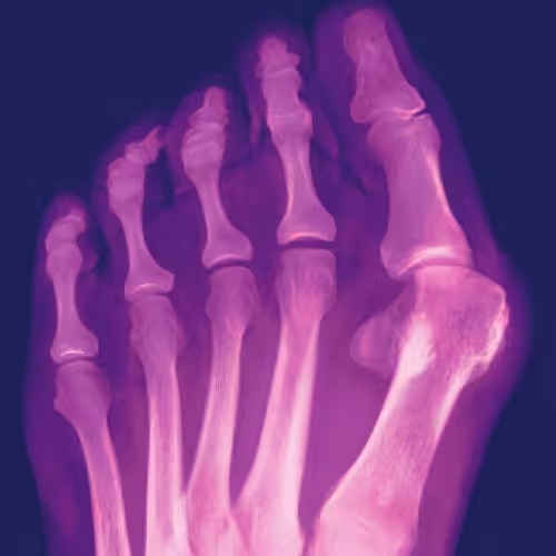
Before surgery, we’ll perform a full exam, take imaging, and explain your treatment plan.
During surgery, we use precise techniques to realign bones and remove excess tissue.
After surgery, you’ll receive a customized recovery plan, including follow-ups, footwear guidance, and physical therapy if needed. Most patients return to walking comfortably within weeks.
Why Choose Balance Foot & Ankle Specialist in Howell & Bloomfield Township?
With board-certified podiatrists, advanced surgical tools, and compassionate care, Balance Foot & Ankle Specialist is your trusted partner in foot health. We tailor every treatment to your needs and support you every step of the way—from consultation to recovery.
Book Your Podiatrist Visit Now
Trusted Footwear Recommendations From
Michigan’s Leading Podiatrists
Dr. Tom Biernacki DPM
(Podiatrist, Wound Care, Foot & Ankle Specialist)
Dr. Tom Biernacki, DPM, is a highly respected and experienced podiatrist proudly serving patients in Howell, Michigan, and throughout Oakland County. With a strong commitment to personalized patient care, Dr. Biernacki specializes in diagnosing and treating a many foot and ankle conditions, including chronic foot pain, sports injuries, diabetic foot care, and structural deformities. His practice offers comprehensive services such as routine foot care, advanced wound care, and both non-surgical and surgical solutions for complex issues affecting the lower extremities. Known for his compassionate approach and attention to detail, Dr. Biernacki works closely with each patient to create a customized treatment plan that ensures the best possible outcome.
Dr. Carl Jay DPM
(Podiatric Surgeon & Foot Doctor)
Dr. Carl Jay is a board-certified foot and ankle surgeon and proud partner at Balance Foot & Ankle. After completing his residency at the Genesys Regional Medical Center program, he took his talents to Texas. There he covered trauma call, treated complex wounds in the Wound Care Center, and cared for a many patients in private practice, from diabetic limb preservation to sports medicine.
Dr. Jay certainly enjoyed practicing in the Lone Star State. (I mean that amazing warm weather and sunshine.)Family brought him back to Michigan.
Now serving the Detroit Metro area, Dr. Jay brings advanced treatment modalities and years of surgical and clinical experience to every patient encounter. He’s passionate about delivering expert, compassionate care that helps people move better and live better.
Over 10,000
Patients Helped
Advanced 3D
Orthotic Scanning

Top Patient Reviews
“I’ve struggled with bunion pain for years, making it difficult to walk or wear shoes comfortably. Dr. Tom Biernacki at Balance Foot & Ankle Specialist in Howell provided excellent care. He explained my options clearly and recommended treatments that eased my pain almost immediately. I can finally walk without discomfort. Highly recommend for seniors dealing with bunions!”
– Agatha, Howell, MI
“My bunions had been worsening, and I was worried about surgery. Dr. Carl Jay at Balance Foot & Ankle Specialist in Bloomfield Township offered non-surgical solutions that really helped. With custom orthotics and gentle care, my pain has decreased, and I can wear my favorite shoes again. The staff is compassionate and patient with seniors—it’s a wonderful experience.”
– Josh, Bloomfield HIlls, MI
“As a senior, bunion pain was limiting my mobility and daily activities. The team at Balance Foot & Ankle Specialist treated me with kindness and expertise. After a combination of orthotics, stretching exercises, and professional guidance, I feel so much more comfortable on my feet. I’m grateful for their thorough and compassionate approach.”
– Mellisa, Bloomfield HIlls, MI
Bunion Surgery FAQ
Yes, it’s a common and safe procedure when performed by a qualified podiatrist.
Most patients begin walking within a few weeks and return to full activity in about 6–8 weeks.
Recurrence is rare with proper foot care and post-surgery support.
Modern techniques minimize scarring. We focus on both function and aesthetics.
Most health insurance plans cover bunion surgery if it’s medically necessary. We can help verify your benefits.
Struggling with Another Foot Problem? We Can Help.
Foot & Ankle Injections (Steroid or Natural Solutions.)
Management of Foot & Ankle Fractures (Walking Boots for broken toes or broken feet).
Senior Toenail Cutting & Toenail Trimming Services.
Insurance & Medicare Friendly
Accepts Medicare
Accepts Most Major Insurances
Affordable Options Available
Don’t let bunions limit your comfort, mobility, or choice of footwear. Bunion Surgery at Balance Foot & Ankle Specialist is designed to correct the deformity, relieve pain, and restore proper alignment.
Our experienced podiatrists in Howell and Bloomfield Township provide personalized surgical care, advanced techniques, and thorough post-operative guidance to ensure a smooth recovery and lasting results.
Book a Foot Examination
(810) 206-1402
Areas We Serve
Insurance Accepted: Medicare · BCBS · United Healthcare · Aetna and more
Sources
- A Multi-Dimensional Systematic Review of Minimally Invasive Bunion Surgery (MIBS). PubMed, 2025. pubmed.ncbi.nlm.nih.gov/40283587
- A New Paradigm for Failed Bunions with Minimally Invasive Methods. PubMed, 2024. pubmed.ncbi.nlm.nih.gov/39550086
- Current concepts of fourth-generation minimally invasive and open hallux valgus surgery. PubMed, 2024. pubmed.ncbi.nlm.nih.gov/39740690
Related: Learn more about our bunion treatment, explore our full list of conditions we treat, or schedule your visit.
Clinical References
- Nix SE, Mathews M, Engstrom B, Arenson T, Adeyi O. The Lapiplasty 3D Bunion Correction System: A Review of Outcomes and Technique. Foot Ankle Orthop. 2023;8(1). doi:10.1177/24730114221148657
- Torkki M, Malmivaara A, Seitsalo S, Hoikka V, Laippala P, Paavolainen P. Surgery vs orthosis vs watchful waiting for hallux valgus: a randomized controlled trial. JAMA. 2001;285(19):2474-2480. doi:10.1001/jama.285.19.2474
- Fraissler L, Konrads C, Hoberg M, Rudert M, Walcher M. Treatment of hallux valgus: a systematic review. EFORT Open Rev. 2016;1(9):295-302. doi:10.1302/2058-5241.1.000005
Clinical Sources
- Nix S, Smith M, Vicenzino B. Prevalence of hallux valgus in the general population: a systematic review and meta-analysis. J Foot Ankle Res. 2010;3:21. PubMed
- Torkki M, Malmivaara A, Seitsalo S, et al. Surgery vs orthosis vs watchful waiting for hallux valgus: a randomized controlled trial. JAMA. 2001;285(19):2474-2480. PubMed
- Easley ME, Trnka HJ. Current concepts review: hallux valgus part II—operative treatment. Foot Ankle Int. 2007;28(6):748-758. PubMed
- American Academy of Orthopaedic Surgeons. Bunions—OrthoInfo. AAOS
Related Conditions & Treatments
Considering bunion surgery? Dr. Tom Biernacki has performed over 3,000 foot and ankle surgeries. Call (810) 206-1402 or book a consultation online.
Also explore custom orthotics for bunion relief, ankle and foot instability, and sports medicine podiatry.
Bunion Surgery Cost and Insurance Coverage
Most insurance plans, including Medicare, Blue Cross Blue Shield, United Healthcare, and Aetna, cover bunion surgery when conservative treatments have been documented as unsuccessful and the procedure is deemed medically necessary. Prior authorization is typically required.
Typical costs with insurance include your specialist copay ($20 to $60 per visit), plus deductible and coinsurance for the surgical procedure itself. Many patients reach their out-of-pocket maximum with bunion surgery, meaning their insurance covers 100 percent of remaining care for the year. For self-pay patients, view our self-pay pricing options.
Our team verifies your insurance benefits and provides a detailed cost estimate before scheduling surgery. Call (810) 206-1402 to verify your coverage.
Watch: Bunion Surgery Explained by Dr. Tom Biernacki
Planning your recovery? Read our detailed Bunion Surgery Recovery: Week-by-Week Timeline to know exactly what to expect after your procedure.
What Our Patients Say About Bunion Surgery
I suffered with bunion pain for years before finally scheduling surgery with Dr. Biernacki. The minimally invasive approach meant less pain and faster recovery than I expected. I was back in my shoes in six weeks and wish I had done it sooner.
— Michelle R., Howell
Dr. Tom explained everything clearly and made me feel completely comfortable about my bunion surgery. The whole team was professional and caring. My foot looks and feels amazing now.
— Patricia L., Brighton
Insurance Coverage & Recovery Resources
Bunion surgery is typically covered by most insurance plans when medically necessary:
Recovery Guide: Read our complete Bunion Surgery Recovery Guide for a detailed week-by-week recovery timeline, exercises, and tips from our surgeons.
Post-surgery footwear guide: Best Shoes After Bunion Surgery — Podiatrist-recommended shoes by recovery phase, plus what to avoid.
️ When to see a podiatrist about your bunion:
- Persistent pain that doesn’t improve with rest or OTC remedies
- Difficulty walking or fitting into shoes comfortably
- Visible toe deformity that’s getting worse
- Numbness, swelling, or redness around the big toe joint
- Bunion pain interfering with daily activities or exercise
Related Reading
- Bunion Prevention: How to Stop Bunions Before They Start
- Best Shoes After Bunion Surgery
- Flat Feet Treatment Guide
- When to See a Podiatrist: 10 Signs You Shouldn’t Ignore
Ready to Get Relief?
Same-day appointments available in Howell & Bloomfield Township, MI
4.9★ | 1,193 Reviews | 3,000+ Surgeries
Or call: (810) 206-1402
Frequently Asked Questions About Bunion Surgery
How long is recovery after bunion surgery?
Recovery time depends on the surgical technique used. With minimally invasive bunion surgery, most patients can bear weight in a surgical shoe within a few days and return to regular shoes within 4 to 6 weeks. Traditional bunionectomy may require 6 to 8 weeks of limited weight-bearing. Full recovery including return to athletic activities typically takes 3 to 4 months.
Can bunions come back after surgery?
Recurrence rates vary by procedure type and patient factors. Modern techniques using internal fixation have recurrence rates below 5 percent. Wearing proper footwear and using custom orthotics after surgery significantly reduces the chance of recurrence. Dr. Biernacki discusses long-term prevention strategies at your follow-up visits.
Does insurance cover bunion surgery in Michigan?
Most health insurance plans, including Medicare and Blue Cross Blue Shield of Michigan, cover bunion surgery when it is medically necessary — meaning the bunion causes significant pain, limits daily activities, or has not improved with conservative treatments such as wider shoes, padding, or orthotics. Our team handles insurance verification and pre-authorization before scheduling.
What is minimally invasive bunion surgery?
Minimally invasive bunion surgery uses small incisions (typically 3 to 5 millimeters) and specialized instruments to realign the bone and correct the deformity. Benefits include less post-operative pain, smaller scars, faster recovery, and the ability to walk sooner after surgery compared to traditional open bunionectomy techniques.
Two Convenient Locations
Same-week appointments available at both offices
Howell Office
4330 E Grand River AveHowell, MI 48843
Phone: (810) 206-1402
Hours: Mon–Fri 9:00 AM–4:30 PM
View Howell Office →Bloomfield Township Office
43494 Woodward Ave #208Bloomfield Township, MI 48302
Phone: (810) 206-1402
Hours: Mon–Fri 9:00 AM–4:30 PM
View Bloomfield Office →WHAT OUR PATIENTS SAY
4.9 ★ Rating · 1,193+ Five-Star Reviews
★★★★★
“Dr. Tom performed my Lapiplasty bunion surgery and the results are amazing. My foot looks normal for the first time in years. Best decision I ever made.”
— Patricia L., Hartland
★★★★★
“I was terrified of bunion surgery but the team put me completely at ease. Recovery was faster than expected and the pain relief is life-changing.”
— Linda K., Bloomfield Township
Related Treatments You May Need
Custom 3D Orthotics
Post-surgical orthotics to maintain alignment and prevent recurrence.
Hammertoe Correction
Often performed alongside bunion surgery to restore toe alignment.
Rehabilitation Exercises
Post-operative stretching and strengthening for full recovery.
Digital X-Rays
Advanced imaging to plan and monitor your surgical outcome.
Get Bunion Treatment That Works
Lapiplasty 3D bunion correction — same-week consultations available.
or call (810) 206-1402
Your Board-Certified Podiatrists



Ready to Get Back on Your Feet?
Same-week appointments available at both locations.
Book Your AppointmentServing Patients Across Southeast Michigan
Balance Foot & Ankle provides expert podiatric care from two convenient locations. Our Howell office serves patients from Brighton, Hartland, Fowlerville, Pinckney, Fenton, Hamburg, Whitmore Lake, South Lyon, and throughout Livingston County. Our Bloomfield Township office serves Birmingham, Troy, West Bloomfield, Pontiac, Farmington Hills, Southfield, Royal Oak, Clarkston, Lake Orion, Rochester Hills, Waterford, Commerce Township, Novi, and Walled Lake across Oakland County.
Board-certified care at both Michigan locations
Book your visit today
Most insurance accepted · On-site X-ray · Board-certified podiatrists
Howell · (810) 206-1402 Bloomfield · (810) 206-1402Frequently Asked Questions
How is Lapiplasty bunion surgery different in Howell?
Dr. Tom Biernacki performs Lapiplasty® 3D bunion correction at our Howell office — the same procedure available at major orthopedic centers. Lapiplasty corrects the bunion in all three dimensions by stabilizing the unstable joint at the root cause, unlike traditional 2D bone cuts that have higher recurrence rates.
Can I walk after bunion surgery in Howell?
With Lapiplasty® performed by Dr. Biernacki at our Howell location, most patients walk (in a surgical boot) within days of surgery — a major advantage over traditional techniques. You’ll be in the boot for 6-8 weeks, then transition to regular shoes. Full recovery typically takes 4-6 months.
Frequently Asked Questions
What is recovery like after Lapiplasty® bunion surgery?
Most patients walk in a boot within days of Lapiplasty® surgery — far faster than traditional bunion procedures. Return to regular shoes typically occurs at 6–8 weeks, with full athletic activity by 4–6 months.
Is bunion surgery covered by insurance?
Bunion surgery (CPT 28296 and related codes) is covered by most major insurance plans when conservative treatment has failed. We verify your specific benefits and obtain prior authorization before scheduling surgery.
How is Lapiplasty® different from traditional bunion surgery?
Traditional bunion surgery cuts and shifts the bone in 2D — the deformity often returns. Lapiplasty® corrects the root cause by rotating the metatarsal bone in all three dimensions and securing it with titanium plates, providing a permanent correction.
Frequently Asked Questions
When should I actually have bunion surgery?+
How is modern 3D bunion correction different from older bunionectomies?+
How long is recovery from bunion surgery?+
Will my bunion come back after surgery?+
Does insurance cover bunion surgery?+
Still have questions? Call us — we can usually fit you in within the week.
Call (810) 206-1402Watch: Bunion Surgery in Howell, Michigan
Dr. Tom on Howell bunion surgery — MIS vs open osteotomy, candidate selection, recovery timeline, local Livingston County same-week scheduling.
Related: Bunion Treatment · MIS Bunion Option · Book Howell Consultation
Frequently Asked Questions
When should I see a podiatrist?
If symptoms persist past 2 weeks, affect your normal activity, or are accompanied by red-flag symptoms (warmth, redness, swelling, inability to bear weight).
What does treatment cost?
Most diagnostic visits and conservative treatments are covered by Medicare and major insurers. Out-of-pocket costs vary by your specific plan.
How quickly can I get an appointment?
Most non-urgent cases see us within 5 business days. Urgent cases (sudden pain, possible fracture) typically same or next business day.
Bunion typically responds best to early podiatrist evaluation, conservative treatments such as supportive footwear and targeted physical therapy, and—when needed—custom orthotics or in-office procedures. Most patients see meaningful improvement within 4-6 weeks of starting a structured treatment plan. Schedule an evaluation at our Howell or Bloomfield Township office for a clinical assessment.
What is Bunion?
Bunion is a common foot/ankle condition that affects mobility and quality of life. Understanding the underlying cause is the first step in successful treatment. Our podiatrists at Balance Foot & Ankle perform a hands-on biomechanical exam, review your activity history, and use diagnostic imaging when appropriate to identify the root cause—not just treat the symptom. Many patients have been told to “rest and ice” without a deeper diagnostic workup; our approach is different.
Symptoms and warning signs
Common signs of bunion include pain that worsens with activity, morning stiffness, swelling, tenderness when palpated, and difficulty bearing weight. If you experience sudden severe pain, inability to walk, visible deformity, numbness or color change, contact our office the same day or visit urgent care—these can signal a more serious injury such as a fracture, tendon rupture, or vascular compromise. Diabetics with any foot wound should seek same-day care.
Conservative treatment options
Most cases of bunion respond to non-surgical care: structured rest, supportive footwear changes, custom orthotics, targeted stretching and strengthening protocols, anti-inflammatory medications when medically appropriate, and in-office procedures such as ultrasound-guided injections. We also offer advanced therapies including MLS laser therapy, EPAT/shockwave, regenerative injections, and image-guided procedures. Treatment is sequenced from least invasive to most invasive, and we explain the rationale at every step.
When is surgery considered?
Surgery is reserved for cases that fail 3-6 months of well-structured conservative care, when there is structural pathology (severe deformity, complete tear, advanced arthritis), or when imaging shows damage that will not heal without intervention. Our surgeons have performed 3,000+ foot and ankle procedures and prioritize minimally-invasive techniques whenever appropriate. We discuss recovery timelines, return-to-activity milestones, and realistic outcome expectations before any procedure is scheduled.
Recovery timeline and prevention
Recovery from bunion varies based on severity and chosen treatment path. Conservative cases often improve within 4-8 weeks with consistent adherence to the protocol. Post-procedural recovery may range from a few days (in-office procedures) to several months (reconstructive surgery). Long-term prevention involves footwear assessment, activity modification, structured strengthening, and regular check-ins with your podiatrist if you have a history of recurrence. We provide written home-exercise plans and digital follow-up support.
Ready to feel better?
Same-week appointments at our Howell office, 4330 E Grand River Ave. Booking is open online around the clock — even after our phones close at 4:30 PM.
📅 Book Online — Howell📞 (810) 206-1402Our podiatrists treat the underlying cause, not just the symptom. Same-week appointments at our Howell and Bloomfield Township, Michigan offices.
Learn More About Bunions
Related
Bunion care at our other office:
Request an appointment
If a bunion is painful or making shoes difficult to wear, we can evaluate it and go over your options. Call (810) 206-1402 or send the form below and our office will get back to you.
We see patients at our Howell and Bloomfield Township offices.