


Medically reviewed by Dr. Tom Biernacki, DPM
Board-certified podiatric surgeon | Balance Foot & Ankle
Last reviewed: May 2026
The most important clinical decision with Posterior Tibial Tendon Dysfunction isn’t which treatment to start with — it’s identifying the correct subtype. That changes everything. Call (810) 206-1402.
Posterior Tibial Tendon Dysfunction (PTTD): Stages, Symptoms & Treatment
Your inner ankle has been aching for months. Your foot looks flatter than it used to. You’ve noticed that your heel seems to tilt outward when you stand. You can’t rise on your tiptoe on that leg without pain — or can’t at all. If this sounds like you, you may have posterior tibial tendon dysfunction — the most important cause of adult-acquired flatfoot, and one of the most treatable conditions we manage when caught early.
In our clinic, PTTD is one of the conditions we’re most emphatic about treating promptly. The difference between Stage I and Stage III is the difference between an orthotic and a major reconstruction — and patients move between stages faster than most realize.
What Is the Posterior Tibial Tendon?
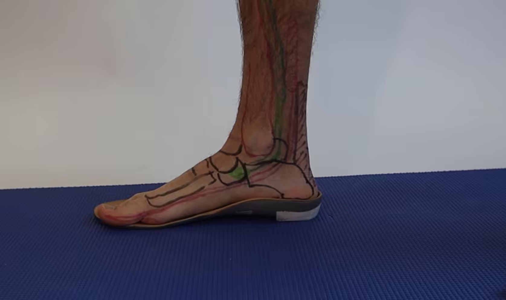
The posterior tibial tendon runs behind the medial malleolus (inner ankle bone) and inserts into the navicular and surrounding midfoot bones. It is the primary dynamic stabilizer of the medial longitudinal arch — responsible for supinating (inverting) the foot and maintaining arch height during walking and running. When this tendon degenerates and fails, the arch progressively collapses, the heel tilts outward (hindfoot valgus), and the forefoot rotates outward (forefoot abduction) — the classic “too many toes sign.”
PTTD predominantly affects women over 40, particularly those who are overweight or have a history of flat feet. Diabetes, hypertension, and steroid use are additional risk factors.
PTTD Stages and What They Mean for Treatment
PTTD is classified into four stages, each reflecting increasing tendon and structural failure:
- Stage I: Tendon inflammation but no deformity. Normal arch height. Tendon is intact but painful and swollen behind the medial ankle. Patient can perform a single-leg heel rise (painful). Best treated with immobilization, orthotics, and PT.
- Stage II: Tendon elongation/partial tearing. Flexible flat foot deformity. Patient cannot perform a pain-free single-leg heel rise. “Too many toes sign” visible. This is the critical stage — flexible deformity can be corrected conservatively if treated aggressively. Surgery is effective when conservative care fails.
- Stage III: Fixed (rigid) flatfoot deformity. The joints are no longer correctable to neutral. Surgery required; joint-sparing procedures often insufficient. Triple arthrodesis (fusion) is frequently necessary.
- Stage IV: Fixed flatfoot with ankle joint involvement (ankle valgus tilt). The most advanced stage; requires reconstruction that may include ankle replacement or fusion.
Key takeaway: The single-leg heel rise test: stand barefoot and rise on tiptoe on only the affected foot. Can’t do it, or severe pain doing it? This is the most reliable clinical indicator of significant PTTD. Get evaluated immediately — you’re in Stage II or beyond.
Treatment of Posterior Tibial Tendon Dysfunction
Conservative Treatment (Stages I–II)
- Immobilization: For acute Stage I, a short-leg walking cast or cam boot for 4–6 weeks allows the inflamed tendon to rest and reduce swelling
- Custom ankle-foot orthosis (AFO) or Arizona brace: The most effective long-term support device for Stage II PTTD. Provides rigid medial support for the collapsed arch. We use the University of California Biomechanics Lab (UCBL) orthosis for Stage II cases — it controls the hindfoot effectively.
- Custom orthotics: Appropriate for Stage I; often insufficient as sole support for Stage II. Used in combination with accommodative footwear.
- Physical therapy: Posterior tibial tendon strengthening (towel scrunch, single-leg heel raises, resistance band inversion), calf stretching, and gait retraining
- NSAIDs / corticosteroid injection: Anti-inflammatory measures reduce acute tendon sheath inflammation. Cortisone injection is used cautiously — there is a theoretical risk of tendon rupture with direct injection into a degenerated tendon.
Surgical Treatment (Stages II–IV)
Surgery for PTTD is tailored to stage and severity. Stage II procedures aim to restore alignment while preserving motion:
- FDL tendon transfer + calcaneal osteotomy: The flexor digitorum longus tendon augments the failed posterior tibial tendon; the calcaneus is cut and shifted medially to correct heel valgus. This is the most common Stage II procedure.
- Lateral column lengthening: A bone graft extends the lateral calcaneus, correcting forefoot abduction
- Triple arthrodesis (Stage III–IV): Fusion of the subtalar, talonavicular, and calcaneocuboid joints into a corrected position. Eliminates pain from arthritic joints; corrects rigid deformity.
⚠️ When to see a podiatrist:
- Inner ankle swelling that has been present for more than 4 weeks
- Inability to perform a single-leg heel rise without pain
- Rapid progression of arch collapse over weeks to months
- New outward drift of the foot when standing (too many toes sign)
- Ankle pain in addition to arch and medial ankle pain (Stage IV)
- Prior PTTD treatment that is no longer controlling symptoms
⭐ 4.6★ | 30K+ Sold
Provides medial ankle support for posterior tibial tendon dysfunction — reduces pronation force on the failing PTT with every step.
PowerStep Pinnacle Arch Support Insoles
⭐ 4.7★ | 50K+ Sold
Controls pronation and supports the arch to reduce PTT stress — the most important conservative treatment for PTTD stages 1 and 2.
Frequently Asked Questions
Can PTTD be treated without surgery?
Yes — Stage I PTTD treated promptly with immobilization and orthotics has an excellent prognosis without surgery. Stage II PTTD can also be managed conservatively with an AFO or Arizona brace, though surgical correction produces better long-term functional outcomes in most patients under 65 who are surgical candidates. The key is acting at Stage II — once Stage III is reached, conservative management can only control symptoms, not correct the deformity.
How quickly does PTTD progress?
Progression varies widely. Some patients remain at Stage II for years with appropriate bracing. Others progress from Stage I to Stage III in 12–18 months without treatment. Risk factors for rapid progression include obesity, diabetes, and untreated inflammation. This unpredictability is exactly why prompt evaluation and treatment is critical — you cannot safely “wait and see” with PTTD.
The Bottom Line
Posterior tibial tendon dysfunction is the most common cause of adult-acquired flatfoot deformity and one of the most important conditions to catch early. Stage I and early Stage II respond excellently to conservative management. Advanced stages require complex surgical reconstruction with longer recovery. The single-leg heel rise test is your at-home screening tool — if you can’t do it painlessly, call us. Early intervention changes outcomes dramatically.
Sources
- Deland JT. “Adult-acquired flatfoot deformity.” J Am Acad Orthop Surg. 2024.
- Kohls-Gatzoulis J, et al. “Tibialis posterior dysfunction: a common and treatable cause of adult acquired flatfoot.” BMJ. 2004 (foundational; updated 2023).
- Nwankwo CD, et al. “Algorithm for the treatment of PTTD.” Foot Ankle Int. 2024.
Dr. Tom’s Tendon & Ligament Recovery Kit
For tendon pain and inflammation. Arnica + menthol + magnesium formula — what I use in our clinic for post-injection soreness. Apply directly 3–4x daily. FSA-eligible.
View on Amazon →
Proper arch support reduces abnormal tendon strain. The OTC insole I recommend most — semi-rigid heel cradle and firm arch hold shape 12+ months.
Read My Full Review →
Graduated compression reduces tendon sheath swelling. Truly graduated — not the cheap OTC kind. Diabetic-friendly knit, 15-20 or 20-30 mmHg, real sizing.
View on Amazon →
As an Amazon Associate and Foundation Wellness affiliate I earn from qualifying purchases at no extra cost to you.
Ready to Get Relief?
Same-day appointments available in Howell & Bloomfield Township, MI
4.9★ | 1,123 Reviews | 3,000+ Surgeries
Or call: (810) 206-1402
Related: are Birkenstocks good for flat feet? A podiatrist weighs in.
Looking for flat feet treatment near you? Balance Foot & Ankle treats flat feet, fallen arches, and posterior tibial tendon dysfunction (PTTD) — with custom orthotics, bracing, and reconstruction when needed — at our Bloomfield Township office (Oakland County) and Howell office (Livingston County). Book online or call (810) 206-1402.
Arch collapsing or inner-ankle pain?
PTTD (adult flatfoot) is very treatable when caught early. In Michigan? our specialists can stop it from progressing. Same-week visits in Howell & Bloomfield Township, most insurance accepted.
📅 Book an Appointment 📞 (810) 206-1402★ 4.9 from 1,123+ Google reviews · Same-week visits · We verify your insurance free — most visits are just a copay.

Dr. Tom Biernacki, DPM is a board-certified foot & ankle surgeon (ABFAS & ABPM) at Balance Foot & Ankle Specialists in Southeast Michigan. With over a decade of clinical experience, he specializes in heel pain, bunions, diabetic foot care, sports injuries, and minimally invasive surgery. Dr. Biernacki is a member of the APMA and ACFAS, and his patient education content on MichiganFootDoctors.com and YouTube has made him one of the most-followed foot & ankle educators on YouTube.