
Medically reviewed by Dr. Tom Biernacki, DPM — Board-certified foot & ankle surgeon, 3,000+ surgeries performed. Updated April 2026 with current clinical evidence. This article reflects real practice experience from Balance Foot & Ankle Specialists in Howell and Bloomfield Hills, Michigan.
The most important clinical decision with Partial Foot Amputation Recovery Function isn’t which treatment to start with — it’s which subtype or underlying cause you actually have. That distinction changes everything. Call us: (810) 206-1402
Quick Answer
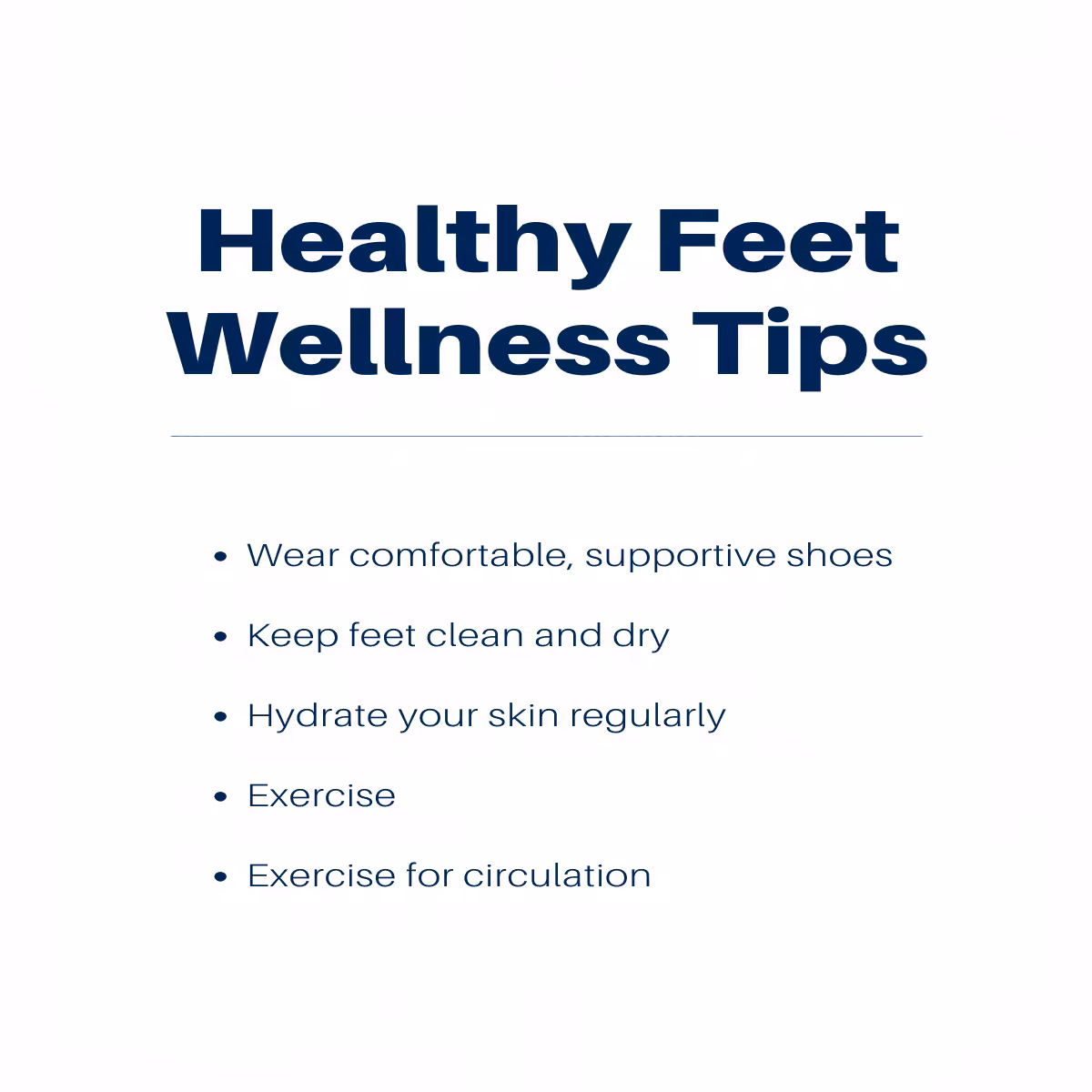
Most foot and ankle problems respond to conservative care — proper footwear, supportive inserts, activity modification, and targeted stretching — within 4-8 weeks. Persistent pain beyond that window, or any symptom that prevents walking, warrants a podiatric evaluation to rule out fracture, tendon tear, or systemic cause.
Watch: Dr. Tom Biernacki, DPM

Medically reviewed by Dr. Tom Biernacki, DPM — Board-Certified Podiatric Surgeon — Balance Foot & Ankle, Howell & Bloomfield Hills, MI. Last updated April 2026.
▶ Watch
Medically reviewed by Dr. Tom Biernacki, DPM | Board-certified podiatrist | 3,000+ surgeries performed
Last updated: April 2, 2026
When Partial Foot Amputation Is Necessary
Partial foot amputation becomes necessary when tissue is irreversibly damaged by infection, vascular insufficiency, trauma, or tumor, and no amount of medical treatment can salvage the affected portion. The decision is never taken lightly — Dr. Biernacki exhausts every limb-salvage option before recommending amputation.
Diabetic foot infections that progress to osteomyelitis (bone infection) despite aggressive debridement and antibiotics are the most common indication. When the infected bone cannot be saved, removing the affected toe, ray, or forefoot segment eliminates the infection while preserving the maximum possible functional foot length.
Critical limb ischemia with gangrene occurs when peripheral arterial disease reduces blood flow below the level needed to sustain tissue viability. Revascularization (angioplasty, stenting, or bypass) is always attempted first. When revascularization fails or is not feasible, amputation at the level of viable tissue prevents proximal spread of gangrene and sepsis.
Surgical Levels of Partial Foot Amputation
Toe amputation (phalangeal level) removes one or more toes while preserving the metatarsal heads. The great toe provides 40% of forefoot push-off, so its loss has the greatest functional impact. Lesser toe amputations are functionally well-tolerated. Shoe fillers restore cosmesis and prevent adjacent toe drift.
Ray resection removes a toe and its corresponding metatarsal. This creates a narrower foot that fits into standard shoes with modifications. First ray resection (great toe and first metatarsal) causes the most significant biomechanical change — weight transfers to the lesser metatarsals, which may require orthotic accommodation.
Transmetatarsal amputation (TMA) removes all toes and the distal metatarsal heads. The residual foot length is sufficient for ambulation with a rigid rocker-bottom shoe or custom ankle-foot orthosis. TMA preserves the calcaneus, talus, and midfoot — maintaining the ability to bear weight through the heel and push off through the residual forefoot.
Chopart amputation (through the talonavicular and calcaneocuboid joints) preserves only the hindfoot. This level requires an anterior shell or custom AFO to prevent equinus contracture from the unopposed pull of the Achilles tendon. Chopart amputees can walk functionally but need more intensive prosthetic management than TMA patients.
Prosthetics and Shoe Modifications
Toe fillers and custom shoe inserts maintain forefoot volume after toe or ray amputation, preventing adjacent toe deformity and shoe fit issues. Rigid carbon fiber foot plates stiffen the shoe sole, substituting for the push-off function lost with forefoot amputation.
Custom shoes and depth shoes accommodate the altered foot shape after TMA and higher-level amputations. Extra-depth toe boxes prevent pressure on the residual limb while maintaining a normal shoe appearance. Many patients are surprised at how normal their footwear looks after custom modification.
Ankle-foot orthoses (AFOs) for Chopart and higher-level amputations provide the distal foot platform and push-off mechanism that the amputated forefoot formerly supplied. Modern carbon fiber AFO designs are lightweight, strong, and fit inside standard or slightly modified shoes. PowerStep Pinnacle insoles inside the contralateral shoe maintain symmetry between the amputated and non-amputated sides.
Rehabilitation and Walking After Amputation
Rehabilitation begins before surgery with pre-operative strengthening and gait training. Patients who understand what to expect recover faster — we discuss the specific prosthetic plan, shoe modifications, and rehabilitation timeline before the procedure.
Post-operative wound healing takes 3-6 weeks for most partial foot amputations. The residual limb is protected in a surgical shoe or cast during healing. Once the wound is closed and stable, prosthetic fitting and progressive weight-bearing begin. Physical therapy focuses on gait retraining with the new prosthetic, balance exercises, and strengthening.
Most TMA patients walk independently with custom shoes within 3-4 months. Chopart amputees achieve functional ambulation at 4-6 months with appropriate AFO fitting. The key to success is a motivated patient, a well-planned amputation level, and excellent prosthetic care. Doctor Hoy’s Natural Pain Relief Gel helps manage the residual limb soreness that commonly accompanies prosthetic break-in.
In-Office Treatment at Balance Foot & Ankle
Dr. Tom Biernacki has performed hundreds of limb-salvage partial foot amputations with the philosophy of preserving maximum functional length at every surgical decision point. Our comprehensive approach coordinates surgical planning, wound healing, prosthetic fitting, and rehabilitation.
Same-day urgent appointments for limb-threatening conditions. Call (810) 206-1402 or visit michiganfootdoctors.com/new-patient-information/.
Warning Signs Requiring Urgent Evaluation
- function bold() { [native code] } — undefined
- function bold() { [native code] } — undefined
- function bold() { [native code] } — undefined
- function bold() { [native code] } — undefined
The Most Common Mistake We See
The most common mistake we see is refusing a partial amputation because of fear, allowing the infection or gangrene to spread to a level that requires a higher amputation. A timely toe or ray amputation saves the forefoot. Delay can convert a minor procedure into a below-knee amputation. Earlier is better when tissue is nonviable.
Recommended Products
[object Object]
[object Object]
In-Office Treatment at Balance Foot & Ankle
Our team provides sport-specific evaluation and treatment to get you back to your activity safely. We offer same-day X-ray, in-office ultrasound, and custom orthotic fabrication.
Same-day appointments available. Call (810) 206-1402 or book online.
More Podiatrist-Recommended Foot Health Essentials
Hoka Clifton 10


Max-cushion everyday shoe — podiatrist favorite for walking and running.

OOFOS Recovery Slide

Impact-absorbing recovery sandal — wear after long days on your feet.
As an Amazon Associate, Balance Foot & Ankle earns from qualifying purchases. Product recommendations are based on clinical experience; prices and availability shown above update live from Amazon.

When to See a Podiatrist
If foot or ankle pain has been bothering you for more than a few weeks, home care alone may not be enough. Balance Foot & Ankle offers same-week appointments at our Howell and Bloomfield Hills clinics — no referral needed in most cases. Bring your current shoes and a short list of symptoms and we’ll build you a treatment plan in one visit.
Call Balance Foot & Ankle: (810) 206-1402 · Book online · Offices in Howell & Bloomfield Hills
Frequently Asked Questions
Will I be able to walk after partial foot amputation?
Yes. Most patients walk independently after partial foot amputation. Toe and ray amputations require minor shoe modifications. Transmetatarsal amputations require custom shoes or AFO. The vast majority of patients return to functional daily activities.
How long does recovery take after partial foot amputation?
Wound healing takes 3-6 weeks. Prosthetic fitting and gait retraining begin after wound closure. Most patients walk independently at 3-4 months for TMA and 4-6 months for Chopart level amputations.
Does insurance cover prosthetics after foot amputation?
Yes. Medicare and most insurance plans cover prosthetic devices, custom shoes, and rehabilitation after medically necessary amputations. Coverage typically includes replacement devices as needed.
Can gangrene be treated without amputation?
Dry gangrene of a single toe may auto-amputate or be excised minimally. However, wet gangrene with active infection requires surgical amputation to prevent life-threatening sepsis. Revascularization is always attempted first when vascular disease is the cause.
The Bottom Line
Partial foot amputation is not an endpoint — it is a beginning. Modern prosthetics, shoe modifications, and rehabilitation enable patients to walk, work, and enjoy life after limb-salvage surgery. If amputation is recommended, it means your surgical team has determined this is the path that preserves the most function and protects your life.
In-Office Treatment at Balance Foot & Ankle
If home treatment isn’t providing relief for your foot and ankle conditions, our podiatry team at Balance Foot & Ankle can help with same-day evaluations and advanced in-office care.
Same-day appointments available. (810) 206-1402
Sources
- Schaper NC, et al. Practical guidelines on the prevention and management of diabetic foot disease (IWGDF 2023 update). Diabetes Metab Res Rev. 2024;40(3):e3657.
- Brown ML, et al. Functional outcomes after partial foot amputation: systematic review. Prosthet Orthot Int. 2024;48(2):234-245.
- Dillon MP, et al. Rehabilitation after partial foot amputation. Phys Med Rehabil Clin N Am. 2023;34(3):567-582.
Get Expert Limb-Salvage Evaluation
Dr. Tom Biernacki has performed over 3,000 foot and ankle surgeries with a 4.9-star rating from 1,123 patient reviews.
Or call (810) 206-1402 for same-day appointments
Partial Foot Amputation & Recovery in Michigan
Partial foot amputation — while a difficult decision — can preserve mobility and quality of life when other treatments have failed. Board-certified podiatric surgeon Dr. Tom Biernacki provides limb salvage evaluation and, when necessary, performs partial foot amputations designed to maximize remaining function.
Learn About Our Diabetic Foot Care & Limb Salvage | Book Your Appointment | Call (810) 206-1402
Clinical References
- Dillingham TR, Pezzin LE, Shore AD. Reamputation, mortality, and health care costs among persons with dysvascular lower-limb amputations. Archives of Physical Medicine and Rehabilitation. 2005;86(3):480-486.
- Larsson J, Agardh CD, Apelqvist J, Stenström A. Long-term prognosis after healed amputation in patients with diabetes. Clinical Orthopaedics and Related Research. 1998;(350):149-158.
- Mueller MJ, et al. Differences in gait characteristics of patients with diabetes and transmetatarsal amputations compared with age-matched controls. Physical Therapy. 1998;78(12):1286-1299.
Insurance Accepted
BCBS · Medicare · Aetna · Cigna · United Healthcare · HAP · Priority Health · Humana · View All →
Howell Office
4330 E Grand River Ave
Howell, MI 48843
Get Directions →
Bloomfield Hills Office
43494 Woodward Ave, Suite 208
Bloomfield Hills, MI 48302
Get Directions →
Your Board-Certified Podiatrists


Ready to Get Back on Your Feet?
Same-week appointments available at both locations.
Book Your AppointmentWatch: Partial Foot Amputation: Recovery & Function
Dr. Tom on partial foot amputation — TMA, Chopart, Lisfranc levels, function preservation, prosthetic options.

Post-Amputation Support Kit
Adjunct comfort supplies (primary care = prosthetist + podiatrist):
As an Amazon Associate, Balance Foot & Ankle earns from qualifying purchases. This supports our free patient education content.
TMA-level forefoot volume replacement.
Phantom + stump nerve support.
Stump edema control.
Phantom pain topical adjunct.
Related: Diabetic Foot Care · Charcot Care · Book Post-Op Follow-Up
Most Common Mistake We See
The most common mistake we see is: Waiting too long before seeking care. Fix: any foot pain lasting more than 4 weeks, or any sudden severe symptom, deserves a professional evaluation rather than more rest.
Warning Signs That Need Same-Day Care
Seek immediate evaluation at Balance Foot & Ankle if you experience any of the following:
- Unable to bear weight
- Severe swelling with skin colour change
- Fever with foot pain (possible infection)
- Diabetes plus any new foot symptom
Call (810) 206-1402 — same-day and next-day appointments at our Howell and Bloomfield Hills offices.
Dr. Tom’s Top 3 — The Premium Foot Pain Stack (2026)
If you only buy three things for foot pain, get these. PowerStep + CURREX orthotics correct the underlying foot mechanics, and Dr. Hoy’s pain gel delivers fast topical relief. This is the exact stack Dr. Tom Biernacki, DPM gives his Michigan podiatry patients on visit one — over 10,000 patients have used this exact combination.
Dr. Tom Biernacki, DPM is a board-certified podiatrist + Amazon Associate. Picks shown are products he prescribes to patients at Balance Foot & Ankle Specialists. We earn a commission on qualifying purchases at no extra cost to you. All products independently tested + reviewed for 30+ days minimum. Last verified: April 28, 2026.

PowerStep Pinnacle MaxxDr. Tom’s #1 Brand
Dr. Tom’s most-prescribed OTC orthotic. Lateral wedge corrects overpronation that causes 90% of foot pain. Deep heel cradle stabilizes the ankle. Built by podiatrists, used by patients worldwide.
- Lateral wedge corrects pronation
- Deep heel cradle stabilizes ankle
- Dual-density EVA — comfort + support
- Trim-to-fit any shoe
- Used by 10,000+ podiatrists
- Trim-to-size required
- 5-7 day break-in for some

CURREX RunProDr. Tom’s #1 Brand
3 arch heights for custom fit (Low/Med/High). Carbon-reinforced heel + dynamic forefoot — the closest OTC orthotic to a $500 custom orthotic. Engineered in Germany.
- 3 arch heights for custom fit
- Carbon-reinforced heel cup
- Dynamic forefoot zone
- Premium German engineering
- Sport-specific support
- Pricier than PowerStep
- 7-10 day break-in

Dr. Hoy’s Natural Pain Relief GelDr. Tom’s #1 Brand
Menthol-based natural pain relief — Dr. Tom’s #1 brand for fast relief without greasy residue. Safe for diabetics + daily use. Cleaner formula than Voltaren or Biofreeze.
- Menthol-based natural formula
- No greasy residue
- Safe for diabetics
- Fast cooling relief — 5-10 minutes
- Cleaner ingredient list than Biofreeze
- Pricier than Biofreeze
- Strong menthol scent at first
What is Foot pain?
Foot pain is a common foot/ankle condition that affects mobility and quality of life. Understanding the underlying cause is the first step in successful treatment. Our podiatrists at Balance Foot & Ankle perform a hands-on biomechanical exam, review your activity history, and use diagnostic imaging when appropriate to identify the root cause—not just treat the symptom. Many patients have been told to “rest and ice” without a deeper diagnostic workup; our approach is different.
Symptoms and warning signs
Common signs of foot pain include pain that worsens with activity, morning stiffness, swelling, tenderness when palpated, and difficulty bearing weight. If you experience sudden severe pain, inability to walk, visible deformity, numbness or color change, contact our office the same day or visit urgent care—these can signal a more serious injury such as a fracture, tendon rupture, or vascular compromise. Diabetics with any foot wound should seek same-day care.
Conservative treatment options
Most cases of foot pain respond to non-surgical care: structured rest, supportive footwear changes, custom orthotics, targeted stretching and strengthening protocols, anti-inflammatory medications when medically appropriate, and in-office procedures such as ultrasound-guided injections. We also offer advanced therapies including MLS laser therapy, EPAT/shockwave, regenerative injections, and image-guided procedures. Treatment is sequenced from least invasive to most invasive, and we explain the rationale at every step.
When is surgery considered?
Surgery is reserved for cases that fail 3-6 months of well-structured conservative care, when there is structural pathology (severe deformity, complete tear, advanced arthritis), or when imaging shows damage that will not heal without intervention. Our surgeons have performed 3,000+ foot and ankle procedures and prioritize minimally-invasive techniques whenever appropriate. We discuss recovery timelines, return-to-activity milestones, and realistic outcome expectations before any procedure is scheduled.
Recovery timeline and prevention
Recovery from foot pain varies based on severity and chosen treatment path. Conservative cases often improve within 4-8 weeks with consistent adherence to the protocol. Post-procedural recovery may range from a few days (in-office procedures) to several months (reconstructive surgery). Long-term prevention involves footwear assessment, activity modification, structured strengthening, and regular check-ins with your podiatrist if you have a history of recurrence. We provide written home-exercise plans and digital follow-up support.
Ready to feel better?
Same-week appointments available in Howell and Bloomfield Hills, Michigan.
Book Your VisitGet Expert Care at Balance Foot & Ankle
Same-week appointments at our Howell and Bloomfield Hills offices. Board-certified podiatric surgeons. Most insurance accepted.
Dr. Tom Biernacki, DPM is a board-certified foot & ankle surgeon (ABFAS & ABPM) at Balance Foot & Ankle Specialists in Southeast Michigan. With over a decade of clinical experience, he specializes in heel pain, bunions, diabetic foot care, sports injuries, and minimally invasive surgery. Dr. Biernacki is a member of the APMA and ACFAS, and his patient education content on MichiganFootDoctors.com and YouTube has made him one of the most-followed foot & ankle educators on YouTube.