

✅ Medically Reviewed by Dr. Tom Biernacki, DPM
Board-certified podiatric physician & surgeon | Balance Foot & Ankle | Updated April 2026
⚡ Quick Answer: What Is a Stress Fracture of the Calcaneus?
Treatment at Balance Foot & Ankle: Foot Emergency Guide →
A calcaneal stress fracture is an overuse injury causing deep heel pain that worsens with activity. Treatment requires non-weight-bearing rest for 6–8 weeks in a walking boot or with crutches.
Calcaneus Stress Fracture: Diagnosis & Recovery (Podiatrist 2026)
A calcaneus (heel) stress fracture is a hairline crack in the heel bone caused by repetitive impact — common in runners, military recruits, and post-menopausal women with low bone density. Symptoms: gradual onset heel pain that worsens with activity, point tenderness when squeezing the heel side-to-side, swelling, sometimes bruising. Initial X-rays often miss it — MRI or bone scan confirms diagnosis at 7-14 days.
In my Michigan podiatry clinic, my calcaneal stress fracture protocol: (1) 4-6 weeks NWB or PWB in a CAM boot, (2) vitamin D + calcium supplementation, (3) bone-stimulator device for slow-healing cases, (4) gradual return to running over 4-8 weeks. About 90% heal completely. Red flag: can’t bear ANY weight + sudden severe pain = possible complete fracture; needs urgent X-ray/MRI. Female athletes with stress fractures need DEXA scan + endocrine workup (RED-S syndrome).
Same-Week Appointments at Balance Foot & Ankle
Three board-certified podiatric surgeons. 950K+ YouTube subscribers. 1,123+ five-star reviews. Howell & Bloomfield Hills, Michigan.
✅ Medically reviewed by Dr. Thomas Biernacki, DPM — Board-Certified Podiatrist · Last updated April 7, 2026
Medically Reviewed

Dr. Carl Jay, DPM — Board-Certified Podiatrist
Balance Foot & Ankle · Howell & Bloomfield Hills, MI

Dr. Daria Gutkin, DPM — Board-Certified Podiatrist
Balance Foot & Ankle · Howell & Bloomfield Hills, MI
Last updated: April 2026 · Evidence-based content
QUICK ANSWER
A calcaneus stress fracture is a hairline crack in the heel bone caused by repetitive impact — most common in runners, military recruits, and people who suddenly increase their activity level. The hallmark symptom is deep heel pain that worsens with activity and improves with rest, unlike plantar fasciitis which is worst with the first steps in the morning. Treatment involves 6–8 weeks of protected weight-bearing in a walking boot, followed by a gradual return to activity. Most stress fractures heal completely without surgery.

Table of Contents
- What Is a Calcaneus Stress Fracture?
- Stress Fracture vs. Plantar Fasciitis — How to Tell the Difference
- Causes & Risk Factors
- Symptoms
- How We Diagnose It
- Treatment Protocol
- Recovery Timeline
- Best Shoes & Supports for Healing
- How to Prevent Recurrence
- Warning Signs — See a Podiatrist Now
- Frequently Asked Questions
- Bottom Line
What Is a Calcaneus Stress Fracture?
Invalid table id.If your heel hurts deep inside the bone — a throbbing ache that gets worse the longer you stand and better when you sit down — you may be dealing with more than just soreness. A calcaneus stress fracture is a small, incomplete crack in the heel bone that develops gradually from repeated impact rather than a single traumatic event.
The calcaneus is the largest bone in the foot and absorbs tremendous force with every step. During running, the heel experiences impact forces of 2–3 times your body weight. When the rate of bone breakdown from this repetitive loading exceeds the body’s ability to repair it, microscopic cracks form — and if activity continues, those cracks can progress into a true stress fracture.
Calcaneal stress fractures account for roughly 2 percent of all stress fractures, but they are frequently misdiagnosed as plantar fasciitis or heel bruises because they share similar pain patterns. The distinction matters because the treatment approaches are very different — plantar fasciitis improves with stretching and activity modification, while a stress fracture requires strict offloading to heal.
Stress Fracture vs. Plantar Fasciitis — How to Tell the Difference
This is one of the most important distinctions in heel pain diagnosis. Both conditions cause significant heel pain, but they behave differently and require different treatment. Here is how we differentiate them in our clinic:
| Feature | Calcaneus Stress Fracture | Plantar Fasciitis |
|---|---|---|
| Pain timing | Worsens progressively throughout the day with activity | Worst with first steps in morning, improves with walking |
| Pain location | Deep inside the heel, often both sides | Bottom of heel at plantar fascia origin |
| Squeeze test | Positive — squeezing sides of heel reproduces pain | Negative — squeezing sides does not hurt |
| Swelling | Often present around the heel | Usually absent |
| Effect of rest | Pain resolves completely with rest | Pain may persist as dull ache even at rest |
| Onset | Often follows sudden increase in activity | Gradual onset without clear trigger |
| X-ray findings | May show fracture line or sclerosis (after 2–3 weeks) | Possible heel spur (not diagnostic) |
| Best initial imaging | MRI (gold standard for early detection) | Ultrasound (shows fascia thickening) |
| Treatment focus | Offloading — walking boot, limited weight-bearing | Stretching, orthotics, physical therapy |
The calcaneal squeeze test is the single most useful clinical test. If squeezing the sides of your heel together between your palms reproduces your pain, there is a significant chance of a stress fracture. Plantar fasciitis rarely causes pain with this maneuver because the fascia is on the bottom of the heel, not the sides.
Causes & Risk Factors
Calcaneal stress fractures develop when repetitive mechanical loading overwhelms the bone’s ability to remodel and repair. Several factors increase risk:
1. Sudden Increase in Activity
This is the most common cause. Running mileage increases of more than 10 percent per week, starting a new high-impact exercise program, or transitioning from a sedentary lifestyle to a physically demanding job all place sudden stress on the calcaneus that the bone is not conditioned to handle. Military recruits in basic training are among the highest-risk populations for this exact reason.
2. Running and High-Impact Sports
Distance runners, basketball players, gymnasts, and dancers are at elevated risk because their activities deliver repetitive high-impact forces directly to the heel. Running on hard surfaces (concrete, asphalt) increases loading on the calcaneus compared to softer surfaces like trails or tracks.
3. Low Bone Density
Osteopenia and osteoporosis weaken the calcaneus, making it vulnerable to stress fractures even with normal activity levels. Postmenopausal women and patients on long-term corticosteroid therapy are particularly at risk. If you develop a calcaneal stress fracture without an obvious increase in activity, bone density testing should be considered.
4. Nutritional Deficiencies
Calcium and vitamin D deficiency impair bone remodeling and repair. The female athlete triad — disordered eating, menstrual irregularities, and low bone density — is an important risk factor in young female athletes. Adequate nutrition is not just about prevention; it directly affects how quickly a stress fracture heals.
5. Inadequate Footwear
Worn-out running shoes lose their shock absorption capacity, transmitting more impact force directly to the heel bone. Shoes should be replaced every 300–500 miles for runners. Minimalist shoes and thin-soled flats also increase calcaneal loading and may contribute to stress fracture development in susceptible individuals.
6. Biomechanical Factors
Patients with rigid high arches (cavus feet) absorb less shock through foot pronation, concentrating impact forces on the heel. Leg length discrepancies, tight calf muscles, and rearfoot varus alignment can also alter load distribution and increase stress on the calcaneus.
Symptoms of a Calcaneus Stress Fracture
Calcaneal stress fractures have a characteristic pattern that differs from other types of heel pain:
Key Symptoms:
- Deep, aching heel pain that feels like it is coming from inside the bone rather than from a specific spot on the surface
- Pain that worsens with weight-bearing activity and improves significantly with rest — this is the most reliable distinguishing feature
- Positive calcaneal squeeze test — pain when the sides of the heel are compressed together
- Diffuse swelling around the heel — the heel may look puffy compared to the other side
- Pain that appeared after increasing activity level — typically develops over days to weeks, not suddenly
- Inability to run or jump — these high-impact activities become too painful to perform
- Pain with prolonged standing — even standing in place becomes uncomfortable
- Night pain in advanced cases — a throbbing ache at night suggests the fracture may be progressing
Many patients recall a specific period when they increased their training volume or started a new activity — often 2–4 weeks before symptoms began. This “loading history” is one of the most important diagnostic clues.
How We Diagnose a Calcaneus Stress Fracture
Diagnosis begins with a thorough clinical examination and is confirmed with imaging when a stress fracture is suspected.
Physical examination — The calcaneal squeeze test is performed by compressing both sides of the heel simultaneously. A positive test (pain with squeezing) has high sensitivity for stress fractures. We also assess for swelling, skin temperature changes, and point tenderness.
X-rays — Standard heel X-rays are often normal in the first 2–3 weeks of a stress fracture because the crack is too small to see. After 2–3 weeks, a sclerotic line (a white line of new bone formation) may become visible on X-ray, confirming the fracture. Because early X-rays can miss the diagnosis, a negative X-ray does not rule out a stress fracture.
MRI — This is the gold standard for diagnosing calcaneal stress fractures. MRI detects bone marrow edema (swelling inside the bone) that appears weeks before any changes show on X-ray. An MRI can also grade the severity of the stress injury — from a mild stress reaction (pre-fracture) to a complete fracture line — which directly guides treatment decisions.
Bone scan — A nuclear bone scan is highly sensitive for stress fractures and shows increased uptake in the calcaneus. While less specific than MRI (it can be positive with other conditions), it is sometimes used when MRI is not available or not tolerated.
Treatment Protocol
The cornerstone of calcaneal stress fracture treatment is reducing mechanical load on the heel bone while optimizing the body’s ability to repair it. Surgery is rarely needed — the vast majority of calcaneal stress fractures heal completely with conservative management.
Acute Phase (Weeks 1–4): Offloading
Walking boot — A CAM (controlled ankle motion) walking boot is the standard treatment. The boot redistributes weight away from the heel and limits ankle motion that stresses the calcaneus. Most patients wear the boot full-time for 4–6 weeks, removing it only for sleeping and bathing.
Activity modification — All high-impact activities must stop completely. This includes running, jumping, prolonged walking, stair climbing, and standing for long periods. Pool running, swimming, and seated cycling are acceptable alternatives for maintaining cardiovascular fitness.
Non-weight-bearing in severe cases — If MRI shows a complete fracture line or if pain is severe with any weight-bearing, a period of crutch-assisted non-weight-bearing may be necessary for the first 1–2 weeks before transitioning to the walking boot.
Nutritional optimization — Ensure adequate calcium intake (1,000–1,200 mg/day from food and supplements) and vitamin D (1,000–2,000 IU/day). We check vitamin D levels in all patients with stress fractures and correct deficiencies aggressively, as low vitamin D significantly delays healing.
Recovery Phase (Weeks 4–8): Gradual Loading
Transition to supportive shoes — Once pain-free in the boot (usually around week 4–6), begin wearing a well-cushioned, supportive shoe with orthotic insoles. The transition should be gradual — alternate between the boot and shoes for the first week.
Gentle walking program — Start with 10–15 minutes of flat-surface walking and increase by 5 minutes every 2–3 days as tolerated. If any heel pain returns during walking, return to the boot for an additional week.
Physical therapy — Begin gentle calf stretching, ankle range-of-motion exercises, and progressive strengthening. Pool-based exercises are excellent during this phase because water reduces impact on the healing bone while allowing movement.
Return to Activity (Weeks 8–12+)
Gradual return to running — Begin with walk-run intervals on a soft surface (track, grass, or treadmill). A typical starting point is alternating 1 minute of jogging with 2 minutes of walking for 20 minutes. Progress by gradually increasing the jogging intervals while decreasing walking intervals over 4–6 weeks.
Follow-up imaging — A repeat X-ray or MRI at 6–8 weeks confirms healing before returning to full activity. The stress fracture should show evidence of new bone formation (callus) and resolution of bone marrow edema.
Recovery Timeline
| Timeframe | Activity Level | What to Expect |
|---|---|---|
| Weeks 1–2 | Walking boot, minimal weight-bearing | Pain with standing, swelling present |
| Weeks 3–4 | Walking boot, normal gait pattern | Pain decreasing, swelling resolving |
| Weeks 5–6 | Transitioning to supportive shoes | Minimal pain with walking, squeeze test improving |
| Weeks 7–8 | Full walking, gentle PT exercises | Pain-free walking, follow-up imaging |
| Weeks 9–12 | Walk-run intervals, sport-specific training | Gradual return to impact activities |
| Weeks 12–16 | Full activity including running and sports | Complete healing confirmed on imaging |
Most patients can return to full running by 10–14 weeks. Returning to activity too quickly is the number one cause of stress fracture recurrence — patience during recovery pays off with a much lower risk of re-injury.
Best Shoes & Supports for Calcaneal Stress Fracture Recovery
Once you transition out of the walking boot, the right shoes and insoles protect the healing bone and reduce the risk of recurrence.
OUR #1 RECOMMENDATION
Hoka Bondi — Best Maximum-Cushion Shoe for Heel Stress Fractures
The Hoka Bondi delivers the most heel cushioning of any running shoe on the market. Its thick, oversized midsole absorbs impact forces before they reach the calcaneus, making it the ideal shoe for transitioning out of a walking boot. The meta-rocker geometry promotes a smooth gait cycle that reduces peak loading on the heel during push-off. Patients recovering from calcaneal stress fractures consistently report that this shoe allows longer, more comfortable walks than any other option.
PowerStep Pinnacle Orthotic Insoles
These semi-rigid orthotics distribute pressure evenly across the foot, reducing the concentration of force on the heel during walking. The built-in heel cradle and arch support work together to optimize foot biomechanics, which helps protect the calcaneus from excessive loading during the recovery phase. They fit well inside the Hoka Bondi and most other athletic shoes.
OOFOS OOriginal Recovery Sandals
OOFOS sandals are made from OOfoam technology that absorbs 37 percent more impact than traditional footwear foam. They are an excellent option for around-the-house wear during recovery — providing significant heel cushioning in a convenient slip-on format. Many patients wear them as their “indoor shoe” while healing from a calcaneal stress fracture.
How to Prevent Calcaneal Stress Fracture Recurrence
Once you have had one stress fracture, the risk of another increases. These evidence-based strategies significantly reduce recurrence risk:
Follow the 10 percent rule — Never increase weekly running mileage, training duration, or exercise intensity by more than 10 percent per week. This gives bone sufficient time to adapt to increasing loads.
Replace shoes regularly — Running shoes should be replaced every 300–500 miles. Track the mileage on your shoes using a running app or simply marking the date you started using them.
Optimize calcium and vitamin D — Aim for 1,000–1,200 mg of calcium daily from food sources (dairy, leafy greens, fortified foods) plus a supplement if needed. Maintain vitamin D levels above 30 ng/mL — many patients in Michigan are deficient and benefit from supplementation, especially during winter months.
Cross-train — Incorporate low-impact activities (cycling, swimming, elliptical) into your routine to maintain fitness while reducing cumulative heel loading. Alternating running days with cross-training days is one of the most effective prevention strategies.
Vary running surfaces — Alternate between softer surfaces (trails, tracks, grass) and pavement. This varies the stress patterns on the calcaneus rather than loading it identically with every run.
Warning Signs — See a Podiatrist Now
⚠ When to Seek Immediate Care
- Heel pain that worsens with every day of continued activity — progressive pain is the hallmark of a stress fracture that is getting worse
- Positive squeeze test — pain when you compress the sides of your heel together with your hands
- Swelling around the heel without a history of direct trauma
- Heel pain that started after a significant increase in training — especially in runners or military recruits
- Night pain or pain at rest — suggests the fracture may be progressing toward a complete break
- History of previous stress fractures, eating disorders, or irregular periods — these risk factors warrant early evaluation
More Podiatrist-Recommended Stress Fracture Essentials
Max-Cushion Walking Shoe

Hoka Bondi 9 — maximum shock absorption during stress fracture recovery.
Foam Roller for Recovery

- Patented foam roller design offers a superior, multi-density exterior constructed over a rigid, hollow core
- Constructed from quality materials that won’t break down or lose shape from repeated use
- Includes access to free online instructional video library on foam rolling best practices from the experts at trigger point
- Trusted foam roller of physical and massage therapists, coaches, trainers and athletes
- Original Grid: Standard density, 13 x 5.5 inches, 500 pound weight limit; 1 year manufacturer's warranty
TriggerPoint foam roller — maintains lower-leg mobility during return to activity.
Supportive Insole

- The Pinnacle Full length insoles for men & women provide maximum cushioning, from high activity to moderate support. The PowerStep arch support shape provides stability to the foot and ankle, helping to relieve foot pain.
- When you spend all day on your feet, every step counts. PowerStep insoles are a podiatrist-recommended orthotic to help relieve & prevent foot pain related to athletes, runners, Plantar Fasciitis, heel spurs & other common foot, ankle & knee injuries
- The Pinnacle plantar fasciitis insoles offer superior heel cushioning and arch support. The dual-layer cushioning is designed to reduce stress and fatigue, while PowerStep premium arch support is designed for plantar fasciitis relief.
- The PowerStep Pinnacle arch support inserts for men & women can be worn in a variety of shoe types such as; athletic, walking, running, work & some casual shoes. Orthotic Inserts are ordered by shoe size, no trimming required.
- Made in the USA & backed by a 30-day money-back guarantee. PowerStep orthotic inserts for men & women are designed for shoes where the factory insole can be removed. HSA & FSA Eligible
PowerStep Pinnacle — distributes impact evenly across the foot.
As an Amazon Associate, Balance Foot & Ankle earns from qualifying purchases. Product recommendations are based on clinical experience; prices and availability shown above update live from Amazon.
When to See a Podiatrist
Most foot stress fractures heal in 6-8 weeks of protected weight-bearing — but rushing back to activity can turn a hairline fracture into a full break. Balance Foot & Ankle confirms stress fractures on X-ray or MRI and guides your return-to-running protocol. Don’t guess — we’ll tell you the exact week you can start jogging again.
Call Balance Foot & Ankle: (810) 206-1402 · Book online · Offices in Howell & Bloomfield Hills
Frequently Asked Questions
How long does it take for a calcaneus stress fracture to heal?
Most calcaneal stress fractures heal within 6–8 weeks of protected weight-bearing in a walking boot. Full return to running and high-impact sports typically takes 10–14 weeks. The healing timeline depends on the severity of the fracture (stress reaction vs. complete fracture line), patient age, nutritional status, and compliance with the offloading protocol. Patients who continue to walk on the fracture without a boot often experience delayed healing that extends recovery to 4–6 months.
Can you walk on a calcaneus stress fracture?
You can walk in a walking boot, which is the standard treatment. Walking without a boot or supportive device puts direct load on the fracture site and slows healing — or can cause the fracture to progress from a hairline crack to a complete break. Most patients are able to walk comfortably in a boot from the beginning of treatment. If pain is too severe even in the boot, a short period of crutch-assisted walking may be needed.
Will an X-ray show a calcaneus stress fracture?
Standard X-rays are often normal in the first 2–3 weeks because stress fractures start as microscopic cracks too small to see on X-ray. After 2–3 weeks, the body’s healing response produces a visible sclerotic (white) line on X-ray that confirms the fracture. If a stress fracture is strongly suspected clinically but the X-ray is normal, an MRI should be obtained — it detects bone marrow edema (the earliest sign of stress injury) weeks before X-ray changes appear.
What is the difference between a stress fracture and a stress reaction?
A stress reaction is the precursor to a stress fracture. It represents bone marrow edema (swelling inside the bone) without a visible fracture line — essentially, the bone is overloaded and inflamed but has not yet cracked. A stress fracture has a visible fracture line on MRI or X-ray. The distinction matters for treatment: stress reactions may heal in 4–6 weeks, while stress fractures typically take 6–8 weeks. Both require offloading, but stress reactions may be managed with a stiff-soled shoe rather than a full walking boot.
Bottom Line
A calcaneus stress fracture is a serious but highly treatable cause of heel pain. The key to a good outcome is early diagnosis — using the calcaneal squeeze test and MRI — followed by disciplined offloading in a walking boot for 6–8 weeks. Nutritional optimization with adequate calcium and vitamin D supports bone healing, and a gradual return to activity following the 10 percent rule prevents recurrence. If your heel pain worsens with activity, improves with rest, and appeared after increasing your training, do not assume it is plantar fasciitis — get it properly evaluated.
Sources
- Boden BP, Osbahr DC. “High-risk stress fractures: evaluation and treatment.” J Am Acad Orthop Surg. 2000;8(6):344-353.
- Pegrum J, et al. “Stress fractures of the foot and ankle.” Clin Sports Med. 2012;31(2):291-306.
- Banal F, et al. “Sensitivity and specificity of ultrasonography in early diagnosis of metatarsal bone stress fractures.” Rheumatology. 2009;48(2):183-185.
- Nattiv A, et al. “Stress injury to bone in the female athlete.” Br J Sports Med. 2013;47(7):486-492.
Heel Pain That Gets Worse With Every Step?
Dr. Carl Jay and Dr. Daria Gutkin diagnose and treat calcaneal stress fractures at Balance Foot & Ankle. Same-week appointments available in Howell & Bloomfield Hills, MI.
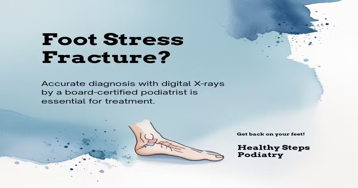
Concerned About a Calcaneus Stress Fracture?
A stress fracture of the heel bone (calcaneus) causes deep, aching heel pain that worsens with activity. Our podiatrists provide accurate diagnosis and effective management for calcaneal stress injuries.
📞 Or call us directly: (810) 206-1402
Clinical References
- Sormaala MJ, Niva MH, Kiuru MJ, et al. Stress injuries of the calcaneus detected with magnetic resonance imaging in military recruits. Journal of Bone and Joint Surgery. 2006;88(10):2237-2242.
- Welck MJ, Hayes T, Sherief T, et al. Stress fractures of the foot and ankle. Injury. 2017;48(8):1722-1726.
- Boden BP, Osbahr DC. High-risk stress fractures: evaluation and treatment. Journal of the American Academy of Orthopaedic Surgeons. 2000;8(6):344-353.
Insurance Accepted
BCBS · Medicare · Aetna · Cigna · United Healthcare · HAP · Priority Health · Humana · View All →

👟 Dr. Tom Also Recommends
Podiatrist Recommended Shoes 2026: Dr. Tom’s Top Picks for Every Condition
The right footwear can make or break your recovery. Dr. Tom’s complete guide to the best shoes for plantar fasciitis, flat feet, neuropathy, bunions & more — with clinical picks for every foot type.
See Dr. Tom’s Top Shoe Picks →Howell Office
3980 E Grand River Ave, Suite 140
Howell, MI 48843
Get Directions →
Bloomfield Hills Office
43700 Woodward Ave, Suite 207
Bloomfield Hills, MI 48302
Get Directions →
Your Board-Certified Podiatrists


Ready to Get Back on Your Feet?
Same-week appointments available at both locations.
Book Your AppointmentIn-Office Treatment at Balance Foot & Ankle
When conservative care isn’t enough, Dr. Tom Biernacki and the team at Balance Foot & Ankle offer advanced, same-day options — including Foot & Ankle Fracture Repair Michigan at our Howell and Bloomfield Hills clinics.
Same-day appointments available. Call (810) 206-1402 or book online.
Stress Fractures & Foot Trauma
Dr. Tom Biernacki, DPM is a double board-certified podiatrist and foot & ankle surgeon at Balance Foot & Ankle Specialists in Southeast Michigan. With over a decade of clinical experience, he specializes in heel pain, bunions, diabetic foot care, sports injuries, and minimally invasive surgery. Dr. Biernacki is a member of the APMA and ACFAS, and his patient education content on MichiganFootDoctors.com and YouTube has reached over one million views.
- Plantar Fasciitis: Diagnosis and Conservative Management (PubMed)
- Plantar Fasciitis (APMA)
- Diagnosis and Treatment of Plantar Fasciitis (PubMed / AAFP)
- Heel Pain (APMA)