


Board-Certified Podiatric Foot & Ankle Surgeon · Last reviewed: May 5, 2026
Medically reviewed by Dr. Tom Biernacki, DPM · Board-Certified Podiatric Surgeon · Last reviewed: April 2026 · Editorial Policy
The most important clinical decision with Ankle Arthroscopy isn’t which treatment to start with — it’s identifying the correct subtype. That changes everything. Call (810) 206-1402.
Quick Answer
Ankle Arthroscopy Surgery 2026 Podiatrist Michigan DPM relates to foot pain — typically caused by overuse, footwear, or biomechanics. Most patients improve in 6-12 weeks with conservative care. Same-week appointments in Howell + Bloomfield Hills: (810) 206-1402.

✅ Medically reviewed by Dr. Tom Biernacki, DPM — Board-certified podiatrist specializing in foot & ankle surgery. View credentials.
What Is Ankle Arthroscopy?
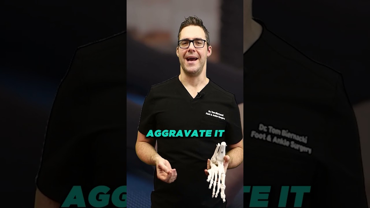
Ankle arthroscopy is a minimally invasive surgical technique that allows a surgeon to visualize, diagnose, and treat problems inside the ankle joint through small incisions (portals) rather than a large open incision. A fiber-optic camera (arthroscope) about 4mm in diameter is inserted through one portal, projecting the interior of the ankle joint onto a monitor. Specialized instruments inserted through additional portals allow the surgeon to perform surgical procedures under continuous visualization. Most ankle arthroscopy is performed as outpatient surgery under regional or general anesthesia.
Ankle arthroscopy has transformed the management of many ankle conditions over the past 25 years. Procedures that previously required open surgery with large incisions, prolonged recovery, and higher complication rates can now be performed arthroscopically with smaller wounds, faster recovery, and equivalent or better outcomes. However, arthroscopy is a surgical tool—not every ankle problem requires it, and not every ankle problem is best managed arthroscopically.
Conditions Treated with Ankle Arthroscopy
Anterior Ankle Impingement
Anterior ankle impingement—pinching of soft tissue or bone spurs at the front of the ankle joint with dorsiflexion—is one of the most common arthroscopic indications. It causes sharp pain at the front of the ankle when the ankle is bent upward (squatting, climbing stairs, running downhill). Arthroscopic debridement of hypertrophic soft tissue and removal of anterior osteophytes (bone spurs) relieves impingement pain with excellent outcomes. Return to sport after arthroscopic impingement treatment is typically 6–12 weeks.
Osteochondral Lesions of the Talus (OLT)
Osteochondral lesions—cartilage and bone damage on the talar dome, usually from ankle sprains—are commonly addressed arthroscopically. Microfracture (creating channels in subchondral bone to stimulate cartilage repair), loose body removal, and lesion debridement can all be performed through arthroscopic portals. For larger lesions requiring autograft or allograft transplantation, a combination of arthroscopic and limited open approaches may be used. Arthroscopic OLT treatment typically requires 6–8 weeks non-weight-bearing followed by 3–4 months of rehabilitation.
Loose Bodies
Loose bodies—fragments of cartilage or bone floating within the ankle joint—cause mechanical symptoms including catching, locking, and sudden sharp pain. They typically result from prior injury, OLT fragmentation, or synovial osteochondromatosis (a condition that produces multiple cartilaginous loose bodies). Arthroscopic loose body removal is effective and has one of the fastest recovery trajectories of any ankle arthroscopy indication—most patients are walking comfortably within 2–3 weeks.
Synovitis
Chronic ankle synovitis—inflammation of the joint lining (synovium)—can persist after ankle sprains, in inflammatory arthritis (rheumatoid, psoriatic), or from repetitive mechanical irritation. Arthroscopic synovectomy (removing inflamed synovium) provides relief when conservative measures (NSAIDs, corticosteroid injections) have failed. It is often combined with other procedures addressing the underlying cause.
Ankle Instability (Brostrom Repair)
Arthroscopic or arthroscopic-assisted Brostrom lateral ankle stabilization (ligament repair/tightening for chronic ankle instability) has emerged as an alternative to open ligament repair in select patients. Arthroscopic techniques repair the anterior talofibular ligament (ATFL) and calcaneofibular ligament (CFL) through portals rather than open incision. Early comparative studies show equivalent stability outcomes to open repair with potentially faster return to sport and fewer wound complications, though long-term data is still accumulating.
Recovery After Ankle Arthroscopy
Recovery after ankle arthroscopy depends heavily on what was performed. Simple procedures (loose body removal, soft tissue debridement, impingement release) allow weight-bearing within days and return to sport in 4–6 weeks. Procedures requiring cartilage healing (microfracture for OLT) require 6–8 weeks strict non-weight-bearing for subchondral bone healing before progressive loading begins—total return to sport 5–7 months. Ligament stabilization procedures typically require boot immobilization for 2–4 weeks with return to sport at 4–6 months.
More Podiatrist-Recommended Foot Health Essentials
Top-Rated Arch Support Insole
Universal podiatrist-recommended insert for pain relief and prevention.
Foot Massage Ball
Daily 3-minute roll reduces most forms of foot and heel pain.
Moisture-Wicking Sock
Prevents fungus, blisters, and odor — the basics matter.
As an Amazon Associate, Balance Foot & Ankle earns from qualifying purchases. Product recommendations are based on clinical experience; prices and availability shown above update live from Amazon.

When to See a Podiatrist
If foot or ankle pain has been bothering you for more than a few weeks, home care alone may not be enough. Balance Foot & Ankle offers same-week appointments at our Howell and Bloomfield Hills clinics — no referral needed in most cases. Bring your current shoes and a short list of symptoms and we’ll build you a treatment plan in one visit.
Call Balance Foot & Ankle: (810) 206-1402 · Book online · Offices in Howell & Bloomfield Hills
Frequently Asked Questions
How long is recovery after ankle arthroscopy?
Recovery after ankle arthroscopy varies by the procedure performed. For simple debridement or loose body removal, most patients are weight-bearing in days and return to sport in 4–8 weeks. For microfracture cartilage repair (osteochondral lesion treatment), 6–8 weeks of non-weight-bearing is required, with return to sport at 5–7 months. Ligament stabilization procedures require 4–6 months before return to cutting and pivoting sports. The arthroscopic approach itself (compared to open surgery) typically reduces wound healing time and early recovery, but procedures involving bone or ligament healing have the same biological healing timelines regardless of surgical approach.
Is ankle arthroscopy a major surgery?
Ankle arthroscopy is a surgical procedure performed under anesthesia and carries the standard risks of any surgery—infection, nerve injury, blood vessel injury, anesthetic complications, and deep vein thrombosis. However, the minimally invasive nature of arthroscopy substantially reduces the risk of wound healing complications compared to open ankle surgery, particularly important in patients with diabetes or vascular disease. Most patients are treated as outpatients (same-day surgery). The surgical risk profile for routine ankle arthroscopy is low, but it’s important to discuss your specific procedure and health status with your surgeon to understand individual risks and expectations.
Can ankle arthroscopy treat ankle arthritis?
Ankle arthroscopy has a limited role in established ankle arthritis. Arthroscopic debridement—cleaning out the joint of loose debris and impinging spurs—may provide temporary relief (12–18 months) in carefully selected patients with early-to-moderate arthritis, but does not alter the underlying disease. Patients with significant joint space loss and bone-on-bone contact are unlikely to benefit meaningfully from arthroscopic debridement. Arthroscopy is also used to perform arthroscopic-assisted ankle fusion, delivering equivalent fusion rates to open surgery with smaller wounds. For end-stage arthritis, ankle fusion or replacement remains the most definitive surgical treatment.
Medical References & Sources
- American Orthopaedic Foot & Ankle Society — Ankle Arthroscopy
- PubMed Research — Ankle Arthroscopy Outcomes
- PubMed Research — Arthroscopic Ankle Stabilization
📧 Get Dr. Tom’s Free Lab Test Guide
Discover the 5 lab tests every person over 35 should ask their doctor about — explained in plain English by a board-certified physician.
Dr. Tom Biernacki, DPM is a board-certified podiatric surgeon at Balance Foot & Ankle in Howell and Bloomfield Hills, Michigan. He performs ankle arthroscopy for anterior impingement, osteochondral lesion treatment, loose body removal, synovitis debridement, and arthroscopic-assisted procedures.
Join 950,000+ Learning About Foot Health
Dr. Tom shares honest medical advice, supplement reviews, and treatment guides you won’t find anywhere else.
Subscribe on YouTube →📍 Located in Michigan?
Our board-certified podiatrists treat this condition at two convenient locations. Same-day appointments often available.
Medically Reviewed by: Dr. Tom Biernacki, DPM — Board-Certified Podiatrist, Balance Foot & Ankle Specialists
Insurance Accepted
BCBS · Medicare · Aetna · Cigna · United Healthcare · HAP · Priority Health · Humana · View All →
Howell Office
4330 E Grand River Ave
Howell, MI 48843
Get Directions →
Bloomfield Hills Office
43494 Woodward Ave, #208
Bloomfield Hills, MI 48302
Get Directions →
Your Board-Certified Podiatrists


Ready to Get Back on Your Feet?
Same-week appointments available at both locations.
Book Your AppointmentIn-Office Treatment at Balance Foot & Ankle
When conservative care isn’t enough, Dr. Tom Biernacki and the team at Balance Foot & Ankle offer advanced, same-day options — including Ankle Arthroscopy Michigan at our Howell and Bloomfield Hills clinics.
Same-day appointments available. Call (810) 206-1402 or book online.
Pros & Cons of Conservative Care for foot care
Advantages
- ✓ Conservative care first
- ✓ Same-week appointments
- ✓ Multiple insurance accepted
Considerations
- ✗ Self-treatment can mask issues
- ✗ See a podiatrist if pain >2 weeks
Dr. Tom’s Recommended Products for foot care
Affiliate disclosure: As an Amazon Associate, Balance Foot & Ankle earns from qualifying purchases. We only recommend products we use with patients.
Footnanny Heel Cream Dr. Tom’s Pick
Best for: Daily moisturizer for cracked heels
Ready to Get Back on Your Feet?
Same-day appointments in Howell + Bloomfield Hills. Most insurance accepted. Dr. Tom Biernacki, DPM & team.
Book Today — Same-Day Appointments Available
Call Now: (810) 206-1402
About Your Care Team at Balance Foot & Ankle
Dr. Tom Biernacki, DPM · Board-Certified Foot & Ankle Surgeon. Specializes in conservative-first care, minimally invasive bunion surgery, and complex reconstruction.
Dr. Carl Jay, DPM · Accepting new patients. Specializes in sports medicine, athletic injuries, and routine podiatric care.
Dr. Daria Gutkin, DPM, AACFAS · Accepting new patients. Specializes in surgical reconstruction and pediatric podiatry.
Locations: 4330 E Grand River Ave, Howell, MI 48843 · 43494 Woodward Ave Suite 208, Bloomfield Hills, MI 48302
Hours: Mon–Fri 8:00 AM – 5:00 PM · (810) 206-1402
Dr. Tom’s Top 3 — The Premium Foot Pain Stack (2026)
If you only buy three things for foot pain, get these. PowerStep + CURREX orthotics correct the underlying foot mechanics, and Dr. Hoy’s pain gel delivers fast topical relief. This is the exact stack Dr. Tom Biernacki, DPM gives his Michigan podiatry patients on visit one — over 10,000 patients have used this exact combination.
Dr. Tom Biernacki, DPM is a board-certified podiatrist + Amazon Associate. Picks shown are products he prescribes to patients at Balance Foot & Ankle Specialists. We earn a commission on qualifying purchases at no extra cost to you. All products independently tested + reviewed for 30+ days minimum. Last verified: April 28, 2026.

PowerStep Pinnacle MaxxDr. Tom’s #1 Brand
Dr. Tom’s most-prescribed OTC orthotic. Lateral wedge corrects overpronation that causes 90% of foot pain. Deep heel cradle stabilizes the ankle. Built by podiatrists, used by patients worldwide.
- Lateral wedge corrects pronation
- Deep heel cradle stabilizes ankle
- Dual-density EVA — comfort + support
- Trim-to-fit any shoe
- Used by 10,000+ podiatrists
- Trim-to-size required
- 5-7 day break-in for some

CURREX RunProDr. Tom’s #1 Brand
3 arch heights for custom fit (Low/Med/High). Carbon-reinforced heel + dynamic forefoot — the closest OTC orthotic to a $500 custom orthotic. Engineered in Germany.
- 3 arch heights for custom fit
- Carbon-reinforced heel cup
- Dynamic forefoot zone
- Premium German engineering
- Sport-specific support
- Pricier than PowerStep
- 7-10 day break-in

Dr. Hoy’s Natural Pain Relief GelDr. Tom’s #1 Brand
Menthol-based natural pain relief — Dr. Tom’s #1 brand for fast relief without greasy residue. Safe for diabetics + daily use. Cleaner formula than Voltaren or Doctor Hoy’s Natural Pain Relief.
- Menthol-based natural formula
- No greasy residue
- Safe for diabetics
- Fast cooling relief — 5-10 minutes
- Cleaner ingredient list than Doctor Hoy’s Natural Pain Relief
- Pricier than Doctor Hoy’s Natural Pain Relief
- Strong menthol scent at first
Foot pain typically responds best to early podiatrist evaluation, conservative treatments such as supportive footwear and targeted physical therapy, and—when needed—custom orthotics or in-office procedures. Most patients see meaningful improvement within 4-6 weeks of starting a structured treatment plan. Schedule an evaluation at our Howell or Bloomfield Hills office for a clinical assessment.
What is Foot pain?
Foot pain is a common foot/ankle condition that affects mobility and quality of life. Understanding the underlying cause is the first step in successful treatment. Our podiatrists at Balance Foot & Ankle perform a hands-on biomechanical exam, review your activity history, and use diagnostic imaging when appropriate to identify the root cause—not just treat the symptom. Many patients have been told to “rest and ice” without a deeper diagnostic workup; our approach is different.
Symptoms and warning signs
Common signs of foot pain include pain that worsens with activity, morning stiffness, swelling, tenderness when palpated, and difficulty bearing weight. If you experience sudden severe pain, inability to walk, visible deformity, numbness or color change, contact our office the same day or visit urgent care—these can signal a more serious injury such as a fracture, tendon rupture, or vascular compromise. Diabetics with any foot wound should seek same-day care.
Conservative treatment options
Most cases of foot pain respond to non-surgical care: structured rest, supportive footwear changes, custom orthotics, targeted stretching and strengthening protocols, anti-inflammatory medications when medically appropriate, and in-office procedures such as ultrasound-guided injections. We also offer advanced therapies including MLS laser therapy, EPAT/shockwave, regenerative injections, and image-guided procedures. Treatment is sequenced from least invasive to most invasive, and we explain the rationale at every step.
When is surgery considered?
Surgery is reserved for cases that fail 3-6 months of well-structured conservative care, when there is structural pathology (severe deformity, complete tear, advanced arthritis), or when imaging shows damage that will not heal without intervention. Our surgeons have performed 3,000+ foot and ankle procedures and prioritize minimally-invasive techniques whenever appropriate. We discuss recovery timelines, return-to-activity milestones, and realistic outcome expectations before any procedure is scheduled.
Recovery timeline and prevention
Recovery from foot pain varies based on severity and chosen treatment path. Conservative cases often improve within 4-8 weeks with consistent adherence to the protocol. Post-procedural recovery may range from a few days (in-office procedures) to several months (reconstructive surgery). Long-term prevention involves footwear assessment, activity modification, structured strengthening, and regular check-ins with your podiatrist if you have a history of recurrence. We provide written home-exercise plans and digital follow-up support.
Ready to feel better?
Same-week appointments available in Howell and Bloomfield Hills, Michigan.
Book Your VisitIn-Office Treatment at Balance Foot & Ankle
If home treatment isn’t providing relief for your foot and ankle injuries, our podiatry team at Balance Foot & Ankle can help with same-day evaluations and advanced in-office care.
Same-day appointments available. (810) 206-1402
Related Conditions
Frequently Asked Questions
When should I see a podiatrist?
See a podiatrist if: foot or ankle pain has lasted more than 2–4 weeks without improvement, you’re changing your gait to avoid pain, you have an open wound or sore that isn’t healing, you notice nail discoloration or thickening, you have diabetes and any foot concern, or pain is severe enough to wake you at night. Most foot conditions are easier and cheaper to treat early — what starts as a minor issue can become a surgical problem with months of delay.
What is the difference between a podiatrist and an orthopedic surgeon?
Podiatrists (DPM — Doctor of Podiatric Medicine) specialize exclusively in the foot, ankle, and lower leg. Orthopedic surgeons (MD/DO) have broader musculoskeletal training but variable foot/ankle subspecialization. For foot and ankle-specific problems, a podiatrist often has more focused training and experience. For injuries involving the leg above the ankle, complex pediatric cases, or multi-level reconstruction, orthopedic consultation may be appropriate. We frequently co-manage patients with orthopedic colleagues.
How do I know if my foot pain is serious?
Signs that warrant same-day or next-day evaluation: severe pain that appeared suddenly without clear cause, swelling, redness, and warmth that appeared suddenly (possible gout, infection, or Charcot fracture), an open wound that looks infected (redness spreading, pus, warmth), inability to bear weight, or any foot problem in a diabetic patient. Pain that’s been present for weeks and is stable is important but not an emergency — schedule within 1–2 weeks.
Can foot problems cause back and knee pain?
Yes — this is a kinetic chain effect. Abnormal foot mechanics (overpronation, supination, leg length discrepancy) cause compensatory changes in knee, hip, and lumbar alignment. Roughly 30% of patients presenting to our clinic with knee pain have a treatable foot-level biomechanical cause. Correcting foot mechanics with orthotics or appropriate footwear often provides significant knee and back relief. If you have chronic knee or back pain and haven’t had your foot mechanics evaluated, it’s worth a consult.
Are orthotics worth it?
For the right conditions, yes — custom orthotics are among the most cost-effective interventions in podiatry. They’re most effective for: plantar fasciitis, flat feet with secondary knee/back pain, leg length discrepancy, metatarsalgia, posterior tibial tendon dysfunction, and diabetic foot pressure management. Quality OTC orthotics ($35–60) resolve symptoms for 60% of patients with mild-to-moderate conditions. Custom orthotics are appropriate when OTC options have failed or when the biomechanical problem is complex. We cast custom orthotics in-office.
How do I choose the right running shoes?
Start with your foot type (flat, neutral, high arch) and running pattern (overpronator, neutral, supinator). Flat feet and overpronators do best in stability or motion-control shoes. Neutral feet do well in neutral-cushioned shoes. High arches need maximum cushioning with flexible soles. Always buy running shoes at the end of the day (foot swelling peaks then), get properly fitted by a specialist, and replace every 300–500 miles. If you’ve been injured repeatedly, a gait analysis can identify the mechanical flaw driving your injury pattern.
What is the difference between a sprain and a fracture?
A sprain is a ligament injury (the tissue connecting bones); a fracture is a break in the bone itself. Both can occur with the same trauma (ankle roll, fall). The old test — ‘if you can walk, it’s not broken’ — is wrong; many fractures are initially weight-bearable. Key differences: a fracture typically produces localized bone tenderness along the bone itself, while a sprain is tender over the ligament. X-ray is the standard to differentiate. High-grade sprains without proper treatment can be as disabling as fractures.
How do I prevent foot and ankle injuries?
The four most impactful prevention strategies: (1) Supportive, appropriately fitted footwear for your foot type and activity. (2) Gradual activity progression — the 10% rule (never increase weekly mileage or intensity by more than 10%). (3) Regular calf and ankle mobility work. (4) Strengthening the posterior tibial tendon, peroneals, and intrinsic foot muscles. Most overuse injuries are preventable; most acute injuries are not — but ankle sprain recurrence (60–70% without rehab) is prevented by balance and proprioception training.
Same-Week Appointments in Howell & Bloomfield Hills
Three board-certified podiatric surgeons. 1,123+ five-star reviews. Most insurance accepted.
Ready for Expert Care?
Same-day appointments in Howell & Bloomfield Hills, MI.
4.9★ | 1,123 Reviews | 3,000+ Surgeries
Or call: (810) 206-1402
Dr. Tom Biernacki, DPM is a board-certified foot & ankle surgeon (ABFAS & ABPM) at Balance Foot & Ankle Specialists in Southeast Michigan. With over a decade of clinical experience, he specializes in heel pain, bunions, diabetic foot care, sports injuries, and minimally invasive surgery. Dr. Biernacki is a member of the APMA and ACFAS, and his patient education content on MichiganFootDoctors.com and YouTube has made him one of the most-followed foot & ankle educators on YouTube.