
★ 4.9 Stars · 1,123+ Reviews · Board-Certified Michigan Podiatrists
Sports Medicine for Foot & Ankle — Howell & Bloomfield Township
Faster Return to Activity with Expert Care
Active patients need a podiatrist who understands performance. Our team treats sprains, stress fractures, Achilles tendinopathy, and turf toe — with treatment plans designed to get you back faster and stronger.
Medically reviewed by Dr. Tom Biernacki, DPM, FACFAS
Board-certified podiatric surgeon | Balance Foot & Ankle | Last reviewed: May 2026
Quick answer: Sports medicine podiatry focuses on diagnosing and treating foot and ankle injuries in athletes and active individuals — from recreational runners to competitive athletes. Common conditions include plantar fasciitis, medial tibial stress syndrome, IT band friction syndrome, stress fractures, Achilles tendinopathy, and lateral ankle instability. Evidence-based treatment uses the PEACE & LOVE framework, biomechanical gait analysis, return-to-sport criterion testing, and — where appropriate — regenerative options including PRP and MLS laser therapy.
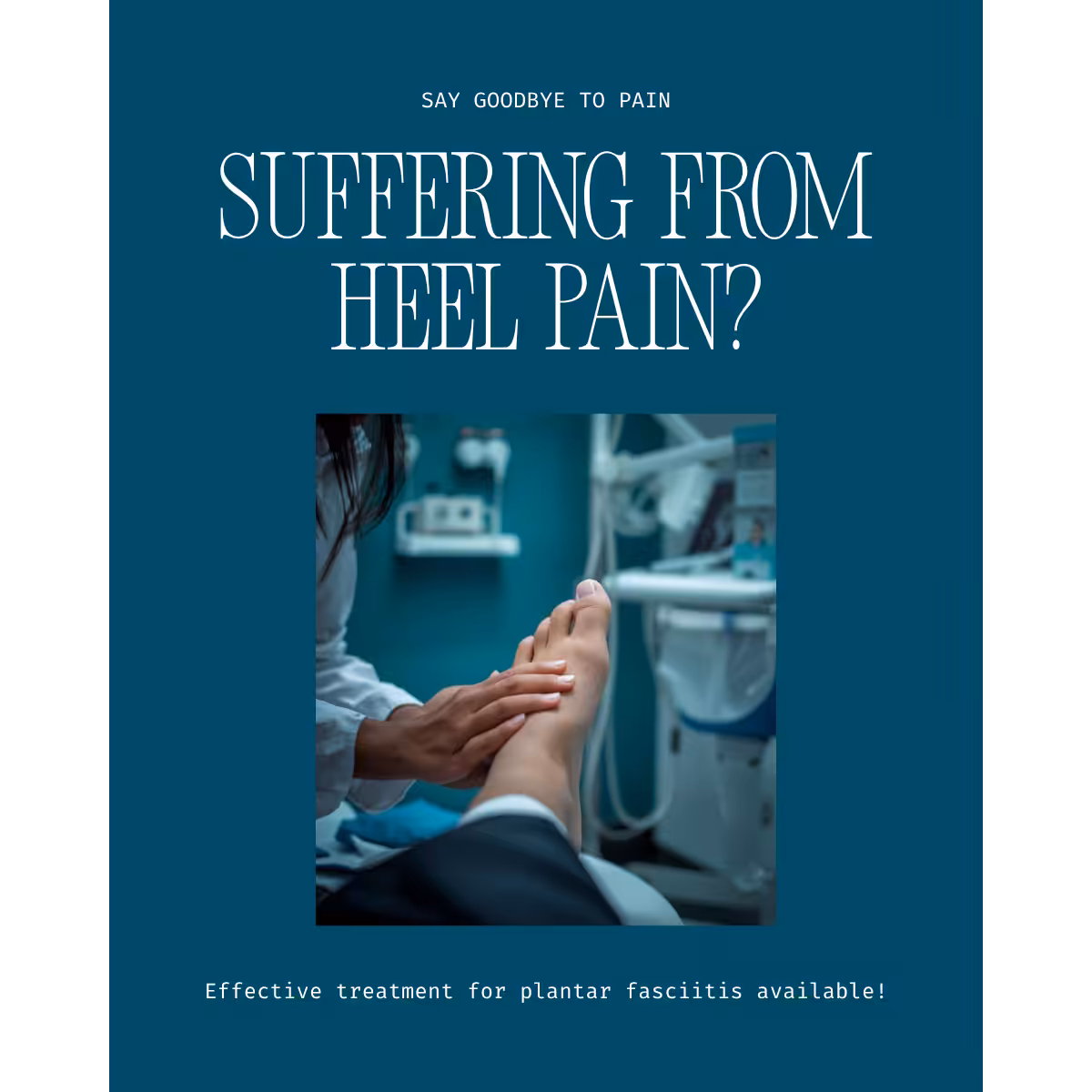
Watch Dr. Tom Biernacki DPM explain sports medicine for foot and ankle injuries — diagnosis, treatment, and return-to-sport strategies at Balance Foot & Ankle.
Why Sports Medicine Podiatry Is Different
Athletic patients have different needs than the general population. “Rest until it stops hurting” is not acceptable when you have a race in six weeks, a season to protect, or a livelihood that depends on performance. Sports medicine podiatry requires understanding not just the injury but the biomechanics that caused it, the tissue healing timeline, and the specific demands of the sport — then building a plan that accomplishes three things simultaneously: healing the current injury, preventing re-injury, and preserving as much training load as safely possible during recovery.
Dr. Tom Biernacki, DPM, FACFAS brings surgical subspecialty training in foot and ankle reconstruction to sports injury care. This means that when conservative treatment is appropriate, we apply it correctly with the right timeline and progressive loading. When it is not — when a Jones fracture needs a screw, when a Grade III ankle sprain has instability that won’t resolve, when a peroneal tendon has a tear that won’t heal without surgical repair — we can provide that care without referral.
Common Running Injuries: Pathophysiology and Management
Plantar Fasciitis (Plantar Fasciopathy)
Plantar fasciitis is the most common cause of heel pain in runners, affecting approximately 10% of all runners over their lifetime. Despite the “-itis” name, histological studies of chronic plantar heel pain demonstrate degenerative collagen disorganization (angiofibroblastic tendinosis) rather than acute inflammation in most cases — hence the preferred term plantar fasciopathy. The plantar fascia is a thick fibrous band originating at the medial calcaneal tubercle and inserting into the plantar plate and bases of the proximal phalanges. It functions as a tension band, storing and releasing energy with each gait cycle through the windlass mechanism.
Risk factors specific to runners: rapid mileage escalation (>10% increase per week), inadequate ankle dorsiflexion (<10° — the equinus-plantar fascia connection is biomechanically critical), high weekly mileage, hard running surfaces, and transitioning to minimal footwear without adequate preparation. The insertional form (pain at the calcaneal tubercle) is distinguished from non-insertional (pain along the midportion of the fascia) — they have different pathophysiology and different treatment responses.
Evidence-based treatment hierarchy:
- Eccentric calf strengthening + intrinsic foot strengthening — the Alfredson-adapted protocol for plantar fascia; performed with a towel scrunching, marble pickup, and step-edge heel drop program
- Ankle dorsiflexion restoration — addressing equinus is the most important modifiable mechanical risk factor; low-dye taping provides immediate symptom relief while addressing this
- Night splints — maintain the plantar fascia in slight dorsiflexion during sleep, reducing the first-step pain of post-static dyskinesia; compliance is the limiting factor
- Custom orthotics with medial arch support and heel cushion — offload the medial calcaneal insertion
- Extracorporeal shockwave therapy (ESWT) — for cases failing 6 months of conservative care; multiple RCTs demonstrate superiority over corticosteroid injection at 12-month follow-up; particularly effective for insertional calcific plantar fasciitis
- Corticosteroid injection — provides short-term relief (6–8 weeks) but associated with plantar fascia rupture risk with repeated injections; limit to 1–2 lifetime injections; avoid in runners with significant training load as rupture causes prolonged disability
- PRP injection — growing evidence base for recalcitrant cases; may provide more durable tissue regeneration than corticosteroid with lower rupture risk
- Surgical fasciotomy — reserved for cases failing 6–12 months of comprehensive conservative care; partial release with concomitant plantar heel spur excision if indicated
Medial Tibial Stress Syndrome (MTSS / Shin Splints)
MTSS is the most common running injury by incidence, accounting for 13–20% of all running injuries. The pathophysiology involves tibial cortex stress reaction from repetitive cyclic loading, exacerbated by the pull of the soleus and deep posterior compartment (flexor digitorum longus, tibialis posterior) on the posteromedial tibial periosteum. The critical distinction from tibial stress fracture (which is a more serious diagnosis requiring imaging and modified weight-bearing):
- MTSS: Pain over the distal two-thirds of the posteromedial tibial border; diffuse tenderness over a 5–13 cm zone; pain worsens at the start of running but may diminish mid-run (until advanced stages); X-ray normal; MRI shows periosteal edema, not cortical signal change
- Tibial stress fracture: Focal, point-tender pain over 1–2 cm; pain that is consistently worse with activity and does not diminish mid-run; X-ray may show periosteal reaction in later cases; MRI shows cortical signal change; cannot run through it
Treatment of MTSS: 50% training load reduction and cross-training (pool running, cycling) to maintain fitness; gait retraining — increasing step rate (cadence) by 10% reduces tibial stress by 10–20% without reducing speed; motion-control footwear or custom orthotics for excessive pronation contribution; progressive return-to-run program over 4–8 weeks. Persistent MTSS beyond 6 months warrants evaluation for chronic exertional compartment syndrome with compartment pressure measurement after exercise.
IT Band Syndrome (Iliotibial Band Friction Syndrome)
Though primarily a knee-level condition, IT band syndrome in runners frequently presents to podiatric sports medicine because of its foot and ankle biomechanical drivers. The IT band is a thickened lateral fascial band extending from the tensor fasciae latae and gluteus maximus to Gerdy’s tubercle on the lateral tibia. At approximately 30° of knee flexion (which occurs during foot strike in distance runners), the IT band compresses a highly innervated fat pad beneath it, producing the characteristic lateral knee pain.
Foot/ankle contribution: excessive foot pronation increases tibial internal rotation during stance, which increases IT band tension. Runners with significant overpronation who develop lateral knee pain are candidates for motion-control orthotic intervention at the foot — a proximal solution to a distal driver. Hip weakness (gluteus medius) is the most common proximate mechanical contributor; a thorough treatment plan addresses both the hip and the foot.
Achilles Tendinopathy
Achilles tendinopathy (non-insertional mid-portion and insertional) is the second most common running injury by incidence. The Achilles tendon absorbs forces of 6–8× body weight during running, and is poorly vascularized in the critical zone 2–6 cm proximal to insertion — precisely where mid-portion tendinopathy occurs.
The Alfredson heavy-load eccentric heel drop protocol remains the most evidence-supported treatment for mid-portion tendinopathy: 3 sets of 15 repetitions twice daily on a step edge, both with straight and bent knee, for 12 weeks. Insertional tendinopathy responds less consistently to Alfredson (because the eccentric load at the insertion may aggravate it) — isometric loading protocols and ESWT have stronger evidence for the insertional form.
PRP injection for Achilles tendinopathy: level 1 evidence (multiple RCTs) has been mixed; pooled analyses suggest benefit for mid-portion over insertional form. MLS laser is used adjunctively to reduce tendon inflammation and accelerate collagen remodeling. Surgical options (open or endoscopic debridement, Haglund’s deformity resection for insertional form) reserved for cases failing 6+ months of optimized conservative care.
Stress Fractures in Runners
Stress fractures in runners require specific anatomical knowledge because treatment and prognosis vary dramatically by location:
- High-risk (require aggressive management): Navicular — strict non-weight-bearing cast 6–8 weeks, CT surveillance; Jones fracture (5th metatarsal metadiaphyseal junction) — intramedullary screw fixation preferred in competitive athletes; anterior tibia (tension side) — surgical fixation; sesamoid — conservative first but high non-union rate
- Lower-risk (respond to conservative rest): 2nd–4th metatarsal shaft — protective boot 4–6 weeks; fibula — relative rest; calcaneus — protected weight-bearing in boot 4–6 weeks
Biomechanical gait analysis is essential after stress fracture recovery to identify and modify the mechanical loading pattern that caused the injury — without this step, recurrence rates are high.
Biomechanical Gait Analysis
Gait analysis in sports medicine serves two functions: identifying the mechanical drivers of existing injury and optimizing movement patterns to prevent re-injury. Our gait assessment includes:
- Static assessment: Foot posture index, navicular drop test, ankle dorsiflexion measurement (weight-bearing lunge test — the most clinically relevant measure of functional ankle dorsiflexion), Q-angle, hip rotation symmetry
- Dynamic assessment: Video gait analysis in shoes and barefoot, at walk and run, from posterior and lateral views. Key variables: foot strike pattern (heel vs. midfoot vs. forefoot), step rate (cadence), crossover gait (a lateral knee and IT band risk factor), excessive contralateral pelvic drop (gluteus medius weakness marker), peak rearfoot eversion velocity
- In-shoe pressure mapping: Pedobarography identifies focal high-pressure regions — particularly relevant for plantar fasciitis, metatarsalgia, and diabetic foot orthotic prescription
Acute Injury Management: PEACE & LOVE Framework
The traditional RICE protocol (Rest, Ice, Compression, Elevation) has been replaced by the more nuanced PEACE & LOVE framework, reflecting current understanding that the inflammatory phase of healing is necessary and suppressing it too aggressively may impair tissue quality:
PEACE (first 1–3 days):
- Protect — offload the injured tissue; use crutches or boot if weight-bearing is painful
- Elevate — above heart level to reduce swelling
- Avoid anti-inflammatory modalities — NSAIDs and ice in the first 72 hours may suppress the inflammatory phase necessary for tissue repair signaling; use with caution
- Compress — reduce swelling and limit hematoma formation
- Educate — set realistic recovery expectations; avoid passive modalities
LOVE (after day 3, subacute and rehabilitation phases):
- Load — controlled progressive loading stimulates collagen remodeling; early controlled loading produces stronger tissue than complete rest
- Optimism — patient mindset and expectation of recovery are significant predictors of return-to-sport outcomes
- Vascularization — aerobic exercise (pain-free cycling, swimming) maintains cardiovascular fitness and drives tissue perfusion without loading the injured structure
- Exercise — progressive strengthening, neuromuscular training, and sport-specific movement patterns; not delayed until pain-free but advanced in parallel with tissue healing
Return-to-Sport Protocol
Return-to-sport (RTS) clearance should be criterion-based, not calendar-based. We use the following framework:
- Pain: 0/10 VAS with full sport-specific training loads
- Strength symmetry: Injured limb within 90% of contralateral on single-leg heel-raise test (30+ repetitions), hop testing, and isokinetic strength testing where available
- Proprioception and balance: Y-Balance Test composite score within published sport-specific normative ranges; Star Excursion Balance Test >90% on injured limb vs. healthy
- Functional sport-specific testing: Completed the sport-specific RTS ladder (walk → jog → run → accelerate/decelerate → cut → full training → return to competition) without pain or compensatory movement
- Psychological readiness: ACL-RSI (psychological readiness to return to sport) score >65/100; athlete confidence in the limb during sport-specific movements
Regenerative Medicine Options
- Platelet-Rich Plasma (PRP): Autologous blood centrifuged to concentrate growth factors (PDGF, TGF-β, VEGF, IGF-1). In-office procedure; collected from patient’s own blood. Strongest evidence base in chronic tendinopathy (Achilles mid-portion, plantar fascia), lateral epicondylitis, and knee osteoarthritis. Variable results in acute ligament injuries. We discuss the evidence base, expected outcomes, and insurance/cost considerations with every patient before recommending PRP.
- MLS Laser Therapy (Multiwave Locked System): Class IV photobiomodulation simultaneously delivering 808 nm continuous and 905 nm pulsed wavelengths. Stimulates mitochondrial ATP production in damaged tissue, reduces neuroinflammation, promotes angiogenesis, and accelerates collagen synthesis. Evidence supports use in Achilles tendinopathy, plantar fasciitis, ankle sprain, and stress fracture management. Non-invasive, typically 6–12 sessions. Available at both our offices.
- Extracorporeal Shockwave Therapy (ESWT): High-energy acoustic pressure waves delivered to chronic tendinopathy and bone pathology. Stimulates neovascularization and growth factor release. Multiple RCTs support use in plantar fasciitis (particularly insertional/calcific), Achilles tendinopathy, and stress reactions. In-office procedure; typically 3 sessions at 1-week intervals.
Most Common Mistakes in Sports Injury Management
- Returning to sport too early: This is the No.1 driver of re-injury and chronic conditions in our practice. Tissue strength recovers much more slowly than pain subsides. A runner who feels no pain at 3 weeks post-Grade II sprain may still have only 40% of normal ligament strength — yet can run without pain. The result is re-injury at a higher grade on the first return to sport. Criterion-based RTS testing, not symptom resolution, is the appropriate threshold.
- Treating the symptom without addressing the mechanism: A runner with recurrent shin splints who is prescribed anti-inflammatories and told to rest will re-injure when they return to running. The overloading mechanism — training error, gait pattern, footwear, equinus — must be identified and modified, not just the pain treated.
Red Flags — Seek Immediate Evaluation
- Fracture deformity, inability to weight-bear, or immediate severe swelling after ankle injury: Ottawa Rules mandate X-ray; fracture-dislocation requires emergency care
- Progressively worsening leg pain during activity that requires stopping: Consider chronic exertional compartment syndrome — compartment pressures must be measured after exercise
- Sudden, severe calf pain with a “pop” during running: Achilles tendon rupture — the Thompson test (squeezing the calf) will not produce plantarflexion; requires urgent surgical or functional bracing management decision
- Foot numbness, weakness, or tingling after ankle or leg injury: Peroneal or tibial nerve injury — requires urgent evaluation
- Stress fracture at a high-risk site (navicular, anterior tibia, Jones fracture) with continued weight-bearing: Continued loading dramatically increases risk of complete fracture and prolonged recovery
Care at Balance Foot & Ankle
Our Howell and Bloomfield Township offices provide comprehensive sports medicine care for foot and ankle injuries. We offer same-week appointments for acute injuries, gait analysis, digital X-ray and diagnostic ultrasound in-office, and advanced treatments including MLS laser therapy and PRP. Dr. Tom Biernacki, DPM, FACFAS completed fellowship-level training in foot and ankle reconstruction and brings surgical capability to sports medicine — ensuring that when surgery is the right answer, you don’t need to be referred out.
Call (810) 206-1402 or book online. Same-week appointments are available.
Howell: 4330 E Grand River Ave, Howell MI 48843 | Bloomfield Township: 43494 Woodward Ave #208, Bloomfield Township MI 48302
Related Treatments at Balance Foot & Ankle
Common conditions we treat in our Howell and Bloomfield Township offices.
Our podiatrists treat the underlying cause, not just the symptom. Same-week appointments at our Howell and Bloomfield Township, Michigan offices.