

★ 4.9 Stars · 1,200+ Reviews · Board-Certified Michigan Podiatrists
Ready to be seen? Book a real appointment time online, 24/7 — Howell or Bloomfield Township. Takes about a minute, 24/7.
Plantar Fascia Surgery Bloomfield Township
Minimally Invasive Endoscopic Release
For the 5-10% of plantar fasciitis cases that fail conservative treatment, our fellowship-trained podiatric surgeons offer minimally invasive endoscopic plantar fascia release. Same-day outpatient procedure. Rapid return to activities.
Board-Certified Podiatrist • Howell & Bloomfield Township, MI • View credentials
Related Treatments You May Need
“After trying every conservative option, Dr. Biernacki performed my plantar fasciitis surgery. The recovery was smooth and I wish I had done it sooner. No more morning heel pain!”
— James W., Plantar Fasciitis Surgery Patient
Serving Patients Across Southeast Michigan
Balance Foot & Ankle provides expert podiatric care from two convenient locations. Our Howell office serves patients from Brighton, Hartland, Fowlerville, Pinckney, Fenton, Hamburg, Whitmore Lake, South Lyon, and throughout Livingston County. Our Bloomfield Township office serves Birmingham, Troy, West Bloomfield, Pontiac, Farmington Hills, Southfield, Royal Oak, Clarkston, Lake Orion, Rochester Hills, Waterford, Commerce Township, Novi, and Walled Lake across Oakland County.
Watch: How to Get Rid of Plantar Fasciitis Fast
![Peroneal Tendonitis Self Treatment [Stretches, Exercises & Massage]](https://www.michiganfootdoctors.com/wp-content/cache/flying-press/c520c31df4e25b56435df883dd00fd12.jpg)
Dr. Tom Biernacki, DPM explains the clinical basics — then covers what we do differently in our Howell and Bloomfield Township clinics. Subscribe on YouTube for weekly walkthroughs.
Your Board-Certified Podiatrists



Board-certified care at both Michigan locations
Book your visit today
Most insurance accepted · On-site X-ray · Board-certified podiatrists
Howell · (810) 206-1402 Bloomfield · (810) 206-1402Anatomy of the Plantar Fascia — What Dr. Tom Sees in the OR
The plantar fascia is a thick fibrous band that runs from the medial tubercle of the calcaneus (the inside bottom edge of your heel bone) out to the base of each toe. It has three bundles — a thick medial band, a central band (the one that hurts in classic plantar fasciitis), and a thin lateral band. Under a microscope it looks less like a muscle tendon and more like a strap: dense, parallel collagen fibers designed to resist tensile load as the arch flattens with every step. In a healthy foot, it is about 2–3 mm thick. In chronic plantar fasciopathy, it thickens to 4–7 mm and the fiber architecture breaks down into disorganized scar. That thickening is what we measure on ultrasound before recommending surgery.
When we operate, the first thing we appreciate is how much the chronically inflamed fascia has changed. Instead of the glossy white tendinous tissue we expect, we see duller, yellowed, scarred, sometimes partially torn fibers with surrounding fatty infiltration. That is the histology of chronic fasciopathy — it stopped being an “itis” (inflammation) years ago and became an “opathy” (degeneration). This distinction matters because anti-inflammatories and ice are poor matches for a degenerative problem. The tissue needs either a targeted injury that restarts biologic healing (the purpose of needle tenotomy, TOPAZ coblation, or PRP) or mechanical release (the purpose of fasciotomy surgery).
The other anatomic reality: the medial calcaneal branch of the tibial nerve runs along the medial edge of the plantar fascia origin. An aggressive open release that strays medial can cause nerve pain that is worse than the fasciitis it was meant to fix. That is why endoscopic and ultrasound-guided percutaneous techniques have largely replaced old-school large open releases — the smaller the wound, the lower the nerve-injury risk, the faster the recovery.
Who Actually Needs Surgery (and Who Definitely Does Not)
The overwhelming majority of plantar fasciitis closes without surgery. Depending on which long-term cohort you read, 80–95% of patients who follow a structured non-surgical program for 6–12 months achieve meaningful, durable relief. Surgery is for the remaining 5–20% — and we are strict about who qualifies. In our clinic, a patient does not get booked for plantar fascia release until they have documented failure of a full 12-month conservative ladder, imaging that confirms the diagnosis (ultrasound thickening ≥ 4.5 mm and/or MRI showing fascial signal change), and no untreated driver that is causing the load (tight Achilles, collapsing arch, poor footwear, uncontrolled weight, or a spondyloarthritis that no one has screened for yet).
If you’re reading a surgery page because nothing has worked yet, start here instead: the overwhelming majority of heel pain resolves without an operation once the treatment is actually right for your foot. Let us confirm which group you’re in first — book our Howell office or Bloomfield Township online before you assume surgery is the answer.
The patient who almost never needs surgery: the one who has had pain for 4–6 months, has not yet tried custom orthotics, has not tried a night splint, and has not completed a structured eccentric loading program. For that patient, surgery is a shortcut that skips the step that would have worked. The patient who genuinely benefits from surgery: the one who has completed 12 months of evidence-based conservative care, still cannot walk to their car without pain, and has imaging that shows thickened, degenerated fascia with no acute inflammatory edema left to treat. Those patients often experience dramatic relief from a well-executed release.
The patient we decline to operate on: any patient we suspect has a systemic driver like seronegative spondyloarthropathy or reactive arthritis masquerading as plantar fasciitis. If both heels hurt, there is morning sickness in multiple joints, or there is a family history of psoriasis or IBD, we screen the patient first with rheumatology labs and defer surgery until the systemic picture is managed. Cutting a plantar fascia in a patient with undiagnosed spondyloarthritis just moves the pain to the next attachment.
The 12-Month Non-Surgical Ladder We Walk Before Any Incision
We do not skip rungs on the conservative ladder. Skipping rungs is how patients end up in an operating room unnecessarily. The structured 12-month ladder we use in our Howell and Bloomfield Township offices has seven distinct phases, each with measurable outcomes before we move on.
Months 1–2 — Load management + daily self-care. PowerStep Pinnacle insole, supportive shoe swap (Brooks Adrenaline or Hoka Bondi in our typical recommendation), calf stretching three times daily, plantar fascia stretch on a frozen water bottle morning and night, NSAID course with gut protection if appropriate. Most brand-new plantar fasciitis resolves at this level alone.
Months 2–4 — Structured eccentric loading. The Patrick Rathleff heavy-load eccentric calf raise protocol is the best-studied physical therapy intervention we have. Three sets of eight repetitions, slow 3-second lower, performed every other day with a towel rolled under the toes and weight in a backpack. We coach the patient through the first two weeks ourselves and then check in at week 6. Patients who complete this protocol correctly show 30–50% pain reduction at 12 weeks in the published data.
Months 3–6 — Night splint + taping + custom orthotic. Strassburg-style night splint keeps the fascia on stretch during sleep so the first morning step is less painful. Low-Dye taping or a KT Tape technique off-loads the fascia during high-demand days. Custom functional orthotic — cast, not scanned from a pressure mat — redistributes peak plantar pressure away from the medial tubercle. These three tools layered on top of phases 1 and 2 move another 20–30% of remaining patients into remission.
Months 4–8 — Cortisone injection (only once). We reserve cortisone for patients who have not responded to the first three phases AND have imaging that confirms active inflammatory edema (MRI T2 signal change) as opposed to chronic degeneration. Ultrasound-guided corticosteroid injection into the deep fascia — never into the superficial fat pad — can break a painful cycle. One injection only. Repeated cortisone increases fat-pad atrophy and fascial rupture risk.
Months 6–9 — Regenerative medicine. Platelet-rich plasma injection, amniotic membrane injection, or extracorporeal shockwave therapy (ESWT) are our preferred tools when cortisone failed or was inappropriate. These treatments try to restart biologic healing in chronic degenerated tissue. Insurance coverage varies. We discuss cost-benefit explicitly before recommending.
Months 9–12 — Minimally invasive in-office release. Ultrasound-guided percutaneous needle tenotomy or TOPAZ microdebridement is our next step before open surgery. Done under local anesthetic in the office, walking the same day, return to full activity in 4–6 weeks. This is the rung that captures most of the remaining non-surgical candidates.
Surgical Technique: Open vs Endoscopic vs TOPAZ Coblation
If conservative care has genuinely failed, we have three surgical options and we pick between them based on the patient’s anatomy, imaging, insurance coverage, and how quickly they need to return to activity. All three release the medial and central bands of the plantar fascia at the calcaneal origin — the difference is how we get there and how much surrounding tissue we disturb.
Open plantar fasciotomy is the legacy gold-standard. A 3–4 cm incision on the medial heel, direct visualization of the fascia, partial release (usually the medial third and half of the central band — never a complete release, which destabilizes the longitudinal arch). Done under monitored anesthesia care. Advantages: direct visualization means the lowest rate of incomplete release and the lowest rate of medial calcaneal nerve injury in experienced hands. Disadvantages: bigger scar, longer recovery, more post-op pain. We reserve this for patients with unusual anatomy or previous failed endoscopic surgery elsewhere.
Endoscopic plantar fasciotomy (EPF) is our workhorse. Two 5 mm portals on either side of the heel, an endoscope to visualize the fascia from below, and a retrograde hook knife to perform a partial medial release under direct camera visualization. Twenty-minute operation, walking out of the surgery center in a surgical shoe the same day, back in sneakers at two weeks, back to full impact at 6–8 weeks. This is the technique the published literature supports most strongly when done by a surgeon who does 20+ per year.
Ultrasound-guided TOPAZ coblation is our minimally invasive alternative for patients who either cannot tolerate general anesthesia or whose imaging shows chronic degeneration without the structural thickening that would benefit from a formal release. A single needle is passed through the fascia under ultrasound guidance and radiofrequency energy is delivered in a grid pattern. The goal is not to cut the fascia but to induce controlled micro-injury that restarts biologic healing. Done in the office under local anesthetic. Recovery is typically faster than endoscopic release but the effect size is smaller — we counsel patients that TOPAZ is a middle option.
We do not perform complete plantar fascia releases. Cutting 100% of the fascia across its full width removes the longitudinal arch tension band and causes midfoot collapse with secondary lateral column pain — a complication that often hurts worse than the plantar fasciitis. Partial release (roughly 40–50% of the width) preserves arch stability while relieving the painful attachment. Any surgeon offering a “complete release” is out of step with contemporary evidence.
Pre-Surgical Workup: What We Do Before the OR
Before we schedule a plantar fascia release, we run a pre-surgical workup that is more thorough than most patients expect. The goal is not to gate-keep the surgery — it is to make sure we are fixing the right problem and that the patient is set up for the best possible outcome. Surgery on the wrong diagnosis is the single most common reason we see second-opinion patients from outside practices who did not improve after their first fasciotomy.
- Diagnostic ultrasound — measures fascia thickness, looks for partial tear or fluid collections, and screens for alternative diagnoses (Baxter’s nerve entrapment, fat pad atrophy).
- MRI — reserved for ambiguous ultrasound or when we need to rule out a stress fracture of the calcaneus, tarsal tunnel compression, or bone edema suggesting seronegative arthritis.
- Gait analysis and functional assessment — identifies driver-level problems (tight Achilles, hallux limitus, first ray insufficiency) that we need to address simultaneously or the fasciitis will recur after surgery.
- Lab workup when indicated — CBC, CRP, HLA-B27 if we suspect spondyloarthritis; HbA1c if diabetes is in play; vitamin D if stress fracture is on the differential.
- Pre-operative medical clearance — PCP clearance for any patient with cardiac, pulmonary, or clotting risk. EKG if over 60 or symptomatic.
- Informed-consent conversation — realistic outcome expectations, published complication rates (roughly 5–10% chance of incomplete relief, 1–2% risk of nerve injury, rare risk of lateral column pain), recovery timeline, and the honest statement that surgery is a supplement to ongoing conservative care — not a one-and-done fix.
Week-by-Week Recovery Timeline After Plantar Fascia Release
Recovery from endoscopic plantar fascia release is faster than most patients expect, but the structured post-op schedule matters. Patients who skip the early protective phase end up with surgical-site pain that outlasts the original fasciitis.
Day 0 (day of surgery): Surgical shoe, weight-bearing as tolerated on the heel only, ice 20 minutes on / 40 off, elevation above heart level whenever sitting. Oral pain medication as prescribed — most patients need it for 2–3 days. Dressing stays dry and on until day 3.
Days 3–7: Dressing change in office, surgical shoe continues, sutures still in place, short kitchen walks only. No driving if the right foot was operated on. Plantar arch stretching is deliberately paused to let the early healing response mature.
Week 2: Suture removal, first post-op visit. Transition from surgical shoe to a supportive sneaker with a fresh PowerStep Pinnacle insole. Gentle calf stretches begin. Short flat-terrain walks resume. No running, jumping, hills, or stairs on the surgical side for 2 more weeks.
Weeks 3–4: Full-day sneakers, return to desk work and most activities of daily living. Structured eccentric calf raises resume at low load. Swimming and stationary cycling are green-lit. Running and jumping remain red-lit.
Weeks 6–8: Return to running, hiking, and low-impact sport if progression milestones are hit. We want pain-free 30-minute walks and pain-free single-leg heel raises before we clear running. Most patients hit this window comfortably.
Months 3–6: Return to full-impact activity including basketball, pickleball, and racquet sports. The fascia continues to remodel for up to 12 months, so minor tenderness on long days in the first 6 months is normal and not a reason to panic.
Red Flags After Plantar Fascia Surgery
Most patients recover from plantar fascia release uneventfully. A small subset encounter post-operative complications that require prompt attention. These are the signals that should prompt an immediate phone call to our office, not a “wait and see”.
- Sudden calf swelling, tenderness, or pain — DVT until proven otherwise. Needs Doppler ultrasound same day.
- Fever over 101°F or expanding redness at the portal site — surgical site infection.
- Pus, foul odor, or increasing drainage — infection needing incision and drainage plus oral or IV antibiotics.
- Numbness, burning, or shooting pain along the medial heel — possible medial calcaneal nerve neuritis. Early treatment prevents chronic nerve pain.
- Sudden sharp pain on the outside of the foot weeks after surgery — may signal lateral column overload from over-release. Addressed with orthotic modification and activity management.
- Pain that suddenly returns to pre-op intensity at week 3 or 4 — could mean a tethered scar, under-release, or a new problem entirely. Evaluation needed.
- Chest pain, shortness of breath, or leg swelling with cough — PE. ER immediately.
Differential: Heel Pain That Looks Like Plantar Fasciitis but Is Not
The fastest way to have a failed plantar fascia surgery is to operate on the wrong diagnosis. Several conditions mimic classic plantar fasciitis in the exam room and require completely different treatment. When a patient presents with heel pain that has not responded to standard care, we actively rule each of these in or out before we even discuss surgical options.
Baxter’s nerve entrapment — compression of the first branch of the lateral plantar nerve under the abductor hallucis. Point tenderness is slightly more medial and distal than classic plantar fasciitis. Percussion over the nerve reproduces shooting pain. Treatment is different — nerve release, not fascia release.
Calcaneal stress fracture — deep bone pain with positive squeeze test and pain with single-leg hopping. X-ray often normal in early weeks; MRI confirms. Treatment is protected weight-bearing for 6–8 weeks, not a fasciotomy.
Fat pad atrophy or displacement — diffuse deep heel ache, especially in older patients or after prior cortisone injections. Imaging shows thin plantar fat pad. Treatment is heel cupping and offloading, not release.
Tarsal tunnel syndrome — burning, numbness, or tingling along the medial arch radiating into the toes. Tinel’s sign positive at the medial ankle. Electrodiagnostic testing confirms. Treatment is nerve release or steroid injection to the tunnel, not plantar fascia surgery.
Seronegative spondyloarthritis (ankylosing spondylitis, psoriatic arthritis, reactive arthritis) — bilateral heel pain, morning stiffness in the spine or other joints, family history, skin findings. Rheumatology referral and systemic treatment. Operating on a spondyloarthritis enthesitis just moves the inflammation to the next enthesis.
Plantar fascia rupture — acute onset pain with audible pop, often after multiple cortisone injections or during forceful push-off. Palpable defect at the origin. Treatment is protected weight-bearing and staged return to activity. This is an already-ruptured fascia — releasing it surgically is both unnecessary and potentially destabilizing.
Three Real Patient Scenarios We See Every Week
Scenario 1 — The correct surgical candidate. A 48-year-old nurse with 14 months of unilateral heel pain. Completed 8 months of custom orthotics, 12 weeks of physical therapy, one diagnostic cortisone injection that gave 6 weeks of relief and then returned. Ultrasound shows 6.2 mm thickened medial fascia with hypoechoic degenerative changes. MRI confirms chronic fasciopathy without active edema. She is the textbook candidate for endoscopic partial release. We operate. She is back in sneakers at 2 weeks, walking her shifts without pain at 6 weeks, and pain-free at 3 months. This patient exists precisely because we waited the full year.
Scenario 2 — The wrong surgical candidate, caught just in time. A 52-year-old schoolteacher referred in for “pre-op clearance for plantar fascia release.” Bilateral heel pain, worse in morning, plus intermittent knee and low-back stiffness. Family history of psoriasis. HLA-B27 positive on our workup. Rheumatology diagnoses early axial spondyloarthritis, starts DMARD therapy, and her heel pain resolves with systemic treatment. No surgery was ever the right answer. Pattern recognition saved her a scar and a failed operation.
Scenario 3 — The salvage patient from a prior surgery elsewhere. A 61-year-old retired electrician who had a complete (not partial) open plantar fascia release two years ago at another practice. He now has severe lateral column pain and a flattening arch on weight-bearing X-ray. We diagnose iatrogenic longitudinal arch insufficiency. Treatment is custom orthotic with deep heel cup and aggressive medial longitudinal arch fill, PowerStep Maxx as an interim, physical therapy for posterior tibial tendon strengthening. No second surgery. This scenario is the reason we never perform complete releases.
Advanced Questions Patients Ask About Plantar Fascia Surgery
Will my heel spur be removed? Not usually. The spur is a radiographic finding, not a pain generator. Ten percent of asymptomatic feet have heel spurs on X-ray. We address the fascia, not the spur. Removing the spur adds operative time and bleeding with no additional benefit in the published data.
What is the success rate? Modern endoscopic partial release in properly selected patients reports 80–90% satisfaction at 1 year in the peer-reviewed literature. The honest caveat: “satisfaction” is not “complete pain elimination.” Most patients describe meaningful relief with occasional mild soreness on long days, not a pain-free foot forever.
Do I still need orthotics after surgery? Almost always yes. The structural problem that loaded the fascia in the first place — pronation, arch insufficiency, tight Achilles, weight, occupation — does not go away because we released a piece of fascia. Custom or premium over-the-counter orthotics (PowerStep Pinnacle is our default recommendation) stay in the rotation indefinitely.
Can both feet be done at once? We do not recommend it. Bilateral release means bilateral non-weight-bearing recovery, which doubles the risk of DVT, deconditioning, and household falls. We stage 3–6 months apart.
Will insurance cover this? Plantar fascia release is a covered CPT code in most major insurance plans when documented conservative care of 6–12 months is exhausted. Our office handles prior authorization and submits all conservative-care documentation on your behalf. We discuss any out-of-pocket surgical facility cost before scheduling so there are no billing surprises.
Ready to schedule a surgical consultation? Our Bloomfield Township office sees plantar fascia surgery patients on dedicated surgical-consult days. Book online or call (810) 206-1402 or book online. Please bring any prior imaging, prior conservative care notes, and a complete list of medications so we can start the pre-op workup the same visit.
Related Conditions We Treat
If you’re researching plantar fasciitis surgery because conservative care hasn’t helped, you may want to review these patient guides on the condition and non-surgical options first:
- Heel Pain: Causes & Treatment
- Best Running Shoes for Plantar Fasciitis
- Best Heel Cups for Plantar Fasciitis
- Best Plantar Fasciitis Socks
- Hoka Bondi vs Clifton for Plantar Fasciitis
Ready for in-office care? Book online or call (810) 206-1402 or book online. Book online 24/7 at our Howell and Bloomfield Township clinics.
Frequently Asked Questions
Who is a candidate for plantar fascia surgery?
Surgery is considered for patients who have failed 9-12 months of aggressive conservative treatment including stretching, custom orthotics, cortisone injections, and shockwave therapy. You must have documented evidence of plantar fasciitis on imaging and persistent pain affecting daily life.
What is the recovery time for plantar fascia surgery?
Minimally invasive endoscopic plantar fascia release allows weight-bearing within days. Most patients return to normal shoes in 4-6 weeks and full activity in 3-4 months. Open surgery has a longer recovery. Dr. Biernacki uses the smallest incisions possible for fastest recovery.
Plantar Fascia Release Surgery — Bloomfield Township
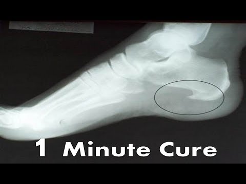
Watch as we explain plantar fasciotomy — endoscopic and open techniques — for patients whose plantar fasciitis has not responded to conservative care after 6+ months.
Frequently Asked Questions
When is plantar fascia surgery necessary?
Surgery is considered after 6–12 months of conservative treatment (orthotics, physical therapy, cortisone, EPAT) without adequate relief. Most plantar fasciitis resolves non-surgically — surgery is a last resort reserved for chronic cases.
What is the success rate of plantar fascia release?
Plantar fasciotomy has an 85–95% success rate for properly selected patients. Endoscopic release offers faster recovery than open surgery, with most patients walking the same day and returning to full activity in 6–8 weeks.
Can plantar fasciitis come back after surgery?
Recurrence after properly performed plantar fasciotomy is uncommon (under 5%). Custom orthotics, maintaining a healthy BMI, and appropriate footwear are recommended post-surgery to reduce any recurrence risk.
Watch Dr. Tom on Plantar Fascia Surgery
Dr. Tom’s complete plantar fasciitis treatment ladder — conservative to surgical. When surgery is the right answer (and when it’s not).

Related from Balance Foot & Ankle
Foot pain — Frequently Asked Questions
When should I see a podiatrist for foot pain?
If symptoms persist beyond 2 weeks of self-care, interfere with daily activity, or worsen suddenly, schedule a podiatrist evaluation. Early intervention typically shortens recovery and prevents chronic compensation patterns.
Will I need imaging or surgery?
Most foot pain cases resolve with conservative care—custom orthotics, supportive shoe changes, anti-inflammatory protocols, and targeted physical therapy. Imaging (X-ray, ultrasound, MRI) is reserved for cases that fail conservative treatment or when structural pathology is suspected. Surgery is rarely the first option.
Does insurance cover foot pain treatment in Michigan?
Most major Michigan insurance plans (BCBS, BCN, Priority Health, HAP, Medicare, Medicaid HMOs, United, Aetna, Cigna) cover medically necessary podiatric care. Custom orthotics may have separate DME coverage rules. Our team verifies your specific benefits before your visit.
Related Treatments at Balance Foot & Ankle
Common conditions we treat in our Howell and Bloomfield Township offices.
Ready to feel better?
Appointments at our Bloomfield Township office, 43494 Woodward Ave #208. Booking is open online around the clock — even after our phones close at 4:30 PM.
📅 Book Online — Bloomfield Township📞 (810) 206-1402Our podiatrists treat the underlying cause, not just the symptom. Appointments at our Howell and Bloomfield Township, Michigan offices.
Learn More About Plantar Fasciitis
Plantar fasciitis that won’t respond to anything?
When months of conservative care fail, a plantar fascia procedure offers lasting relief. In Michigan? our surgeons will review your options. Appointments in Howell & Bloomfield Township, most insurance accepted.
📅 Book an Appointment 📞 (810) 206-1402★ 4.9 from 1,200+ Google reviews · Book online 24/7 · We’ll check your insurance benefits before your visit.
Clinical sources
- Endoscopic plantar fasciotomy for plantar fasciitis: a systematic review & network meta-analysis. PubMed
- Endoscopic fasciotomy vs non-operative treatment for plantar fasciitis: a randomized controlled trial. PubMed
- Outcomes of endoscopic treatment for plantar fasciitis: a systematic review. PMC
Request an appointment
If conservative care has not resolved your heel pain, we can review whether surgery is appropriate for you. Book online or call (810) 206-1402 or send the form below and our office will get back to you.
We see patients at our Howell and Bloomfield Township offices.
Book online — pick the office that’s closer
Real-time scheduling is open 24/7. Choose your office below and you’ll see live appointment times without waiting for a callback:
- Book online at our Howell office →
4330 E Grand River Ave, Howell, MI 48843 (Livingston County) — closest for Brighton, Hartland, Fenton, Pinckney and Fowlerville. Howell office details. - Book online at our Bloomfield Township office →
43494 Woodward Ave #208, Bloomfield Township, MI 48302 (Oakland County) — closest for Birmingham, Troy, West Bloomfield, Royal Oak and Rochester Hills. Bloomfield Township office details.
Appointments are usually available at both offices. Prefer to talk to someone first? Book online or call (810) 206-1402.