

Ready to be seen? Book a real appointment time online, 24/7 — Howell or Bloomfield Township. Takes about a minute, 24/7.
You are in the right place. Dr. Tom Biernacki, DPM, FACFAS — board-certified foot & ankle surgeon with 3,000+ surgeries — explains exactly what diabetic foot ulcer care guide means and what works. Call (810) 206-1402 to book at Howell or Bloomfield Township.
![Diabetes Peripheral Neuropathy Treatment [Diabetic Nerve Pain Remedy]](https://www.michiganfootdoctors.com/wp-content/cache/flying-press/83886306c119511909f4e50d00e25908.jpg)
Watch: Diabetes Peripheral Neuropathy Treatment [Diabetic Nerve Pain Remedy] — MichiganFootDoctors YouTube
Medically reviewed by Dr. Tom Biernacki, DPM, FACFAS
Board-certified podiatric surgeon | Balance Foot & Ankle | Last reviewed: May 2026
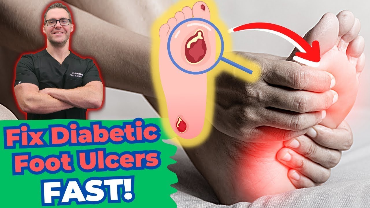
Watch Dr. Tom Biernacki DPM explain diabetic foot ulcer causes, wound grading, and treatment at Balance Foot & Ankle.
A diabetic ulcer will not close without two things, whatever dressing goes on it: pressure taken off the wound, and enough blood supply reaching it. Both get checked before anything else. Book online in Howell or Bloomfield Township — the booking form takes about a minute, 24/7.
What Is a Diabetic Foot Ulcer?
A diabetic foot ulcer (DFU) is a full-thickness wound penetrating through the dermis in a person with diabetes mellitus. The pathophysiology is a convergence of three interacting disease processes: peripheral neuropathy (loss of protective sensation allows repetitive microtrauma to go unnoticed), peripheral arterial disease (reduced arterial perfusion impairs healing and immune response), and immunopathy (hyperglycemia impairs leukocyte chemotaxis and phagocytosis, slowing the inflammatory phase of wound healing).
The typical sequence: neuropathy causes the patient not to feel a blister, callus, or foreign body; continued ambulation creates pressure necrosis; bacteria colonize the moist wound environment; ischemia prevents healing; and what began as a corn or blister evolves into a limb-threatening infection within days to weeks. Roughly 15–25% of people with diabetes will develop a foot ulcer during their lifetime, and DFU is the leading cause of non-traumatic lower-limb amputation in the United States — accounting for approximately 80,000 amputations annually.
Wagner Wound Classification System
The Wagner Classification is the most widely used system for grading DFU severity and guiding treatment decisions. Each grade demands a different management strategy:
- Grade 0: No open ulcer; pre-ulcerative lesion (callus over bony prominence, healed prior ulcer site). Treatment: off-loading, debridement of callus, diabetic footwear.
- Grade 1: Superficial ulcer involving skin and subcutaneous tissue only; no tendon, capsule, or bone involvement. Treatment: aggressive off-loading (TCC or removable cast walker), moist wound care, debridement, glycemic optimization.
- Grade 2: Deep ulcer penetrating to tendon, capsule, or joint. Bone not yet exposed. Treatment: same as Grade 1 plus probing to confirm depth, MRI to rule out osteomyelitis, systemic antibiotics if infection signs present.
- Grade 3: Deep ulcer with osteomyelitis or abscess. Treatment: urgent surgical debridement, parenteral antibiotics, possible bone resection, inpatient management often required.
- Grade 4: Partial foot gangrene (toes, forefoot). Treatment: vascular surgery consultation for revascularization assessment, partial amputation likely required.
- Grade 5: Full foot gangrene. Treatment: below-knee or above-knee amputation; vascular surgery co-management.
Clinical pearl: The probe-to-bone test has a positive predictive value of 89% for osteomyelitis. If a sterile metal probe touches bone at the ulcer base, treat as osteomyelitis until proven otherwise. MRI is the gold-standard imaging modality when osteomyelitis is suspected.
Who Gets Diabetic Foot Ulcers?
Any patient with diabetes is at risk, but several factors dramatically increase ulcer likelihood:
- Peripheral neuropathy confirmed by Semmes-Weinstein 10g monofilament — inability to detect the monofilament at plantar sites correlates with 7× increased ulcer risk
- Peripheral arterial disease — ankle-brachial index (ABI) below 0.9 indicates significant arterial compromise
- History of prior ulceration or amputation — strongest single predictor of recurrence
- HbA1c > 9% — chronic hyperglycemia impairs every phase of wound healing
- Charcot neuroarthropathy — midfoot collapse creates rocker-bottom deformity and abnormal plantar pressure distribution
- Foot deformities (hammertoes, bunions, prominent metatarsal heads) — create focal high-pressure zones
- Nephropathy — CKD patients have compounded immune dysfunction and poor healing
- Tobacco use — accelerates peripheral arterial disease and impairs microvascular perfusion
Diagnosis: How We Assess a Diabetic Foot Ulcer
Thorough wound assessment at every visit drives treatment decisions. Our evaluation includes:
- Wound measurement: Length × width × depth in centimeters; tunneling or undermining noted. Serial photography at each visit.
- Wound bed assessment: Percentage of granulation vs. slough vs. necrotic tissue; exudate character and volume; wound edge condition (maceration, epiboly, rolled edges).
- Infection assessment: IDSA/IWGDF criteria — two or more of: warmth, erythema, edema, purulent discharge, pain (unreliable in neuropathic patients), induration. Probe to bone performed.
- Vascular assessment: Ankle-brachial index (ABI), toe-brachial index (TBI — more sensitive in calcified vessels), transcutaneous oxygen (TcPO₂) if revascularization decision pending. TcPO₂ <30 mmHg predicts poor healing.
- Neuropathy assessment: Semmes-Weinstein 10g monofilament at 10 plantar sites; vibration perception threshold.
- Imaging: Plain X-ray first (gas in soft tissue = emergency); MRI for osteomyelitis assessment (sensitivity 90%, specificity 85%); bone biopsy with culture is the gold standard for definitive osteomyelitis diagnosis.
- Lab work: CBC (WBC elevation suggests infection), ESR/CRP (elevated with osteomyelitis), HbA1c (overall glycemic control), albumin (nutritional status — below 3.0 g/dL impairs healing).
Diabetic Foot Ulcer vs. Similar Conditions
Not every foot wound in a diabetic patient is a classic neuropathic DFU. Key differential diagnoses:
- Venous stasis ulcer: Located medially above the medial malleolus (gaiter region), not plantar. Associated with varicosities, lipodermatosclerosis, brown hemosiderin staining. Shallow, irregular borders. Improves with compression — contraindicated if ABI <0.5.
- Arterial (ischemic) ulcer: Typically on tips of toes, heel, lateral foot. Punched-out appearance, dry base, minimal exudate. Extremely painful (unless neuropathy masks it). Associated with cold feet, pallor on elevation, dependent rubor. Requires urgent vascular referral.
- Pressure injury: Over bony prominences (heel, lateral malleolus) in immobile patients. NPUAP staging (I–IV) used. Prevention is key; off-loading with heel float devices.
- Charcot neuroarthropathy: Acute Charcot presents as a hot, red, swollen foot without an open wound, often mistaken for cellulitis or gout. Bone fragmentation and collapse on X-ray are diagnostic. Critical not to miss — walking on an acute Charcot foot causes irreversible deformity.
Treatment Protocol: Wound Bed Preparation and Off-Loading
Off-Loading — The Foundation of Healing
Pressure reduction is the single most important intervention for neuropathic plantar ulcers. Without adequate off-loading, even perfect wound care will fail.
- Total Contact Cast (TCC): Gold standard. A well-molded plaster or fiberglass cast distributes plantar pressure across the entire foot, reducing forefoot and midfoot pressures by 84–92%. Healing rates of 72–100% reported in trials. Disadvantage: difficult to inspect the wound; must be changed weekly.
- Instant Total Contact Cast (iTCC): A removable cast walker made irremovable by wrapping with cast tape. Provides TCC-equivalent pressure distribution while solving the compliance problem — equivalent outcomes to TCC in trials.
- Removable Cast Walker (RCW): Only as effective as patient compliance; studies show patients wear RCWs only 28% of steps when told to use them. Used when wound inspection frequency is essential or patient cannot tolerate TCC.
- Surgical off-loading: Achilles tendon lengthening (ATL) reduces forefoot pressure by 35% and significantly reduces recurrence rates in forefoot ulcers. Metatarsal head resection for chronic non-healing forefoot ulcers.
Wound Bed Preparation (WBP)
The TIME framework guides wound bed optimization:
- Tissue (T): Sharp debridement of all non-viable tissue at every visit — devitalized tissue harbors biofilm and impedes healing. Enzymatic or autolytic debridement as adjuncts. Callus around the wound must be removed; it transfers pressure directly to the wound base.
- Infection/Inflammation (I): Superficial infections treated with topical antimicrobials (silver-based dressings, iodine); mild-moderate cellulitis with oral antibiotics targeting gram-positive organisms (S. aureus most common). Severe or limb-threatening infections require hospitalization with IV antibiotics and surgical debridement; cover gram-negative and anaerobic organisms.
- Moisture (M): Moist wound healing accelerates re-epithelialization. Goal is moisture balance — not wet (maceration) and not dry (desiccation). Foam dressings, hydrocolloids, alginates selected based on exudate level.
- Edge (E): Non-advancing wound edges after 4 weeks despite optimal care warrant escalation to advanced therapies — bioengineered skin substitutes (e.g., Apligraf, Dermagraft), negative pressure wound therapy (NPWT/VAC), hyperbaric oxygen (HBO).
Vascular Assessment and Referral
Any DFU with ABI <0.6, TcPO₂ <30 mmHg, or absent pedal pulses requires urgent vascular surgery consultation. Revascularization (endovascular angioplasty/stenting or bypass surgery) before wound closure is attempted is critical — attempting to heal an ischemic wound without restoring perfusion is predictably futile. At Balance Foot & Ankle, we maintain a direct referral relationship with vascular surgery for limb-threatening cases.
Advanced Wound Therapies
- Negative Pressure Wound Therapy (NPWT): Continuous or intermittent subatmospheric pressure (125 mmHg) stimulates granulation tissue, reduces edema, and draws wound edges together. Particularly useful post-debridement for deep cavitary wounds.
- Bioengineered Skin Substitutes: Bilayered skin equivalents (Apligraf) or dermal matrices (Dermagraft, OASIS) applied after wound bed preparation. DMERC-covered with appropriate documentation; requires no advance in wound area >50% in 4 weeks as threshold for escalation.
- Hyperbaric Oxygen (HBO): 100% oxygen at 2.0–2.5 atmospheres increases tissue oxygen tension, promotes angiogenesis, and enhances leukocyte killing. Medicare covers 30–40 dives for Wagner Grade III–IV DFU with documented vascular assessment.
- REMY Laser Therapy: class IV laser photobiomodulation at our office reduces wound-margin inflammation and accelerates granulation — used as adjunct to standard WBP protocol.
Most Common Mistakes in Diabetic Wound Management
- Inadequate off-loading: Prescribing a removable boot and trusting patient compliance. Studies consistently show that patients who are given a removable device walk without it most of the time. Making the device irremovable (iTCC technique) doubles healing rates versus removable walker alone.
- Delaying vascular referral: Attempting wound care on an ischemic foot for weeks before checking ABI or referring to vascular surgery. Every week of delay with an ischemic ulcer risks progression to gangrene. ABI should be obtained on the first visit for any DFU.
Red Flags — Seek Care Immediately
- Spreading redness or red streaking: Ascending lymphangitis — represents rapidly spreading infection; same-day emergency evaluation required
- Fever, chills, or confusion with a foot wound: Signs of systemic sepsis from wound; emergency department immediately
- Black or dark-colored tissue in or around the wound: Dry or wet gangrene; urgent vascular surgery consultation
- Wound that suddenly becomes painless after being painful: May indicate progressive ischemia or necrosis of surrounding nerve endings
- Gas bubbles or crepitus in soft tissue: Necrotizing fasciitis — surgical emergency with life-threatening implications
- No improvement in wound area after 4 weeks of standard care: Time to escalate to advanced therapies and specialist co-management
Care at Balance Foot & Ankle
Our Howell and Bloomfield Township offices operate a structured diabetic limb-preservation program. Every DFU patient receives: Wagner grading with documented wound measurement and photography, Semmes-Weinstein monofilament and ABI vascular assessment at the first visit, evidence-based off-loading (TCC or iTCC as first-line for plantar wounds), serial sharp debridement, appropriate dressing selection, glycemic optimization coordination with your primary care or endocrinology team, and direct vascular surgery referral when indicated. Our goal is simple: heal the wound, prevent recurrence, and keep you walking.
Call (810) 206-1402 or book online📅 Book Online — Howell📅 Book Online — Bloomfield for a wound evaluation. Both offices are accepting new patients.
Ready to fix this for good?
Reading goes so far. The fastest path is a 30-minute office visit. Book online for Howell or Bloomfield Township. Call (810) 206-1402.
In-Office Treatment at Balance Foot & Ankle
If home treatment isn’t providing relief for your diabetic foot condition, our podiatry team at Balance Foot & Ankle can help with expert evaluations and advanced in-office care.
Book online 24/7. (810) 206-1402
VitaMedica Arnica Gel
Arnica gel plus 15 plant compounds for sore feet, plantar fasciitis flares and post-activity soreness. Apply a thin layer to the sore area.
Shop VitaMedica Arnica Gel →Related: how wound VAC (negative-pressure) therapy helps stubborn foot wounds heal.
Diabetic foot wound or ulcer?
Early, expert wound care prevents serious complications — this is urgent. In Michigan? we offer same-day visits for active wounds. Appointments in Howell & Bloomfield Township, most insurance accepted.
📅 Book an Appointment 📞 (810) 206-1402★ 4.9 from 1,200+ Google reviews · Book online 24/7 · We’ll check your insurance benefits before your visit.
Getting Care at Balance Foot & Ankle
Treated at our Howell and Bloomfield Township offices — book online 24/7.
- Vitamin D Deficiency & Diabetic Foot Problems — A Michigan Problem
- Diabetic Peripheral Neuropathy: Screening, Staging, and Protective Footwear Prescription
- Diabetic Peripheral Neuropathy Foot Care: Screening, Prevention, and the Annual Exam
- Shoe Selection Guide for Diabetic and Neuropathic Feet
- Medicare Therapeutic Shoe Bill: The Diabetic Shoe Benefit Explained
- Wound Dressings for Chronic Foot Ulcers Guide
- Wound Offloading Devices: Total Contact Casting, Removable Cast Walkers, and Heel Relief Shoes
- Toe Amputation: What to Expect 2026 | DPM

Dr. Tom Biernacki, DPM is a board-certified foot & ankle surgeon (ABFAS & ABPM) at Balance Foot & Ankle Specialists in Southeast Michigan. With over a decade of clinical experience, he specializes in heel pain, bunions, diabetic foot care, sports injuries, and minimally invasive surgery. Dr. Biernacki is a member of the APMA and ACFAS, and his patient education content on MichiganFootDoctors.com and YouTube has made him one of the most-followed foot & ankle educators on YouTube.
More questions patients ask
How often should diabetics check their feet?
Diabetic patients should perform daily self-examinations of both feet, checking for cuts, blisters, redness, swelling, calluses, and temperature changes. Professional diabetic foot exams should occur at least annually, or every 3-6 months for patients with neuropathy, peripheral artery disease, or history of foot ulcers. Dr. Biernacki provides comprehensive diabetic foot care programs.
Why do diabetics have foot problems?
Diabetes causes peripheral neuropathy (nerve damage reducing sensation), peripheral artery disease (reduced blood flow), and impaired immune function. This combination means injuries go unnoticed, heal slowly, and are prone to infection. Approximately 15% of diabetics will develop a foot ulcer, and diabetic foot complications are the leading cause of non-traumatic amputation. Prevention through regular podiatric care is essential.
What are the signs of diabetic neuropathy in feet?
Early signs include tingling, burning, or numbness starting in the toes and progressing upward. Other symptoms include sensitivity to touch, muscle weakness, balance problems, and loss of reflexes. If you experience any of these symptoms, schedule an evaluation with Dr. Biernacki at 810-206-1402 for nerve conduction testing and a comprehensive neuropathy management plan.
Still have a question about coverage or cost? Book online and we will check your benefits before your visit: Book in Howell · Book in Bloomfield Township. Prefer to talk it through first? Call (810) 206-1402.