
Quick answer: Knot On Top Of The Foot affects roughly 1 in 4 adults in our practice. Effective treatment starts with a targeted diagnosis, conservative-first treatment, and escalation only when needed. We treat this regularly at our Howell and Bloomfield Hills practices. Call (810) 206-1402.
Medically reviewed by Dr. Tom Biernacki, DPM · Board-Certified Podiatric Surgeon · Last reviewed: April 2026 · Editorial Policy
Knot on Top of the Foot [What are foot knots on top of the foot?]
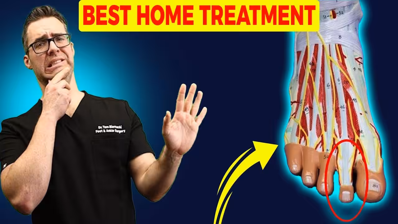
Do you have a foot knot on top of the foot? We review the most common top of the foot knot causes, symptoms and BEST treatment options!![Swollen Top of Foot or Top of Foot Tendonitis? [FIX Top of Foot Pain]](https://www.michiganfootdoctors.com/wp-content/cache/flying-press/ab2350551cc3e0cd3b339a587d8c8018.jpg)
What are Foot Knots?
A knot on top of the foot appears as a protuberance or localized area of swelling on the foot. Knot on top of the foot is also known as a bump, nodule, contusion, tumor, or cyst. Knots on the foot can result from trauma, injury, cyst, infections, or rarely tumors.What are the symptoms of Knot on Top of the Foot?
Depending upon the underlying cause, the knot on top of the foot can vary in:- Size (may grow rapidly or may remain same)
- Shape
- Colour
- Number (single or multiple)
- Consistency (Hard or soft)
- Pain
- Tenderness
- Discomfort
- Swelling
- Stiffness
- Bleeding or bruising
- Difficulty walking
- Difficulty wearing shoes or socks due to rubbing and irritation
- Rough skin/calluses
- Signs of infection such as fever, chills, warmth, redness, pus, or discharge
- Fever and chills
- Open wounds
- Bleeding
- Changes in heart rate (Tachycardia or Bradycardia)
- Red streaks extending up to the leg or foot
What are the causes of the knot on the Foot?
Foot knots can be due to several conditions. Some of the potential causes include:-
Trauma:
- Minor & severe injuries and trauma can cause localized swelling and lumps. These include:
- Broken bone
- Hematoma (collection of blood in body tissue)
- Thrombosis(Blood clots)
- Sting or bite injuries
-
Infections:
- Infection can also produce a lump or knot in an abscess or diffuse lymph node enlargement in the foot. These causes include:
- Abscesses
- Cellulitis ( infection of skin and tissue beneath)
- Ingrown toenail
- Warts caused by Human Papilloma Virus
-
Inflammatory causes:
- Knots can form on the foot as a result of swelling and inflammation. Some potential causes include:
- Ganglion cyst
- Gout
- Osteoarthritis
- Rheumatic fever
- Rheumatoid arthritis
-
Tumors:
- Foot knots can form both by benign or malignant tumors of the skin, soft tissue, or bones such as:
- Fibroma (Benign tumor of fibrous or connective tissue)
- Lipoma (Benign tumor of fatty tissue)
- Lymphoma (cancer of lymphatic system)
- Melanoma(cancer caused by melanocytes/pigment-producing cells of the skin)
- Nevi (moles of the skin)
- Non-melanoma skin cancers
- Sarcoma (a malignant tumor of bone or cartilage)
-
Life-threatening/Serious causes of foot knots include:
- Abscess with spreading infection
- Cancer
- Fracture of bone
- Foot ulcers in diabetics
When to seek medical help?
Seeking medical aid is advised to avoid the possible complications when:- Symptoms aren’t managed at home
- Worsening with time
- Difficulty in walking and carrying out daily life activities
- Bump Increasing in size
- Associated infection
- Associated with a medical condition
- Fracture
- Bleeding
Whom to seek medical help?
Medical aid is required if the foot knot worsens with time or conservative methods fail to treat the cause. You should see a doctor, orthopedic, or a foot and ankle specialist (podiatrist). Podiatrists are the leading experts in problems involving feet and ankles. They are uniquely qualified and specifically trained in treating foot and ankle conditions ranging from simple to complex among all ages.How is a knot on top of the Foot diagnosed?
Your doctor may take a careful history and perform a physical examination to make a differential diagnosis. History: The doctor may ask the following questions to make a differential diagnosis:- When did you first notice the knot?
- What made you notice it?
- Is it painful?
- Is it soft or hard?
- Is there any change in skin color?
- Is there any change in the size of the knot since you noticed it?
- Do you have lumps anywhere else on the body?
- What are the associated symptoms?
- Have you had any recent injuries to the area?
- Is there any other similar bump on your body?
- Do you have any medical illnesses?
- What is your occupation?
- What is the level of your daily physical activity?
- The doctor may apply pressure and check for tenderness
- Size
- Shape
- Surface whether smooth or rough
- Mobility, whether it is freely movable on the skin or not. A ganglion cyst is usually moveable.
- Consistency Whether it is soft or firm
- Margins, whether they are regular or irregular
- Light may also be shined through the cyst to check if it passes through it.
- Complete blood count
- X-ray
- Ultrasound
- Fluid aspiration for evaluation
- Biopsy
What is the treatment of knot on top of the foot?
Home treatment:
- Rest: Avoiding the trauma or repetitive stress that is causing inflammation
- Icing: Application of ice reduces pain, swelling, and inflammation in arthritis, bursitis, Gout, and bunions
- Foot Elevation: Elevation of the foot to avoid fluid buildup and swelling
- Avoid heels so that arch doesn’t bend as the joint will open more in high heel shoes
Medical treatment:
Your doctor may recommend:- Pain-killers: For the treatment of pain and discomfort, over-the-counter pain-relieving and anti-inflammatory medications such as ibuprofen, acetaminophen, etc. might help
- Foot-wears: Wearing wide-spaced footwear or walking aids to help in mobility. Select comfortable shoes to avoid pressure over the knot and to avoid the symptoms
- Orthotics: Use orthotic devices to avoid pressure on the foot
- Pads: Padding over the bump to prevent irritation against the footwear
- Physiotherapy
Surgical treatment:
Bump surgery is very effective as the recurrence rate is rare, but the wound takes time to heal. It may take around 10-21 days to get back into your shoes. You may feel numbness or local sensitivity after surgery. After surgeries, shoes can be worn again once the foot has healed. Six months might be required to wear all kinds of shoes. Top of Form What are the possible complications of the knot on top of the foot? If left untreated, it could worsen and cause complications. Some of the potential complications include:- Limited mobility
- Joint destruction leading to joint deformity
- Spread of infection
- Ulceration
- Necrosis of tissue/Gangrene
- Amputation
- It may spread or be life-threatening depending on the type and stage of cancer if it is due to cancer.
Common causes of the knot on top of the Foot are:
-
Ganglion Cyst:
What is a Ganglion cyst?
Ganglion cysts are fluid-filled bumps formed near joints or tendons, mostly on the wrist but can also form on top of the foot, ankle, or anywhere on the body. They are formed near the joints or tendons when synovial fluid (which cushions and lubricates joints and tendons during movement) leaks into the joint or tendon and is collected in a sac beneath the skin. They are non-cancerous and harmless and are the most common benign soft-tissue masses. The cause is unknown or sometimes form as a result of repetitive micro-trauma.What are the signs and symptoms of a Ganglion cyst?
A ganglion cyst can disappear on its own. It can vary in size from being invisible to an inch or more in diameter. The shape can be round and soft or hard. It is freely movable on the skin. Signs and symptoms include:- Sometimes a noticeable knot/lump may be the only symptom
- Pain if cyst pushes on a joint
- Dull pain if cyst presses against a joint or tendon
- Burning
- Tingling if the nerve is pressed.
- Numbness
- Loss of mobility
- Irritation and difficulty wearing shoes
How is a Ganglion cyst diagnosed?
Your doctor may ask the following questions to rule out the causative factors:- Since when you noticed a bump on your foot?
- Is it painful or painless?
- Is there any change in size?
- Is there any change in the skin color of your foot?
- Are there any associated symptoms?
- Do you have any such bump elsewhere on the body?
- Are you suffering from any other medical condition?
- What is your occupation?
- Level of physical activity?
- The doctor may apply pressure and check for tenderness.
- Size
- Shape
- Surface whether smooth or rough
- Mobility, whether it is freely movable on the skin or not. A ganglion cyst is usually moveable.
- Consistency Whether it is soft or firm
- Margins, whether they are regular or irregular
- Light may also be shined through the cyst to check if it passes through it
- Aspiration of a small amount of fluid for evaluation
- An X-ray might also be taken
What is the treatment of Ganglion cyst?
If a Ganglion cyst is painless and doesn’t interfere with daily life activities, it is monitored regularly for any change and usually doesn’t require any treatmentAt-home treatment:
Home care includes:- Rest
- Foot-wears: Wear shoes that do not irritate the area or rub against the cyst.
- Padding: Pad can also be placed inside the shoe to reduce pressure and rub against the cyst.
- Icing: By putting ice in a towel and placing it on the cyst to reduce swelling and inflammation
- Pain-killers: pain-relieving medications.
- Never try to pop up the ganglion cyst, as it can cause infections.
Medical treatment:
If the cyst is large and painful, medical treatment is required:- Aspiration and injection: If the cyst is large, painful, and causing limitation in movement, it requires drainage, which involves removing the fluid with a syringe, and a steroid is injected into the mass. It may require more than one session.
Surgical treatment:
If the treatments mentioned above fail, surgical removal is advised in which cyst is removed along with a part of the attached joint capsule or tendon sheath. After surgery, the recurrence rate is much lower as compared to other treatment options.-
Osteoarthritis:
What are Bone Spurs/Osteophytes?
Osteophytes or bone spurs are small bony growths formed near damaged joints or on top of the foot and may become visible through the skin. Bone spurs typically form when there’s extra bone growth to repair damage caused by repeated stress or pressure on the bone for a prolonged period. A common cause of bone spur is osteoarthritis. Bone spurs can form anywhere on the body but are most common in the joints.What are the signs and symptoms of bone spurs?
Bone spur/Osteophytes may present with the following signs and symptoms:- Pain
- Discomfort
- Difficulty in walking
- Difficulty in wearing shoes
- Limited motion
- Callus or blister formation around the affected area
How are Bone spurs diagnosed?
The doctor makes take careful history regarding any foot injury, medical illness, or daily physical activity. Osteophytes are usually diagnosed on X-rays.How are Bone spurs treated?
Treatment includes:- Pain-relieving medications such as ibuprofen, acetaminophen, etc. to help ease the pain.
- Rest
- Physiotherapy
- If the underlying cause of bone spurs is osteoarthritis, the treatment also includes medications to relieve joint pain and stiffness.
- Surgical treatment includes the placement of a screw or metal brace (plate) across the joint. But the disadvantage is a limitation of movement across the joint or joint instability due to existing degeneration in the joint movement.
-
Bunions:
What are Bunions?
Growth of bone on the side of the foot, usually at the base of the big toe, due to rotation and sideways angling of the bone that makes up the great toe is called bunion/hallux valgus. It occurs when the joint between the big toe and the long metatarsal bones become misaligned, or additional bone structure appears. The enlarged joint can become inflamed and painful. Bunions can also develop at the base of the fifth toe called Tailor’s bunion or bunionettes Bunions are more common in females due to wearing high heels that crowd the toe but can also occur in males.What are the causes of Bunion formation?
Common causes include:- Structural abnormalities such as flattened arches, missing bones, or short first metatarsal.
- Leg length discrepancy and uneven gait
- Pregnancy: Relaxin is a hormone that widens the pelvis in pregnancy, can soften foot ligaments causing the bones in the feet to spread out and arches to fall
- Wearing high heels as walking on high heels tightens the calf muscles and forces weight-bearing to the foot’s front. Gradually the arch can collapse.
What are the signs and symptoms of Bunions?
Signs and Symptoms include:- Bump on the top of the foot
- Pain
- Swelling
- Mal-aligned toe, toe leans towards other toes
- Difficulty walking
- Difficulty wearing shoes
- Callus formation
How is a Bunion diagnosed?
A bunion can be diagnosed by a simple physical examination of the foot and X-ray.How is bunion treated?
Treatment can be medical or surgical, depending upon the severity of the condition.Medical treatment:
- Wearing wide shoes
- Padding the area
- Arch support
- Icing the area
- Wearing a toe splint
- Pain-relieving medications such as ibuprofen, acetaminophen, etc
Surgical treatment:
If the bunion is progressing after medical treatment or symptoms are worsening with time, surgery is required.-
Bursitis:
What is meant by Bursitis:
Bursa is small fluid-filled sacs (for lubrication) between the joints, allowing the bone to move in the opposite direction and reduce friction/absorb shock between the bone, muscle, tendon, and skin near the joint. Inflammation of this fluid-filled sac is called bursitis, causing pain and limited motion. The foot contains one bursa, located between the heel bone and Achilles tendon. Bursitis may occur due to excessive rubbing or strain on the Achilles tendon. It can occur anywhere on the body, including the base of the big toe, side of feet, or on the heels. It occurs due to trauma or repetitive stress that causes inflammation of the bursae.What are the signs and symptoms of Bursitis?
Signs and Symptoms of bursitis include:- Pain especially during walking or running
- Intense pain when standing on tiptoes
- Warm
- Flushed skin
- Bursitis doesn’t improve in 2 weeks
- Worsening symptoms
- Severe pain
- Excessive swelling
What is the treatment of Bursitis?
Management is based on treating the underlying cause. It includes:- Rest
- Avoiding the trauma or repetitive stress that is causing inflammation
- Apply ice
- Elevation of the foot
- Taking pain-killers and anti-inflammatory medications such as ibuprofen, acetaminophen, etc
- Physiotherapy
- Wearing wide shoes
- Wearing orthotics or padded shoes
- Drainage
- If still not controlled, the doctor may advise steroid injections.
-
Gout:
What is Gout?
Gout is a type of arthritis caused by excessive buildup of uric acid in the joints, most commonly the big toe.What are the Risk Factors of Gout?
Risk factors include- Male gender
- Obesity
- Alcohol intake
- Diet rich in purines and fructose
- People with diabetes, metabolic syndrome, cardiovascular diseases, poor kidney function, etc
What are the signs and symptoms of Gout?
Signs and Symptoms of Gout include:- Sudden Pain
- Burning sensation
- Joint stiffness
- Swelling
- Warmth
- Tenderness
- Flushing
How is Gout diagnosed?
The doctor may take careful history regarding your diet, drinking habits, lifestyle, or any associated medical condition. Diagnosis is made by- physical examination
- blood tests
- X-rays
- ultrasound
What is the treatment of Gout?
Management of Gout includes: Lifestyle modifications:- Eat a healthy diet such as fruits, vegetables, etc
- Limit food rich in purines such as meat, seafood, and beer
- Eat low-fat dairy products
- Maintain healthy weight
- Drink plenty of fluids to flush uric acid out of the body
- Avoid alcohol and smoking
- Anti-inflammatory and pain-relieving medications
- Colchicine
- Steroids (tablets or injections)
- Allopurinol
- Febuxostat
- Uricosuric acid
-
Lipoma:
What is meant by Lipoma?
Lipoma is a non-cancerous growth of fatty tissue formed under the skin. It is usually painless, moveable with fingers, soft to touch, and rubbery. It can form anywhere on the body, including the top of the feet.How is a Lipoma diagnosed?
Lipoma is diagnosed with a simple physical examination of the foot or a biopsy.What is the treatment of Lipoma?
It is harmless, and no treatment is needed. However, surgical excision might be required if it becomes large or impinges on the surrounding nerves, tendons, or ligaments.-
Cutaneous horn:
What is a Cutaneous Horn?
Cutaneous horns are growths composed of keratin (a protein found in the top layer of the skin). It forms a bumpy, spiked-shaped appearance; that’s why called a cutaneous horn. It is found mostly on the face, neck, or shoulders but may appear on the foot. It may be a sign of cancer, so medical consultation is required as early as possible. Seek your doctor immediately if you develop:- Inflammation around the area
- Growth increase
- Hardening of the horn base
-
Hallux Rigidus:
What is Hallux Rigidus?
It is a form of arthritis that occurs when the cartilage is damaged or lost at the base of the big toe. It occurs most commonly between 30 and 60 years of age.What are the signs and symptoms of Hallux Rigidus?
Signs and Symptoms include:- Pain
- Stiffness
- Limited motion of the big toe
- Difficulty in walking
What is the treatment of Hallux Rigidus?
Treatment includes:- Alternate soaking of feet in warm and cold water
- Shoes that prevent the big toe from bending
- If the condition worsens with time, surgery is recommended
-
Rheumatoid nodule:
What is a Rheumatoid nodule?
If you have Rheumatoid arthritis, you may develop Rheumatoid nodules, which are firm lumps under the skin with varying sizes ( as large as walnut or as small as a pea). They occur mostly on the extensor surfaces or joints affected by arthritis. Rheumatoid nodules are painless unless they press on nerves or are inflamed.What is the treatment of Rheumatoid nodule?
Treatment includes:- steroid injection directly into the nodule
- For Rheumatoid arthritis: DMARDs: Disease-Modifying Anti-Rheumatic Drugs
- If the nodule limits motion or compresses a nerve, surgical removal is advised.
-
Sebaceous Cyst:
What is a Sebaceous cyst?
Sebaceous cysts are non-cancerous and closed cyst sacs under the skin and are formed due to blockage of glands or swollen hair follicles in the skin. They are found usually on the face or neck but may occur on the foot.What is the treatment of a Sebaceous cyst?
Treatment includes steroid injections into the cyst, or surgical removal is required if it worsens with time.-
Accessory Navicular syndrome:
What is meant by Accessory Navicular Syndrome?
Bump on the inner side of the foot, just above the arch, maybe due to Accessory Navicular. It is an extra bone or piece of cartilage, and this is congenital. It is usually dormant and doesn’t cause any medical problems. But if it is incorporated within the posterior tibial tendon, it may interfere with the functioning of the tendon.What are the signs and symptoms of Accessory Navicular Syndrome?
Signs and Symptoms include:- The bony protrusion on the inner side of the foot
- Pain
- Redness
- Swelling after significant physical activity
- Throbbing after physical activity
What is the treatment of Accessory Navicular Syndrome?
Treatment includes:- Ice
- Pain-relieving and anti-inflammatory medications such as Ibuprofen, Acetaminophen, etc
- Physiotherapy to strengthen and enlarge the muscle around the navicular and decrease inflammation
- Orthotic device to accommodate extra bone
Related Treatment Guides
- Plantar Fasciitis & Heel Pain Treatment
- Custom 3D Orthotics
- Sports Foot & Ankle Injury Treatment
- Bunion Treatment
In-Office Treatment at Balance Foot & Ankle
If home treatment isn’t providing relief for your foot and ankle conditions, our podiatry team at Balance Foot & Ankle can help with same-day evaluations and advanced in-office care.
Same-day appointments available. (810) 206-1402
Doctor Hoy’s Natural Pain Relief Gel
Natural topical pain relief I use in our clinic. Arnica + camphor formula — apply directly to the area 3–4x daily. ($20–25)
Shop Doctor Hoy’s →Medical References & Sources
- American Podiatric Medical Association — Patient Education
- American Orthopaedic Foot & Ankle Society — Foot Conditions
Insurance Accepted
BCBS · Medicare · Aetna · Cigna · United Healthcare · HAP · Priority Health · Humana · View All →
Howell Office
4330 E Grand River Ave
Howell, MI 48843
Get Directions →
Bloomfield Hills Office
43494 Woodward Ave, #208
Bloomfield Hills, MI 48302
Get Directions →
Your Board-Certified Podiatrists


Ready to Get Back on Your Feet?
Same-week appointments available at both locations.
Book Your AppointmentMore Podiatrist-Recommended Foot Health Essentials
Hoka Clifton 10


Max-cushion everyday shoe — podiatrist favorite for walking and running.

OOFOS Recovery Slide

Impact-absorbing recovery sandal — wear after long days on your feet.
As an Amazon Associate, Balance Foot & Ankle earns from qualifying purchases. Product recommendations are based on clinical experience; prices and availability shown above update live from Amazon.

When to See a Podiatrist
If foot or ankle pain has been bothering you for more than a few weeks, home care alone may not be enough. Balance Foot & Ankle offers same-week appointments at our Howell and Bloomfield Hills clinics — no referral needed in most cases. Bring your current shoes and a short list of symptoms and we’ll build you a treatment plan in one visit.
Call Balance Foot & Ankle: (810) 206-1402 · Book online · Offices in Howell & Bloomfield Hills
Pros & Cons of Conservative Care for foot care
Advantages
- ✓ Conservative care first
- ✓ Same-week appointments
- ✓ Multiple insurance accepted
Considerations
- ✗ Self-treatment can mask issues
- ✗ See a podiatrist if pain >2 weeks
Dr. Tom’s Recommended Products for foot care
Affiliate disclosure: As an Amazon Associate, Balance Foot & Ankle earns from qualifying purchases. We only recommend products we use with patients.
Footnanny Heel Cream Dr. Tom’s Pick
Best for: Daily moisturizer for cracked heels
Ready to Get Back on Your Feet?
Same-day appointments in Howell + Bloomfield Hills. Most insurance accepted. Dr. Tom Biernacki, DPM & team.
Book Today — Same-Day Appointments Available
Call Now: (810) 206-1402
About Your Care Team at Balance Foot & Ankle
Dr. Tom Biernacki, DPM · Board-Certified Foot & Ankle Surgeon. Specializes in conservative-first care, minimally invasive bunion surgery, and complex reconstruction.
Dr. Carl Jay, DPM · Accepting new patients. Specializes in sports medicine, athletic injuries, and routine podiatric care.
Dr. Daria Gutkin, DPM, AACFAS · Accepting new patients. Specializes in surgical reconstruction and pediatric podiatry.
Locations: 4330 E Grand River Ave, Howell, MI 48843 · 43494 Woodward Ave Suite 208, Bloomfield Hills, MI 48302
Hours: Mon–Fri 8:00 AM – 5:00 PM · (810) 206-1402
What is Foot pain?
Foot pain is a common foot/ankle condition that affects mobility and quality of life. Understanding the underlying cause is the first step in successful treatment. Our podiatrists at Balance Foot & Ankle perform a hands-on biomechanical exam, review your activity history, and use diagnostic imaging when appropriate to identify the root cause—not just treat the symptom. Many patients have been told to “rest and ice” without a deeper diagnostic workup; our approach is different.
Symptoms and warning signs
Common signs of foot pain include pain that worsens with activity, morning stiffness, swelling, tenderness when palpated, and difficulty bearing weight. If you experience sudden severe pain, inability to walk, visible deformity, numbness or color change, contact our office the same day or visit urgent care—these can signal a more serious injury such as a fracture, tendon rupture, or vascular compromise. Diabetics with any foot wound should seek same-day care.
Conservative treatment options
Most cases of foot pain respond to non-surgical care: structured rest, supportive footwear changes, custom orthotics, targeted stretching and strengthening protocols, anti-inflammatory medications when medically appropriate, and in-office procedures such as ultrasound-guided injections. We also offer advanced therapies including MLS laser therapy, EPAT/shockwave, regenerative injections, and image-guided procedures. Treatment is sequenced from least invasive to most invasive, and we explain the rationale at every step.
When is surgery considered?
Surgery is reserved for cases that fail 3-6 months of well-structured conservative care, when there is structural pathology (severe deformity, complete tear, advanced arthritis), or when imaging shows damage that will not heal without intervention. Our surgeons have performed 3,000+ foot and ankle procedures and prioritize minimally-invasive techniques whenever appropriate. We discuss recovery timelines, return-to-activity milestones, and realistic outcome expectations before any procedure is scheduled.
Recovery timeline and prevention
Recovery from foot pain varies based on severity and chosen treatment path. Conservative cases often improve within 4-8 weeks with consistent adherence to the protocol. Post-procedural recovery may range from a few days (in-office procedures) to several months (reconstructive surgery). Long-term prevention involves footwear assessment, activity modification, structured strengthening, and regular check-ins with your podiatrist if you have a history of recurrence. We provide written home-exercise plans and digital follow-up support.
Ready to feel better?
Same-week appointments available in Howell and Bloomfield Hills, Michigan.
Book Your VisitDr. Tom Biernacki, DPM is a board-certified foot & ankle surgeon (ABFAS & ABPM) at Balance Foot & Ankle Specialists in Southeast Michigan. With over a decade of clinical experience, he specializes in heel pain, bunions, diabetic foot care, sports injuries, and minimally invasive surgery. Dr. Biernacki is a member of the APMA and ACFAS, and his patient education content on MichiganFootDoctors.com and YouTube has made him one of the most-followed foot & ankle educators on YouTube.