
| Classification | Location | X-ray | CT / MRI | Treatment | Risk of Nonunion |
|---|---|---|---|---|---|
| Stress Reaction (Pre-fracture) | Central 1/3 navicular body | Normal | MRI: bone marrow edema; no cortical break | NWB cast 6 weeks; excellent prognosis | Very low if caught early |
| Type I (Cortical Break) | Dorsal cortex break; incomplete | May be negative; subtle lucency | CT: dorsal cortical crack; incomplete | NWB cast 6–8 weeks; 90%+ union | Low with strict NWB |
| Type II (Complete, Non-displaced) | Complete fracture; medial to lateral | Fracture line on X-ray; often subtle | CT: complete non-displaced fracture; no comminution | NWB cast 6–8 weeks vs. ORIF — debate; ORIF for athletes | Moderate with conservative; lower with ORIF |
| Type III (Complete + Comminuted / Displaced) | Complete; displaced or comminuted | Displacement visible; comminution | CT: comminuted; possible avascular necrosis of navicular body | ORIF required; bone grafting for AVN; longest recovery | High; AVN risk with displaced/comminuted |
| Treatment | Indication | Protocol | Union Rate | Return to Sport |
|---|---|---|---|---|
| NWB Short Leg Cast | Type I–II; non-athletes; first-line; any stress reaction | Strict NWB cast 6–8 weeks; X-ray + CT to confirm union before WB; then graduated PT | 70–90% Type I; lower for Type II in high-demand athletes | 4–6 months (conservative Type I–II) |
| ORIF (Screw Fixation) | Type II–III; athletes wanting faster return; any displaced or comminuted fracture | 1–2 cannulated screws (4.0–4.5 mm) across fracture; bone graft for Type III / AVN; partial WB at 6 weeks if healing confirmed | 85–95% with rigid fixation | 3–5 months (ORIF Type II); 6–9 months (Type III) |
| Bone Stimulator (Adjunct) | Delayed union; high-risk patient (poor bone density, smoker); adjunct to either treatment | Low-intensity pulsed ultrasound (LIPUS) daily; or pulsed electromagnetic field (PEMF) device | Improves union rate in delayed union cases | Adjunct; extends total timeline 4–6 weeks |
| Metabolic Optimization | All navicular stress fractures — mandatory workup | Check vitamin D (goal >40 ng/mL); calcium intake; bone density (if recurrent or bilateral); female athlete triad screen | Reduces recurrence and improves healing biology | Ongoing; prevent future stress fractures |

Watch: Calcaneus Stress Fracture Treatment [Heel Stress Fracture RECOVERY!] — MichiganFootDoctors YouTube
Foot pain isn't resolving?
Same-week appointments at Howell & Bloomfield Hills
Treatment at Balance Foot & Ankle: Foot Emergency Guide →
Medically Reviewed | Dr. Tom Biernacki, DPM | Board-Certified Podiatric Surgeon | Balance Foot & Ankle, Michigan
Quick Answer:
Quick Answer: A navicular stress fracture is a fatigue fracture through the central third of the tarsal navicular — a bone with notoriously poor blood supply in its middle zone. It is classified as a ‘high-risk’ stress fracture because non-operative treatment has unacceptably high non-union rates (up to 56%) compared to surgical fixation. Athletes — particularly runners and jumpers — are most affected. Diagnosis requires MRI (plain X-rays miss up to 80% of cases). Treatment depends on fracture type: Type I (cortical break, no displacement) and Type II (propagating into cancellous bone) may be managed with non-weight-bearing cast immobilization for 6–8 weeks; Type III (complete, displaced fracture) requires surgical fixation with cannulated screws. Return to sport averages 4–5 months with surgery vs. 6–8+ months with conservative care for higher-grade fractures.
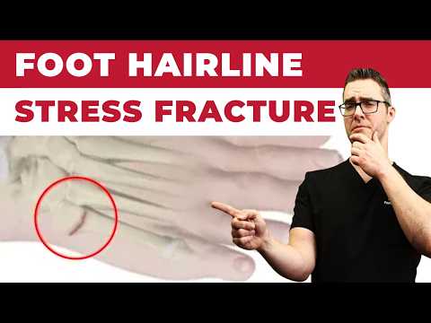

The navicular stress fracture is one of the most consequential athletic foot injuries — and one of the most commonly misdiagnosed. Because plain X-rays miss up to 80% of cases, athletes with dorsal midfoot pain are often treated incorrectly for weeks or months before MRI reveals the true diagnosis. At Balance Foot & Ankle, Dr. Biernacki maintains a low threshold for MRI in athletes with the classic presentation: insidious midfoot pain at the “N-spot” (proximal dorsal navicular) that worsens with activity and often resolves with rest, only to return with resumed training.
Why the Navicular Is High-Risk
The central third of the tarsal navicular has a watershed blood supply — vessels from medial and lateral nutrient arteries converge in the middle, leaving this zone relatively avascular. Repetitive compressive and tensile loading from running and jumping concentrates stress in exactly this watershed zone. Without adequate blood supply to repair microdamage, the bone progressively fractures. Once a complete fracture occurs, union is jeopardized. This vascular anatomy explains why navicular stress fractures are categorized alongside femoral neck and fifth metatarsal stress fractures as requiring aggressive management.
Torg Classification and Treatment Protocol
Dr. Biernacki uses the Torg classification system to guide treatment decisions. Type I: incomplete cortical break — non-weight-bearing short-leg cast for 6–8 weeks achieves union in most cases. Type II: fracture propagates into cancellous bone — non-weight-bearing cast immobilization is standard, though some surgeons advocate early fixation given higher non-union risk. Type III: complete fracture with or without displacement, including sclerotic margins indicating chronicity — surgical fixation with cannulated cancellous screws through the dorsal cortex into the fracture site is the recommended approach. Bone grafting is added in displaced, comminuted, or sclerotic non-union cases. Return to sport is protocolized with CT confirmation of trabecular bridging before full athletic loading resumes.
Conservative vs. Surgical Outcomes
Classic literature (Khan et al., American Journal of Sports Medicine) reported 86% return to sport with non-weight-bearing cast immobilization for complete fractures, but the average time was 5.6 months. Subsequent studies consistently show surgical fixation achieves faster return to sport — 4–5 months average — with lower re-fracture rates in high-level athletes. For competitive athletes who cannot afford a prolonged rehabilitation or who have Type III fractures, Dr. Biernacki discusses the surgical option directly and thoroughly so athletes can make informed decisions about their recovery timelines and career demands.
Returning to Training: Graduated Protocol
Whether treated surgically or conservatively, return to sport follows a graduated loading protocol: pool running and swimming during immobilization phase → walking without pain → low-impact cross-training → sport-specific running on softer surfaces → full training load. CT scan confirmation of bony union is required before progressive loading begins. Custom orthotics addressing any underlying biomechanical risk factors (supinated foot type with rigid midfoot, excessive forefoot adduction, leg length discrepancy) are prescribed to reduce re-injury risk. Calcium and vitamin D status are assessed and corrected in all stress fracture patients.
Dr. Tom's Product Recommendations

CURREX RunPro Insoles — High Arch
⭐ Highly Rated | Foundation Wellness Partner | 30% Commission
Biomechanically engineered running insoles that reduce midfoot impact loading. CURREX uses dynamic arch technology to distribute stress away from high-risk navicular zone — ideal for return-to-run protocols.
Dr. Tom says: “”After my stress fracture healed, CURREX insoles helped me return to marathons without any midfoot pain.””
Runners returning to sport after navicular stress fracture with high arch type
Acute phase — no insoles during cast immobilization or non-weight-bearing period
Disclosure: We earn a commission at no extra cost to you.

Vive Walking Boot — Short Leg Cam Walker
⭐ Highly Rated | Foundation Wellness Partner | 30% Commission
Pneumatic cam walker boot for controlled weight-bearing during stress fracture rehabilitation. Adjustable air cells provide customized compression and support during the protected weight-bearing phase.
Dr. Tom says: “”My doctor prescribed this boot after my navicular stress fracture — it was lightweight and allowed me to stay mobile at work.””
Protected weight-bearing phase of navicular stress fracture treatment (as directed by physician)
Acute Type III fractures requiring strict non-weight-bearing — a boot is not sufficient immobilization
Disclosure: We earn a commission at no extra cost to you.
✅ Pros / Benefits
- MRI diagnosis catches fractures plain X-rays miss — critical for proper treatment staging
- Surgical fixation allows faster return to sport (4–5 months) vs. conservative care for high-grade fractures
- Graduated return-to-sport protocol with CT confirmation ensures safe full athletic loading
❌ Cons / Risks
- High-grade fractures have significant non-union risk with conservative treatment alone
- Total recovery — regardless of treatment — takes 4–8+ months with strict protocol adherence
- Re-fracture risk requires ongoing biomechanical management and load monitoring
Dr. Tom Biernacki’s Recommendation
Navicular stress fractures are the injury I worry most about missing. I’ve seen athletes limp through weeks of ‘midfoot strain’ treatment before an MRI revealed a Type III fracture with sclerotic margins — meaning it had been there for months. My rule is simple: runner or jumper with dorsal midfoot pain at the N-spot gets an MRI, not an X-ray. Once we have the diagnosis, I walk through the Torg classification and explain the real timeline differences between surgical and conservative options. Most competitive athletes opt for fixation when they understand the data. My goal is to get them back to full training as safely and quickly as possible.
— Dr. Tom Biernacki, DPM | Board-Certified Podiatric Surgeon | Balance Foot & Ankle
Frequently Asked Questions
How do I know if I have a navicular stress fracture vs. regular midfoot pain?
The classic presentation is pinpoint tenderness on the dorsal (top) surface of the midfoot — specifically at the ‘N-spot’ in the central proximal navicular. Pain typically builds gradually with training, improves with rest, and returns with resumed activity. Plain X-rays are almost always negative early on. If this description matches your symptoms, request an MRI.
Can a navicular stress fracture heal without surgery?
Yes — Type I and some Type II fractures can heal with strict non-weight-bearing cast immobilization for 6–8 weeks. However, Type III (complete) fractures have up to 56% non-union rates with conservative treatment. Most competitive athletes with complete fractures choose surgical fixation for better and faster outcomes.
What happens if a navicular stress fracture goes untreated?
Untreated or inadequately treated navicular stress fractures can progress to complete fracture, displacement, non-union, and eventually avascular necrosis of the navicular — a severe complication requiring complex reconstruction. Early diagnosis and appropriate treatment are critical.
When can I run again after a navicular stress fracture?
Timeline depends on fracture grade and treatment chosen. With non-weight-bearing cast treatment for early fractures: return to running typically 3–4 months. With surgical fixation of complete fractures: 4–5 months is typical, with CT confirmation of union required before full training resumes.
Michigan Foot Pain? See Dr. Biernacki In Person
4.9★ rated | 1,123 Reviews | 3,000+ Surgeries
Same-week appointments · Howell & Bloomfield Hills
📞 (810) 206-1402 Book Online →Frequently Asked Questions
When should I see a podiatrist?
If symptoms persist past 2 weeks, affect your normal activity, or are accompanied by red-flag symptoms (warmth, redness, swelling, inability to bear weight).
What does treatment cost?
Most diagnostic visits and conservative treatments are covered by Medicare and major insurers. Out-of-pocket costs vary by your specific plan.
How quickly can I get an appointment?
Most non-urgent cases see us within 5 business days. Urgent cases (sudden pain, possible fracture) typically same or next business day.
Dr. Tom Biernacki, DPM is a double board-certified podiatrist and foot & ankle surgeon at Balance Foot & Ankle Specialists in Southeast Michigan. With over a decade of clinical experience, he specializes in heel pain, bunions, diabetic foot care, sports injuries, and minimally invasive surgery. Dr. Biernacki is a member of the APMA and ACFAS, and his patient education content on MichiganFootDoctors.com and YouTube has reached over one million views.
- Plantar Fasciitis: Diagnosis and Conservative Management (PubMed)
- Plantar Fasciitis (APMA)
- Diagnosis and Treatment of Plantar Fasciitis (PubMed / AAFP)
- Heel Pain (APMA)
Related Treatments at Balance Foot & Ankle
Our board-certified podiatrists offer advanced treatments at our Bloomfield Hills and Howell locations.