Board Certified Podiatrists | Expert Foot & Ankle Care
(810) 206-1402 Patient Portal

Plantar Fibromatosis (Ledderhose Disease)
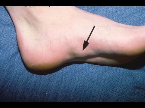
: Nodules on the Arch and When They Need Treatment

Medically reviewed by Dr. Tom Biernacki, DPM | Board-certified podiatrist | 3,000+ surgeries performed
Last updated: April 2, 2026

Quick Answer

Plantar fibromatosis, also called Ledderhose disease, causes firm nodules to develop within the plantar fascia along the arch of the foot. Dr. Tom Biernacki at Balance Foot & Ankle diagnoses and treats these benign but sometimes painful growths with targeted conservative therapy and surgical excision when needed.

Treatment at Balance Foot & Ankle: EPAT Shockwave for Heel Pain →

What Is Plantar Fibromatosis?

Plantar fibromatosis is a benign fibroproliferative disorder in which firm nodules of excess collagen develop within the plantar fascia — the thick band of tissue running along the bottom of the foot from heel to toes. These nodules grow slowly within the tissue rather than on top of it, creating palpable lumps along the medial arch.

The condition is part of a family of fibromatoses that includes Dupuytren’s contracture (hand) and Peyronie’s disease. Approximately 25% of patients with plantar fibromatosis have concurrent Dupuytren’s disease, suggesting a shared genetic predisposition for fibroblast overactivity.

Plantar fibromatosis affects approximately 1-2% of the population, with higher prevalence in Northern European descent populations. Men are affected more commonly than women, and the condition typically presents between ages 30-60. While always benign (non-cancerous), the nodules can grow large enough to cause significant pain and disability.

Symptoms and When Nodules Become Problematic

Early plantar fibromas are often painless — patients notice a firm lump in the arch during self-examination or when a new shoe presses against the area. At this stage, many patients require no treatment beyond monitoring and reassurance that the growth is benign.

Nodules become symptomatic when they grow large enough to create pressure against the ground during weight-bearing, press on adjacent nerves or tendons, or when shoe insoles compress the nodule. Pain is typically described as an aching or burning sensation in the arch that worsens with prolonged standing and walking.

Multiple nodules are common — approximately 30% of patients develop more than one fibroma, and bilateral involvement occurs in 20-25% of cases. Nodules can merge into larger masses called plantar fibromatosis cords that create more extensive arch involvement.

Size progression is variable. Some nodules remain stable for years. Others grow gradually over months. Rapid growth (doubling in weeks) is unusual for plantar fibromatosis and warrants biopsy to confirm the diagnosis and exclude rare soft tissue tumors.

Diagnosis of Plantar Fibromatosis

Clinical examination reveals a firm, fixed nodule within the plantar fascia that does not transilluminate (unlike fluid-filled ganglion cysts). The nodule is adherent to the plantar fascia and moves with the fascia rather than sliding independently. Location within the medial arch band of the plantar fascia is characteristic.

Ultrasound is the first-line imaging modality, demonstrating a hypoechoic (dark) lesion within the plantar fascia with characteristic spindle or fusiform shape. Ultrasound accurately measures nodule size for monitoring growth and can differentiate fibromas from other soft tissue masses.

MRI is obtained when the diagnosis is uncertain, when multiple lesions are present, when surgical planning requires detailed soft tissue mapping, or when the clinical presentation is atypical. MRI shows a well-defined mass within the plantar fascia with low T1 and variable T2 signal intensity.

Biopsy is reserved for atypical presentations — rapidly growing masses, masses with unusual imaging characteristics, or lesions that don’t respond as expected to treatment. Needle biopsy or incisional biopsy confirms the diagnosis histologically.

Conservative Treatment Options

Offloading with accommodative orthotics is the first-line treatment for symptomatic nodules. Custom orthotics with a cutout or depression over the nodule redistribute pressure away from the fibroma, reducing pain with weight-bearing. PowerStep Pinnacle insoles modified with a felt accommodation provide immediate relief while custom devices are fabricated.

Corticosteroid injections can reduce pain and may temporarily shrink nodules by suppressing the inflammatory component of fibromatosis. However, nodules typically recur to their original size after the steroid effect wears off. Serial injections risk plantar fascia weakening and are limited to 2-3 injections.

Verapamil gel (topical calcium channel blocker) has been studied for plantar fibromatosis with moderate evidence suggesting it can slow growth and reduce nodule firmness. Applied directly over the nodule twice daily for 6-12 months, verapamil works by inhibiting fibroblast proliferation and collagen synthesis. Results are variable but the treatment is low-risk.

Radiation therapy using low-dose superficial radiation (orthovoltage) has emerging evidence for early-stage plantar fibromatosis. Studies from European centers report 70-80% symptom improvement and growth arrest. This option is considered for progressive nodules before surgical excision becomes necessary.

Surgical Excision: When and How

Surgery is recommended for nodules that cause significant pain despite conservative treatment, are large enough to impair walking, or are growing progressively. The goal is complete excision of the fibroma and any involved plantar fascia to minimize recurrence.

Limited fasciectomy removes only the nodule and a small margin of surrounding fascia. This approach preserves most of the plantar fascia and has a shorter recovery, but carries a higher recurrence rate of 40-60% because microscopic disease may extend beyond visible margins.

Subtotal plantar fasciectomy removes the nodule along with the entire affected segment of plantar fascia. This more extensive procedure reduces recurrence to 10-25% but creates a larger surgical defect and longer recovery. A plantar incision is required, which takes 4-6 weeks to heal sufficiently for comfortable walking.

Post-operative recovery involves 2-3 weeks of limited weight-bearing followed by gradual return to regular shoes over 4-6 weeks. Custom orthotics are essential after fasciectomy to support the arch that has lost some of its fascial reinforcement. Full recovery takes 2-3 months.

Recurrence Prevention and Long-Term Management

Recurrence after surgical excision is the primary challenge in plantar fibromatosis management. Rates vary from 10-60% depending on excision extent and individual disease activity. Patients with multiple nodules, bilateral disease, and concurrent Dupuytren’s contracture have higher recurrence risk.

Post-operative strategies to reduce recurrence include radiotherapy to the surgical bed, verapamil gel application to the scar, and long-term monitoring with serial ultrasound. Early detection of recurrence when nodules are small allows conservative management that may avoid repeat surgery.

Long-term management focuses on symptom control rather than cure. Even with recurrence, many patients maintain comfortable function with accommodative orthotics, shoe modifications, and periodic interventions. Dr. Biernacki develops individualized long-term management plans that balance intervention with quality of life.

Warning Signs Requiring Urgent Evaluation

  • function bold() { [native code] } — undefined
  • function bold() { [native code] } — undefined
  • function bold() { [native code] } — undefined
  • function bold() { [native code] } — undefined

The Most Common Mistake We See

The most common mistake is having a plantar fibroma excised by a surgeon unfamiliar with the condition’s high recurrence rate. Limited excision (removing just the visible lump) has a 40-60% recurrence rate. Patients benefit from seeing a foot specialist who understands the full spectrum of treatment options — from observation through subtotal fasciectomy — and can match the intervention to the individual disease pattern.

Recommended Products

[object Object]

[object Object]

In-Office Treatment at Balance Foot & Ankle

Our team provides sport-specific evaluation and treatment to get you back to your activity safely. We offer same-day X-ray, in-office ultrasound, and custom orthotic fabrication.

Same-day appointments available. Call (810) 206-1402 or book online.

Frequently Asked Questions

Are plantar fibromas cancerous?

No. Plantar fibromas are always benign. They are composed of excess collagen produced by overactive fibroblasts within the plantar fascia. While they can grow and cause pain, they do not metastasize or become malignant. Rapid growth or atypical features warrant biopsy to confirm the diagnosis.

Will plantar fibromatosis go away on its own?

Plantar fibromas rarely resolve spontaneously. Most remain stable or grow slowly over time. Conservative treatments including orthotics, verapamil gel, and corticosteroid injections can manage symptoms. Surgical excision is reserved for nodules that cause significant pain despite conservative measures.

What causes plantar fibromatosis?

The exact cause is unknown, but genetic predisposition plays a significant role — 25% of patients have concurrent Dupuytren’s contracture. Risk factors include Northern European ancestry, male sex, chronic liver disease, diabetes, and certain anti-seizure medications. Trauma to the plantar fascia may trigger nodule formation in predisposed individuals.

How is plantar fibromatosis different from plantar fasciitis?

Plantar fasciitis is inflammation of the plantar fascia causing heel pain, while plantar fibromatosis is abnormal tissue growth creating nodules in the arch. Fasciitis causes diffuse heel pain worst with first morning steps. Fibromatosis creates palpable lumps with localized arch pain during weight-bearing. Different conditions require different treatments.

The Bottom Line

Plantar fibromatosis requires expert evaluation to determine the appropriate treatment approach — from observation and orthotics through surgical excision. Dr. Tom Biernacki at Balance Foot & Ankle provides comprehensive management for this challenging condition at our Howell and Bloomfield Hills offices, serving patients throughout Southeast Michigan.

Sources

  1. Foot & Ankle International (2024) — Treatment algorithm for plantar fibromatosis
  2. Journal of Foot and Ankle Surgery (2024) — Subtotal fasciectomy outcomes and recurrence rates
  3. Dermatologic Therapy (2023) — Verapamil gel for superficial fibromatoses
  4. Radiotherapy and Oncology (2024) — Low-dose radiation for early plantar fibromatosis

Expert Diagnosis for Arch Lumps — Get Answers Today

Dr. Tom Biernacki has performed over 3,000 foot and ankle surgeries with a 4.9-star rating from 1,123 patient reviews.

Book Your Evaluation

Or call (810) 206-1402 for same-day appointments

Insurance Accepted

BCBS · Medicare · Aetna · Cigna · United Healthcare · HAP · Priority Health · Humana · View All →

Ready to Get Back on Your Feet?

Same-week appointments available at both locations.

Book Your Appointment

(810) 206-1402

Recommended Products for Flat Feet
Products personally used and recommended by Dr. Tom Biernacki, DPM. All available on Amazon.
Structured arch support that provides the structure flat feet are missing.
Best for: All shoe types
Dynamic arch support designed for runners with flat or low arches.
Best for: Running, high-impact sports
These products work best with professional treatment. Book an appointment with Dr. Tom for a personalized treatment plan.
Medical References
  1. Plantar Fasciitis: Diagnosis and Conservative Management (PubMed)
  2. Plantar Fasciitis (APMA)
  3. Diagnosis and Treatment of Plantar Fasciitis (PubMed / AAFP)
  4. Heel Pain (APMA)
This article has been reviewed for medical accuracy by Dr. Tom Biernacki, DPM. References are provided for informational purposes.
Balance Foot & Ankle surgeons are affiliated with Trinity Health Michigan, Corewell Health, and Henry Ford Health — three of Michigan’s largest health systems.