
The most important decision with flat foot surgery isn’t which procedure to pick — it’s confirming the deformity stage (flexible vs. rigid) and the underlying cause first. That changes everything. Call (810) 206-1402.
Quick Answer
Flat foot surgery is reserved for painful, progressive flatfoot that has failed an adequate trial of conservative care — typically 3–6 months minimum of orthotics, bracing, and physical therapy. Procedures range from tendon reconstruction and calcaneal osteotomy (flexible deformity) to fusion (rigid, arthritic deformity). Expect 6–8 weeks non-weight-bearing after bony procedures and 9–12 months for full recovery. Most flat feet — even severe ones — never need surgery. Same-week surgical consultations in Howell & Bloomfield Hills: (810) 206-1402.

Related Conditions
In This Article
When Does Flat Foot Require Surgery?
New to flat feet? Start with our complete guide to flat feet treatment, then come back here when surgery is on the table.

The vast majority of flat feet—including many severely flat feet—do not require surgery. Flexible flatfoot (where an arch forms when standing on tiptoe) in adults without symptoms rarely needs intervention. Surgery for flatfoot is reserved for specific scenarios: adult acquired flatfoot deformity (AAFD) that has progressed to a rigid, painful deformity causing functional disability despite adequate conservative treatment; pediatric flatfoot causing progressive deformity or symptoms that fails non-operative management; or flatfoot associated with specific structural pathology such as posterior tibial tendon dysfunction (PTTD) that is progressing despite conservative care.
The decision to pursue flatfoot surgery requires careful staging of the deformity (flexible vs. rigid, degree of deformity, joint involvement), assessment of the underlying cause (PTTD, tarsal coalition, Charcot arthropathy, hypermobility syndrome), and genuine failure of conservative measures—orthotics, physical therapy, bracing with ankle-foot orthosis, and activity modification—over a sufficient trial period (typically 3–6 months minimum).
Types of Flat Foot Surgery
Posterior Tibial Tendon Reconstruction
Adult acquired flatfoot deformity most commonly results from failure of the posterior tibial tendon (PTT)—the primary dynamic stabilizer of the arch. Stage II PTTD (tendon degeneration with flexible deformity) is treated with a combination of procedures: flexor digitorum longus (FDL) tendon transfer to augment the failed PTT, medial calcaneal osteotomy (shifting the heel bone inward) to correct hindfoot valgus alignment, and often a lateral column lengthening (calcaneal lengthening osteotomy) to restore forefoot coverage of the hindfoot. This combination addresses both the tendon failure and the resulting structural deformity.
Subtalar Joint Arthroereisis
Arthroereisis involves placing a small implant (typically silicone or titanium sinus tarsi implant) into the sinus tarsi space—the cavity on the outer side of the hindfoot—to physically limit excessive subtalar joint pronation. This minimally invasive procedure is primarily used in pediatric and adolescent flexible flatfoot with hindfoot valgus. It can be performed through a small incision with rapid recovery, and is reversible if the implant causes problems. Results in appropriate pediatric patients are good, though long-term data is limited and controversy exists regarding its routine use.
Triple or Selective Subtalar Arthrodesis
Stage III and IV adult acquired flatfoot (rigid deformity with subtalar, talonavicular, or ankle joint involvement) typically requires joint fusion procedures. Triple arthrodesis—fusion of the subtalar, talonavicular, and calcaneocuboid joints simultaneously—corrects the rigid deformity in a fixed position. Selective fusions target only the joints most involved. While fusion eliminates motion in fused joints and places additional demand on adjacent joints over time, it reliably corrects deformity and eliminates arthritis pain in patients who have exhausted conservative options.
Tarsal Coalition Resection
Tarsal coalition—an abnormal bony, cartilaginous, or fibrous connection between two hindfoot bones (most commonly calcaneonavicular or talocalcaneal coalition)—causes a rigid flatfoot with limited subtalar motion, often presenting with pain in adolescence. Resection of the coalition (removal of the abnormal bridge of tissue) in appropriate candidates restores motion and resolves pain. Cases with significant secondary arthritic change typically require fusion rather than simple resection.
Minimally Invasive Flatfoot Surgery (MIS Calcaneal Osteotomy)
A growing number of flatfoot corrections can now be performed through minimally invasive (percutaneous) techniques. The medializing calcaneal displacement osteotomy — traditionally performed through an open lateral incision — can often be done through a small stab incision using a specialized burr (a MICA-style/MIS calcaneal osteotomy), with less soft-tissue disruption, smaller scars, and a lower wound-complication risk. Not every deformity is a candidate: rigid deformities and fusion procedures still generally require open approaches. At Balance Foot & Ankle, we evaluate every surgical candidate for a minimally invasive option first.
Recovery from Flat Foot Surgery
Flatfoot reconstruction is among the more involved foot and ankle surgical procedures, and recovery reflects this complexity. Most bony procedures (osteotomies, fusions) require 6–8 weeks non-weight-bearing in a cast, followed by progressive weight-bearing in a boot over 4–8 weeks. Physical therapy for range of motion and strength begins once weight-bearing is established. Full functional recovery—return to normal activity without significant restriction—takes 9–12 months for complex reconstructions. Swelling often persists for 12–18 months. The timeline for return to sport or high-demand activity is typically 9–12 months after major reconstructive procedures.
In-Office Treatment at Balance Foot & Ankle
If home care isn’t resolving your flat foot deformity, a visit with a board-certified podiatrist is the fastest path to accurate diagnosis and a personalized plan. At Balance Foot & Ankle Specialists, Dr. Tom Biernacki, Dr. Carl Jay, and Dr. Daria Gutkin offer same-day and next-day appointments at both our Howell and Bloomfield Hills offices. We perform on-site diagnostic ultrasound, digital X-ray, conservative care, advanced regenerative treatments, and minimally invasive surgery when indicated.
Call (810) 206-1402 or request an appointment online. Most insurance plans accepted, including Medicare, Blue Cross Blue Shield, Aetna, Cigna, and United Healthcare.
Recovery Essentials After Flat Foot Surgery
These are the items I tell my surgical patients to have ready before surgery day. The first 6–8 weeks are non-weight-bearing — a knee scooter (available at most pharmacies, often covered by insurance) plus the items below make that stretch dramatically easier.
Affiliate disclosure: As an Amazon Associate, Balance Foot & Ankle earns from qualifying purchases. These are products we recommend to our own surgical patients.
| Item | Recovery Phase | Why You Need It | Link |
|---|---|---|---|
| Waterproof Cast & Boot Cover | Weeks 0–8 (cast/boot) | Keeps the incision and cast dry in the shower — water inside a cast causes skin maceration and healing delays. | View on Amazon |
| Seamless Cast Socks | Weeks 0–12 | Cushions skin against the cast or boot — prevents pressure points and blisters during long wear. | View on Amazon |
| EVENup Shoe Leveler | Weeks 8–14 (boot walking) | Levels your other shoe to the boot height — prevents the hip, knee, and back pain caused by walking lopsided. | View on Amazon |
| OOFOS OOahh Recovery Slide | Boot-to-shoe transition | Impact-absorbing slide for around the house once you’re cleared out of the boot — gentle on a healing foot. | View on Amazon |
| Hoka Bondi 9 | Return to walking (3+ months) | Max-cushion, rocker-sole walking shoe — the easiest transition back to full days on your feet. | View on Amazon |
| PowerStep Pinnacle Insoles | Long-term support | Arch support for your non-surgical foot during recovery, and everyday support once you’re back in regular shoes. | View on Amazon |
When to See a Podiatrist
Foot and ankle surgery in 2026 is dramatically different than a decade ago — many procedures are now minimally invasive and outpatient, and recovery protocols are far faster than they were even five years ago. Balance Foot & Ankle surgeons have performed 3,000+ foot/ankle surgeries with modern techniques. If another surgeon has recommended a traditional open procedure, a second opinion may reveal a faster, less-invasive option.
Call Balance Foot & Ankle: (810) 206-1402 · Book online · Offices in Howell & Bloomfield Hills
Differential Diagnosis: What Else Could It Be?
Several conditions share symptoms with Flat Feet (Pes Planus) and are commonly misdiagnosed in the first office visit. Considering these alternatives is part of every Balance Foot & Ankle exam:
- Posterior tibial tendon dysfunction (PTTD). Acquired adult flatfoot with single-leg heel-rise weakness.
- Tarsal coalition. Rigid flatfoot in an adolescent — bone bridge between hindfoot bones.
- Charcot foot (diabetic). Sudden warm, swollen, collapsing midfoot in a diabetic — urgent off-loading.
If your symptoms don’t fit the textbook pattern, ask your podiatrist which differentials they ruled out — that conversation often shortcuts months of trial-and-error treatment.
In Our Clinic
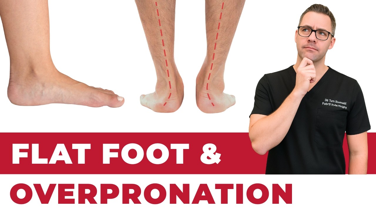
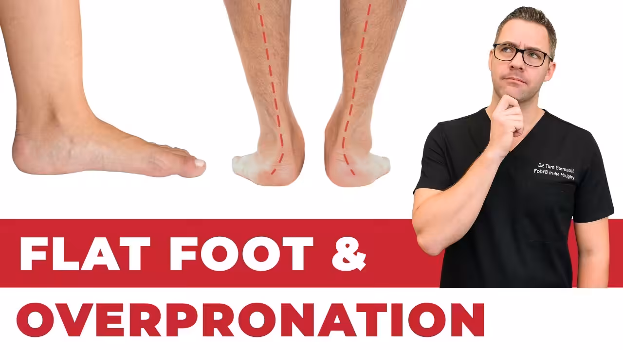
In our clinic, the flat-footed patient who actually needs intervention is the one whose arch is collapsing progressively in adulthood — not the person who was born flat-footed and has been running 5Ks pain-free for 20 years. We evaluate for posterior tibial tendon dysfunction (PTTD) with single-heel-rise testing, check for the “too many toes” sign from behind, and get weight-bearing X-rays. Early PTTD responds well to a custom orthotic with a medial heel skive + short course of boot immobilization. Stage 2+ PTTD is a different conversation — we discuss tendon transfers and calcaneal osteotomy candidates.
Most Common Mistake We See
The most common mistake we see is: Buying motion-control shoes without a gait assessment. Fix: get a pressure-plate analysis or wet-foot test first to confirm overpronation and arch height.
Warning Signs That Need Same-Day Care
Seek immediate evaluation at Balance Foot & Ankle if you experience any of the following:
- Rapid collapse of an arch on one foot (possible PTT rupture)
- Walking becoming impossible
- Redness or warmth along the inner arch
- Diabetes plus progressive arch collapse
Call (810) 206-1402 — same-day and next-day appointments at our Howell and Bloomfield Hills offices.
Insurance Accepted
BCBS · Medicare · Aetna · Cigna · United Healthcare · HAP · Priority Health · Humana · View All →
Howell Office
4330 E Grand River Ave
Howell, MI 48843
Get Directions →
Bloomfield Hills Office
43494 Woodward Ave, #208
Bloomfield Hills, MI 48302
Get Directions →
Your Board-Certified Podiatrists


Frequently Asked Questions
Can flat feet be corrected with surgery?
Yes—flatfoot deformity can be substantially corrected with surgical intervention in appropriately selected patients. Modern flatfoot reconstruction techniques can restore near-normal alignment and function in many patients. For flexible deformities with posterior tibial tendon dysfunction, the combination of tendon reconstruction and bony procedures corrects alignment reliably. For rigid deformities requiring fusion, the goal is correction to a functional, plantigrade position rather than recreating a normal arch. Success is measured in pain relief and functional improvement, not simply arch height restoration. Not all flatfoot requires or benefits from surgical correction—the severity and impact on quality of life must justify the recovery demands before proceeding.
Is flat foot surgery painful?
Flatfoot reconstruction involves significant surgical work (bone cuts, hardware placement, tendon transfer), and postoperative pain management is an important part of the care plan. Modern techniques—regional nerve blocks, multimodal analgesia, anti-inflammatory medications—have substantially reduced postoperative pain compared to earlier approaches. Most patients describe manageable pain in the first 1–2 weeks that decreases significantly over the following weeks. The non-weight-bearing period requires patience and appropriate pain management but is not typically described as severely painful once past the first week. Pain that is out of proportion, worsening, or associated with unusual swelling or fever should prompt immediate contact with the surgical team.
What happens if flat feet are not treated?
Asymptomatic flat feet in adults often require no treatment and may remain stable for life. However, progressive adult acquired flatfoot deformity—particularly when driven by posterior tibial tendon dysfunction—can progress through stages from flexible, correctable deformity to rigid, arthritic deformity over years. Early stage disease is more amenable to joint-preserving procedures; late-stage rigid deformity requires fusion. Progressive deformity can also cause pain and disability in adjacent joints (ankle, knee) as abnormal biomechanics alter loading patterns throughout the lower extremity. Symptomatic progressive flatfoot warrants evaluation and treatment to prevent advancement to more complex and less reversible stages.
Are flat feet always painful?
No — most people with flat feet never develop symptoms. The arch height alone doesn’t predict pain; what matters is whether the foot compensates effectively and how much load it handles. Flat feet become problematic when they cause excessive pronation that stresses the plantar fascia, posterior tibial tendon, knees, or lower back. We see flat-footed patients who run marathons without pain alongside flat-footed patients disabled by daily walking. The biomechanics matter more than the arch height.
Can flat feet be corrected without surgery?
For most people, yes — symptom control without structural correction is the goal. Custom orthotics, motion-control shoes, and targeted physical therapy (posterior tibial strengthening, calf stretching) manage flat foot symptoms effectively in 85–90% of cases. Surgical correction (calcaneal osteotomy, subtalar arthroereisis, or flatfoot reconstruction) is reserved for cases where conservative care has failed despite an adequate trial (typically 3–6 months minimum, often longer) or the deformity is severe enough to cause joint damage.
What’s the difference between flat feet and fallen arches?
‘Fallen arches’ describes acquired adult flatfoot — when an arch that was once normal collapses over time, usually due to posterior tibial tendon dysfunction (PTTD). ‘Flat feet’ typically refers to a lifelong flexible flatfoot present since childhood. The distinction matters for treatment: acquired adult flatfoot is more urgent because active tendon degeneration is involved, and it can progress to a rigid, arthritic deformity if not treated. Flexible childhood flat feet are usually asymptomatic and don’t require intervention.
Do orthotics fix flat feet?
Orthotics don’t structurally fix flat feet — they manage the biomechanical consequences. A custom orthotic holds your foot in a corrected position while weight-bearing, reducing strain on the plantar fascia, posterior tibial tendon, and medial knee. For flexible flat feet (the most common type), a well-fitted orthotic plus motion-control footwear is often sufficient for lifelong symptom control. Rigid flat feet with arthritis may need additional intervention.
Are flat feet genetic?
Both genetic and environmental factors contribute. Flexible flat feet (most common type) have a strong hereditary component — if one or both parents have flat feet, children are significantly more likely to as well. However, obesity, prolonged standing on hard surfaces, and high-impact activity can accelerate collapse in genetically predisposed individuals. Posterior tibial tendon dysfunction — the most common cause of adult acquired flatfoot — has risk factors including age, female sex, hypertension, and diabetes.
Can flat feet cause knee and back pain?
Yes — this is one of the most common presentations we see. Overpronation from flat feet causes internal tibial rotation, which stresses the medial knee and hip. This kinetic chain effect can produce knee pain (patellofemoral syndrome), hip pain, and low back pain in patients with no direct foot symptoms. In our clinic, roughly 30% of patients presenting with knee pain have flat feet as a contributing cause. Correcting the pronation with orthotics often resolves upstream joint pain.
What shoes are best for flat feet?
Motion control and stability categories — specifically those with a medial post (a denser foam section under the arch) and a firm heel counter. New Balance 860, Brooks Adrenaline GTS, and Asics Kayano are consistently strong performers. Avoid neutral-cushioned shoes (they’re designed for efficient gaits that don’t pronate) and minimalist shoes entirely. The goal is to limit the inward collapse of the foot at midstance.
Should children with flat feet wear special shoes?
Only if symptomatic. Flexible flat feet in children are extremely common before age 6 and often resolve naturally as the arch develops. Routine shoe inserts for asymptomatic flat-footed children are not evidence-based and may actually impair natural arch strengthening. If your child complains of foot or leg pain, is walking awkwardly, or fatigues unusually quickly, bring them in for an evaluation. Symptomatic pediatric flat feet do benefit from supportive footwear and sometimes custom orthotics.
Can I strengthen my way out of flat feet?
Strengthening the posterior tibial tendon, intrinsic foot muscles, and peroneals can improve dynamic arch control and reduce symptoms — but won’t change bone structure. Short-foot exercises, single-leg calf raises, and resistance band eversion work are the best evidence-based options. In our experience, strengthening works best when combined with orthotic support rather than as a replacement. Pure strengthening programs without load management often stall.
When does flat foot pain require surgery?
Surgery is considered when: conservative treatment has failed despite an adequate trial (typically 3–6 months minimum, often longer), the deformity is rigid (arthritic), the posterior tibial tendon has ruptured or is severely degenerated (Stage III/IV PTTD), or significant collapse has occurred in the lateral column. About 10–15% of adult acquired flatfoot patients eventually need surgery. Modern reconstructive procedures — calcaneal osteotomy with tendon transfer — have excellent outcomes when timing is right. Delaying too long allows joint damage that makes reconstruction less effective.
Is flat foot a disability?
Flat foot alone rarely constitutes a disability, but severe symptomatic flatfoot with associated PTTD or arthritis can significantly limit function. For workers in physically demanding jobs — standing 8+ hours, climbing ladders — a symptomatic flatfoot can genuinely impact employment. We document severity and functional limitation for patients pursuing VA disability claims, workers’ comp cases, or FMLA paperwork. Schedule an appointment and we’ll provide clinical documentation of your specific case.
Medical References & Sources
- American Orthopaedic Foot & Ankle Society — Adult Acquired Flatfoot
- PubMed Research — PTTD Flatfoot Reconstruction Outcomes
- PubMed Research — Adult Flatfoot Surgery Review
Dr. Tom Biernacki, DPM is a board-certified podiatric surgeon at Balance Foot & Ankle in Howell and Bloomfield Hills, Michigan. He evaluates and stages adult flatfoot deformity and coordinates comprehensive management including orthotics, bracing, tendon reconstruction, osteotomies, and fusion procedures.
Ready for Expert Care?
Same-day appointments in Howell & Bloomfield Hills, MI.
4.9★ | 1,123 Reviews | 3,000+ Surgeries
Or call: (810) 206-1402
Dr. Tom Biernacki, DPM is a board-certified foot & ankle surgeon (ABFAS & ABPM) at Balance Foot & Ankle Specialists in Southeast Michigan. With over a decade of clinical experience, he specializes in heel pain, bunions, diabetic foot care, sports injuries, and minimally invasive surgery. Dr. Biernacki is a member of the APMA and ACFAS, and his patient education content on MichiganFootDoctors.com and YouTube has made him one of the most-followed foot & ankle educators on YouTube.