

You are in the right place. Dr. Tom Biernacki, DPM, FACFAS — board-certified foot & ankle surgeon with 3,000+ surgeries — explains exactly what ankle fracture types/treatment/recovery means and what actually works. Call (810) 206-1402 for a same-day appointment at our Howell or Bloomfield Hills office.
Quick answer: Treatment for ankle fracture types treatment recovery follows a stepwise approach: 1) conservative care first (rest, ice, supportive footwear, OTC anti-inflammatories), 2) physical therapy and targeted exercises, 3) in-office treatments (injections, custom orthotics) if conservative fails at 4-6 weeks, 4) surgery for refractory cases. Most patients resolve at step 1 or 2. Call (810) 206-1402.
Medically reviewed by Dr. Tom Biernacki, DPM · Board-Certified Podiatric Surgeon · Last reviewed: April 2026 · Editorial Policy
The most important clinical decision with Ankle Fracture Types Treatment Recovery isn’t which treatment to start with — it’s identifying the correct subtype. That changes everything. Call (810) 206-1402.
Quick Answer
Ankle Fractures: Types, Treatment Options & What to Exp relates to foot/ankle injury — typically caused by trauma or twist. Most patients improve in 4-8 weeks with conservative care. Same-week appointments in Howell + Bloomfield Hills: (810) 206-1402.

Medically reviewed by Dr. Tom Biernacki, DPM — Board-Certified Podiatric Surgeon — Balance Foot & Ankle, Howell & Bloomfield Hills, MI. Last updated April 2026.
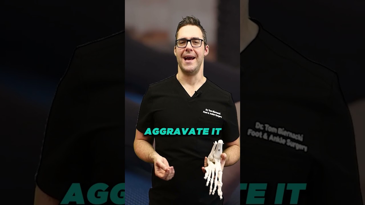
▶ Watch

👟 Dr. Tom Also Recommends
Podiatrist Recommended Shoes 2026: Dr. Tom’s Top Picks for Every Condition
The right footwear can make or break your recovery. Dr. Tom’s complete guide to the best shoes for plantar fasciitis, flat feet, neuropathy, bunions & more — with clinical picks for every foot type.
See Dr. Tom’s Top Shoe Picks →Medically Reviewed by Dr. Tom Biernacki, DPM — Board-Certified Podiatrist, Balance Foot & Ankle Specialists, Michigan. Last updated April 2026.
Ankle fractures are among the most common orthopedic injuries — accounting for roughly 10% of all fractures treated in emergency departments. But not all ankle fractures are alike. Treatment ranges from a simple walking boot to complex surgical reconstruction with plate and screw fixation, and the right approach depends on factors that aren’t visible on a basic X-ray.
Ankle Anatomy and Fracture Patterns
The ankle joint (mortise) is formed by three bones: the tibia (medial malleolus and posterior malleolus), the fibula (lateral malleolus), and the talus. Stability depends on the integrity of the bony mortise and the ligamentous complex surrounding it.
Ankle fractures are classified by which bones are broken and the degree of instability created:
- Isolated lateral malleolus fracture (most common): Fracture of the distal fibula without associated medial or ligamentous injury. Many are stable and can be managed in a boot with weight-bearing as tolerated.
- Bimalleolar fracture: Fractures of both the lateral and medial malleolus. The mortise is disrupted — most require surgery to restore stability.
- Trimalleolar fracture: All three malleoli (lateral, medial, and posterior tibial) are fractured — representing severe instability with significant ligamentous disruption. Typically requires surgical fixation of multiple fragments.
- High fibula fracture with syndesmotic injury (Maisonneuve fracture): A proximal fibula fracture combined with disruption of the syndesmotic ligament complex between the tibia and fibula. The proximal fibula fracture may not be on the initial ankle X-ray — missed if the entire fibula is not imaged. Requires syndesmotic stabilization.
- Syndesmotic injury (high ankle sprain): Injury to the tibiofibular ligament complex without fracture — but with widening of the ankle mortise that compromises stability. Treated similarly to fractures requiring stabilization.
The Critical Question: Stable or Unstable?
The fundamental treatment decision for ankle fractures is stability assessment. A stable fracture — where the mortise remains congruent (talus centered between the malleoli) under weight-bearing stress — can typically be managed non-surgically. An unstable fracture — where the mortise gaps or the talus shifts under load — requires surgical fixation to prevent malunion, post-traumatic arthritis, and chronic functional instability.
Stability is assessed with stress X-rays (gravity stress view or manual stress) and sometimes CT scan for complex fractures. The difference between a stable lateral malleolus fracture managed in a boot vs. an unstable bimalleolar equivalent requiring surgery can sometimes be subtle on initial X-rays.
Non-Surgical Treatment
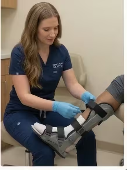
Stable isolated lateral malleolus fractures are managed with a walking boot and weight-bearing as tolerated, progressing to normal footwear within 6–8 weeks as pain allows. Physical therapy for proprioception and strength restoration is recommended after immobilization. Follow-up X-rays confirm maintained reduction.
Surgical Treatment
Unstable ankle fractures require open reduction and internal fixation (ORIF) — usually within 5–10 days after injury, once swelling has subsided enough for safe soft tissue handling. Surgery typically involves:
- Lateral fibula: plate and screw fixation to restore fibular length, alignment, and rotation
- Medial malleolus: screw or tension band fixation
- Posterior malleolus: screw fixation if the fragment is large (greater than 25% of the articular surface)
- Syndesmotic screws or suture-button fixation if the syndesmosis is disrupted
Recovery Timeline
Non-surgical fractures: Progressive weight-bearing over 6–8 weeks, return to normal activity at 10–12 weeks. Surgical fractures: Non-weight-bearing for 2–4 weeks, then boot for 4–6 more weeks, physical therapy starting at 6–8 weeks. Return to full activity at 4–6 months, though full strength and proprioception restoration takes up to a year. Syndesmotic screws may require removal at 3 months depending on hardware type.
Ankle Fracture Evaluation — Southeast Michigan
Dr. Biernacki evaluates ankle fractures with on-site digital X-ray at our Bloomfield Hills and Howell offices. Same-week appointments for urgent injuries.
📞 (810) 206-1402 |
📧 Get Dr. Tom’s Free Lab Test Guide
Discover the 5 lab tests every person over 35 should ask their doctor about — explained in plain English by a board-certified physician.
📍 Located in Michigan?
Our board-certified podiatrists treat this condition at two convenient locations. Same-day appointments often available.
Insurance Accepted
BCBS · Medicare · Aetna · Cigna · United Healthcare · HAP · Priority Health · Humana · View All →
Howell Office
4330 E Grand River Ave
Howell, MI 48843
Get Directions →
Bloomfield Hills Office
43494 Woodward Ave, #208
Bloomfield Hills, MI 48302
Get Directions →
Your Board-Certified Podiatrists


Ready to Get Back on Your Feet?
Same-week appointments available at both locations.
Book Your AppointmentMore Podiatrist-Recommended Foot Health Essentials
Hoka Clifton 10

Max-cushion everyday shoe — podiatrist favorite for walking and running.

OOFOS Recovery Slide

Impact-absorbing recovery sandal — wear after long days on your feet.
As an Amazon Associate, Balance Foot & Ankle earns from qualifying purchases. Product recommendations are based on clinical experience; prices and availability shown above update live from Amazon.
When to See a Podiatrist
If foot or ankle pain has been bothering you for more than a few weeks, home care alone may not be enough. Balance Foot & Ankle offers same-week appointments at our Howell and Bloomfield Hills clinics — no referral needed in most cases. Bring your current shoes and a short list of symptoms and we’ll build you a treatment plan in one visit.
Call Balance Foot & Ankle: (810) 206-1402 · Book online · Offices in Howell & Bloomfield Hills
Watch: Dr. Tom explains
Podiatrist-recommended products
As an Amazon Associate, Dr. Tom earns from qualifying purchases.
Immobilization for stable ankle fractures.
View on Amazon →Post-healing arch support for return to activity.
View on Amazon →Acute ankle fracture swelling control.
View on Amazon →Topical comfort during ankle fracture healing.
View on Amazon →Related resources
Ready to solve this? Book today.
Same-week appointments · Howell & Bloomfield Hills · 4.9★ (1,123+ reviews)
☎ (810) 206-1402Book Online →In-Office Treatment at Balance Foot & Ankle
When conservative care isn’t enough, Dr. Tom Biernacki and the team at Balance Foot & Ankle offer advanced, same-day options — including Foot & Ankle Fracture Repair Michigan at our Howell and Bloomfield Hills clinics.
Same-day appointments available. Call (810) 206-1402 or book online.
Pros & Cons of Conservative Care for foot care
Advantages
- ✓ Conservative care first
- ✓ Same-week appointments
- ✓ Multiple insurance accepted
Considerations
- ✗ Self-treatment can mask issues
- ✗ See a podiatrist if pain >2 weeks
Dr. Tom’s Recommended Products for foot care
Affiliate disclosure: As an Amazon Associate, Balance Foot & Ankle earns from qualifying purchases. We only recommend products we use with patients.
Footnanny Heel Cream Dr. Tom’s Pick
Best for: Daily moisturizer for cracked heels
Ready to Get Back on Your Feet?
Same-day appointments in Howell + Bloomfield Hills. Most insurance accepted. Dr. Tom Biernacki, DPM & team.
Book Today — Same-Day Appointments Available
Call Now: (810) 206-1402
About Your Care Team at Balance Foot & Ankle
Dr. Tom Biernacki, DPM · Board-Certified Foot & Ankle Surgeon. Specializes in conservative-first care, minimally invasive bunion surgery, and complex reconstruction.
Dr. Carl Jay, DPM · Accepting new patients. Specializes in sports medicine, athletic injuries, and routine podiatric care.
Dr. Daria Gutkin, DPM, AACFAS · Accepting new patients. Specializes in surgical reconstruction and pediatric podiatry.
Locations: 4330 E Grand River Ave, Howell, MI 48843 · 43494 Woodward Ave Suite 208, Bloomfield Hills, MI 48302
Hours: Mon–Fri 8:00 AM – 5:00 PM · (810) 206-1402
🩺 Dr. Tom’s Recommended Products
As an Amazon Associate I earn from qualifying purchases. These are products I personally use and recommend to patients.
Natural menthol + arnica for post-injury soreness. FSA-eligible, plant-based formula.
View on Amazon →
Post-injury graduated compression for swelling. Truly graduated — most OTC socks are not.
View on Amazon →
Ready to fix this for good?
Reading goes only so far. The fastest path to relief is a 30-minute office visit with Dr. Biernacki — same-day Howell or Bloomfield Hills. Call (810) 206-1402 or use our online booking.
In-Office Treatment at Balance Foot & Ankle

If home treatment isn’t providing relief for your ankle sprain or injury, our podiatry team at Balance Foot & Ankle can help with same-day evaluations and advanced in-office care.
Same-day appointments available. (810) 206-1402
Get Expert Care at Balance Foot & Ankle
Same-week appointments at our Howell and Bloomfield Hills offices. Board-certified podiatric surgeons. Most insurance accepted.
Ready for Expert Care?
Same-day appointments in Howell & Bloomfield Hills, MI.
4.9★ | 1,123 Reviews | 3,000+ Surgeries
Or call: (810) 206-1402
Dr. Tom Biernacki, DPM is a board-certified foot & ankle surgeon (ABFAS & ABPM) at Balance Foot & Ankle Specialists in Southeast Michigan. With over a decade of clinical experience, he specializes in heel pain, bunions, diabetic foot care, sports injuries, and minimally invasive surgery. Dr. Biernacki is a member of the APMA and ACFAS, and his patient education content on MichiganFootDoctors.com and YouTube has made him one of the most-followed foot & ankle educators on YouTube.