

Medically reviewed by Dr. Tom Biernacki, DPM · Board-Certified Podiatric Surgeon · Last reviewed: April 2026 · Editorial Policy
The most important clinical decision with Ankle Ligament Anatomy Atfl Cfl Ptfl Deltoid isn’t which treatment to start with — it’s identifying the correct subtype. That changes everything. Call (810) 206-1402.
Quick Answer
Ankle Ligament Anatomy: ATFL, CFL, PTFL, and the Deltoid Com relates to foot pain — typically caused by overuse, footwear, or biomechanics. Most patients improve in 6-12 weeks with conservative care. Same-week appointments in Howell + Bloomfield Hills: (810) 206-1402.

Medically reviewed by Dr. Tom Biernacki, DPM — Board-certified foot & ankle surgeon, 3,000+ surgeries performed. Updated April 2026 with current clinical evidence. This article reflects real practice experience from Balance Foot & Ankle Specialists in Howell and Bloomfield Hills, Michigan.
Quick Answer
Most foot and ankle problems respond to conservative care — proper footwear, supportive inserts, activity modification, and targeted stretching — within 4-8 weeks. Persistent pain beyond that window, or any symptom that prevents walking, warrants a podiatric evaluation to rule out fracture, tendon tear, or systemic cause.
Watch: Dr. Tom Biernacki, DPM

Medically reviewed by Dr. Tom Biernacki, DPM — Board-Certified Podiatric Surgeon — Balance Foot & Ankle, Howell & Bloomfield Hills, MI. Last updated April 2026.
▶ Watch

Medically Reviewed by Dr. Tom Biernacki, DPM — Board-Certified Podiatrist, Balance Foot & Ankle Specialists, Michigan. Last updated April 2026.
Understanding ankle ligament anatomy is fundamental to diagnosing and treating ankle sprains, instability, and syndesmotic injuries. Each ankle ligament has a specific biomechanical role, injury mechanism, physical examination test, and treatment implication. The majority of ankle sprains (85%) involve the lateral ligament complex, but the severity of injury across the spectrum of lateral, syndesmotic, and medial ligament injuries varies enormously — and misclassifying injury severity leads to inappropriate treatment and chronic instability.
Lateral Ligament Complex
Anterior talofibular ligament (ATFL): the most commonly sprained ligament in the body; courses anteriorly from the anterior fibula to the talar neck; taut in plantarflexion and inversion; resists anterior talar translation and internal rotation; tested by the anterior drawer test (examiner translates the heel anteriorly with one hand while stabilizing the tibia — >5mm displacement or asymmetry compared to the contralateral side indicates ATFL laxity). Calcaneofibular ligament (CFL): runs inferiorly from the fibular tip to the lateral calcaneal wall; taut in dorsiflexion and inversion; resists combined inversion and dorsiflexion stress; tested by the talar tilt test (inversion stress applied in dorsiflexion — >9° talar tilt or 3° asymmetry indicates CFL laxity); isolated CFL injury is uncommon — CFL tears usually accompany complete ATFL tears. Posterior talofibular ligament (PTFL): the strongest of the three lateral ligaments; rarely injured in typical ankle sprains; injured only in severe ankle dislocations; resists posterior talar translation. Combined ATFL + CFL tear = Grade III lateral ankle sprain (complete lateral complex disruption).
Deltoid and Syndesmotic Ligaments
Deltoid ligament (medial): a broad, fan-shaped ligament complex comprising superficial (tibiospring, tibionavicular, tibiocalcaneal) and deep (anterior and posterior tibiotalar) components; the deep deltoid is the primary restraint against lateral talar shift — its injury is the critical element in bimalleolar equivalent ankle fractures; isolated deltoid sprains occur from eversion injuries and are less common than lateral sprains; medial ankle pain, tenderness, and eversion stress pain indicate deltoid injury; the deep deltoid must be assessed in all ankle fractures (widened medial clear space >4mm on X-ray = deep deltoid incompetence). Syndesmotic ligaments (high ankle ligaments): AITFL, PITFL, transverse tibiofibular ligament, interosseous membrane — assessed separately from the lateral complex; injured by external rotation and hyperdorsiflexion mechanisms; produces high ankle (syndesmotic) sprain with anterolateral ankle and proximal fibular pain (squeeze test, external rotation stress test); requires longer recovery and occasional surgical stabilization for unstable injuries. Dr. Biernacki at Balance Foot & Ankle uses clinical examination and stress imaging to accurately classify ankle ligament injuries and direct appropriate treatment from conservative management to surgical reconstruction. Call (810) 206-1402 at our Bloomfield Hills or Howell office.
📧 Get Dr. Tom’s Free Lab Test Guide
Discover the 5 lab tests every person over 35 should ask their doctor about — explained in plain English by a board-certified physician.
📍 Located in Michigan?
Our board-certified podiatrists treat this condition at two convenient locations. Same-day appointments often available.
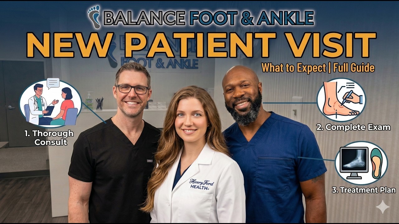
In-Office Treatment at Balance Foot & Ankle
If home care isn’t resolving your your foot or ankle concern, a visit with a board-certified podiatrist is the fastest path to accurate diagnosis and a personalized plan. At Balance Foot & Ankle Specialists, Dr. Tom Biernacki, Dr. Carl Jay, and Dr. Daria Gutkin offer same-day and next-day appointments at both our Howell and Bloomfield Hills offices. We perform on-site diagnostic ultrasound, digital X-ray, conservative care, advanced regenerative treatments, and minimally invasive surgery when indicated.
Call (810) 206-1402 or request an appointment online. Most insurance plans accepted, including Medicare, Blue Cross Blue Shield, Aetna, Cigna, and United Healthcare.
More Podiatrist-Recommended Foot Health Essentials
Hoka Clifton 10


Max-cushion everyday shoe — podiatrist favorite for walking and running.

OOFOS Recovery Slide

Impact-absorbing recovery sandal — wear after long days on your feet.
As an Amazon Associate, Balance Foot & Ankle earns from qualifying purchases. Product recommendations are based on clinical experience; prices and availability shown above update live from Amazon.

When to See a Podiatrist
If foot or ankle pain has been bothering you for more than a few weeks, home care alone may not be enough. Balance Foot & Ankle offers same-week appointments at our Howell and Bloomfield Hills clinics — no referral needed in most cases. Bring your current shoes and a short list of symptoms and we’ll build you a treatment plan in one visit.
Call Balance Foot & Ankle: (810) 206-1402 · Book online · Offices in Howell & Bloomfield Hills
Frequently Asked Questions
How do I know if I sprained or broke my ankle?
Both cause pain, swelling, and difficulty walking. Key differences: fractures often cause more immediate severe pain, tenderness directly over bone (not just ligament), and inability to bear any weight. X-rays and the Ottawa Ankle Rules help determine if imaging is needed.
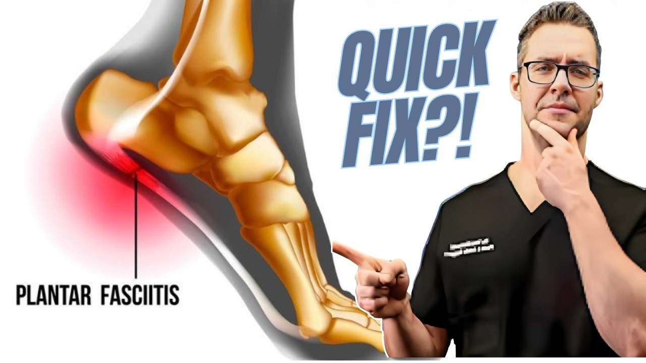
How long does an ankle sprain take to heal?
Grade I (mild): 1–2 weeks. Grade II (moderate): 3–6 weeks. Grade III (complete tear): 2–3 months. Chronic instability from improperly treated sprains can persist and may require surgery.
What is the best treatment for a sprained ankle?
RICE protocol (Rest, Ice, Compression, Elevation) for the first 48–72 hours, followed by protected weight-bearing as tolerated. Physical therapy rehabilitation is critical for high-grade sprains to restore strength and proprioception and prevent chronic instability.
Need Treatment at Balance Foot & Ankle?
Dr. Tom Biernacki, Dr. Carl Jay, and Dr. Daria Gutkin see patients at our Howell and Bloomfield Hills offices.
Book Online or call (810) 206-1402
Insurance Accepted
BCBS · Medicare · Aetna · Cigna · United Healthcare · HAP · Priority Health · Humana · View All →
Howell Office
4330 E Grand River Ave
Howell, MI 48843
Get Directions →
Bloomfield Hills Office
43494 Woodward Ave, #208
Bloomfield Hills, MI 48302
Get Directions →
Your Board-Certified Podiatrists


Ready to Get Back on Your Feet?
Same-week appointments available at both locations.
Book Your AppointmentMost Common Mistake We See
The most common mistake we see is: Waiting too long before seeking care. Fix: any foot pain lasting more than 4 weeks, or any sudden severe symptom, deserves a professional evaluation rather than more rest.
Warning Signs That Need Same-Day Care
Seek immediate evaluation at Balance Foot & Ankle if you experience any of the following:
- Unable to bear weight
- Severe swelling with skin colour change
- Fever with foot pain (possible infection)
- Diabetes plus any new foot symptom
Call (810) 206-1402 — same-day and next-day appointments at our Howell and Bloomfield Hills offices.
Watch: Dr. Tom explains
Podiatrist-recommended products
As an Amazon Associate, Dr. Tom earns from qualifying purchases.
Proper immobilization during ankle ligament healing prevents chronic instability.
View on Amazon →Acute ligament injury icing reduces swelling in the first 72 hours.
View on Amazon →Post-sprain arch support during return-to-activity reduces re-injury risk.
View on Amazon →Topical relief during rehab from acute ligament injuries.
View on Amazon →Related resources
Ready to solve this? Book today.
Same-week appointments · Howell & Bloomfield Hills · 4.9★ (1,123+ reviews)
☎ (810) 206-1402Book Online →Pros & Cons of Conservative Care for foot care
Advantages
- ✓ Conservative care first
- ✓ Same-week appointments
- ✓ Multiple insurance accepted
Considerations
- ✗ Self-treatment can mask issues
- ✗ See a podiatrist if pain >2 weeks
Dr. Tom’s Recommended Products for foot care
Affiliate disclosure: As an Amazon Associate, Balance Foot & Ankle earns from qualifying purchases. We only recommend products we use with patients.
Footnanny Heel Cream Dr. Tom’s Pick
Best for: Daily moisturizer for cracked heels
Ready to Get Back on Your Feet?
Same-day appointments in Howell + Bloomfield Hills. Most insurance accepted. Dr. Tom Biernacki, DPM & team.
Book Today — Same-Day Appointments Available
Call Now: (810) 206-1402
About Your Care Team at Balance Foot & Ankle
Dr. Tom Biernacki, DPM · Board-Certified Foot & Ankle Surgeon. Specializes in conservative-first care, minimally invasive bunion surgery, and complex reconstruction.
Dr. Carl Jay, DPM · Accepting new patients. Specializes in sports medicine, athletic injuries, and routine podiatric care.
Dr. Daria Gutkin, DPM, AACFAS · Accepting new patients. Specializes in surgical reconstruction and pediatric podiatry.
Locations: 4330 E Grand River Ave, Howell, MI 48843 · 43494 Woodward Ave Suite 208, Bloomfield Hills, MI 48302
Hours: Mon–Fri 8:00 AM – 5:00 PM · (810) 206-1402
What is Foot pain?
Foot pain is a common foot/ankle condition that affects mobility and quality of life. Understanding the underlying cause is the first step in successful treatment. Our podiatrists at Balance Foot & Ankle perform a hands-on biomechanical exam, review your activity history, and use diagnostic imaging when appropriate to identify the root cause—not just treat the symptom. Many patients have been told to “rest and ice” without a deeper diagnostic workup; our approach is different.
Symptoms and warning signs
Common signs of foot pain include pain that worsens with activity, morning stiffness, swelling, tenderness when palpated, and difficulty bearing weight. If you experience sudden severe pain, inability to walk, visible deformity, numbness or color change, contact our office the same day or visit urgent care—these can signal a more serious injury such as a fracture, tendon rupture, or vascular compromise. Diabetics with any foot wound should seek same-day care.
Conservative treatment options
Most cases of foot pain respond to non-surgical care: structured rest, supportive footwear changes, custom orthotics, targeted stretching and strengthening protocols, anti-inflammatory medications when medically appropriate, and in-office procedures such as ultrasound-guided injections. We also offer advanced therapies including MLS laser therapy, EPAT/shockwave, regenerative injections, and image-guided procedures. Treatment is sequenced from least invasive to most invasive, and we explain the rationale at every step.
When is surgery considered?
Surgery is reserved for cases that fail 3-6 months of well-structured conservative care, when there is structural pathology (severe deformity, complete tear, advanced arthritis), or when imaging shows damage that will not heal without intervention. Our surgeons have performed 3,000+ foot and ankle procedures and prioritize minimally-invasive techniques whenever appropriate. We discuss recovery timelines, return-to-activity milestones, and realistic outcome expectations before any procedure is scheduled.
Recovery timeline and prevention
Recovery from foot pain varies based on severity and chosen treatment path. Conservative cases often improve within 4-8 weeks with consistent adherence to the protocol. Post-procedural recovery may range from a few days (in-office procedures) to several months (reconstructive surgery). Long-term prevention involves footwear assessment, activity modification, structured strengthening, and regular check-ins with your podiatrist if you have a history of recurrence. We provide written home-exercise plans and digital follow-up support.
Ready to feel better?
Same-week appointments available in Howell and Bloomfield Hills, Michigan.
Book Your VisitGet Expert Care at Balance Foot & Ankle
Same-week appointments at our Howell and Bloomfield Hills offices. Board-certified podiatric surgeons. Most insurance accepted.
Ready for Expert Care?
Same-day appointments in Howell & Bloomfield Hills, MI.
4.9★ | 1,123 Reviews | 3,000+ Surgeries
Or call: (810) 206-1402
Dr. Tom Biernacki, DPM is a board-certified foot & ankle surgeon (ABFAS & ABPM) at Balance Foot & Ankle Specialists in Southeast Michigan. With over a decade of clinical experience, he specializes in heel pain, bunions, diabetic foot care, sports injuries, and minimally invasive surgery. Dr. Biernacki is a member of the APMA and ACFAS, and his patient education content on MichiganFootDoctors.com and YouTube has made him one of the most-followed foot & ankle educators on YouTube.