

Foot pain typically responds to early podiatrist evaluation, conservative treatments like supportive footwear and targeted stretching, and—when needed—custom orthotics. Most patients see improvement within 4-6 weeks of starting a treatment plan. Severe or persistent symptoms warrant in-person assessment to rule out structural issues. Contact our Howell or Bloomfield Hills office for a same-week evaluation.
★ 4.9 Stars · 1,123+ Reviews · Board-Certified Michigan Podiatrists
Foot Wound Care Michigan
Advanced Healing for Diabetic Ulcers & Wounds
Foot wounds — especially in diabetic patients — require specialized podiatric care. Our board-certified Michigan podiatrists use advanced wound care protocols to promote healing, prevent infection, and help you avoid amputation. Same-week urgent evaluations available.
👟 Dr. Tom Also Recommends
Podiatrist Recommended Shoes 2026: Dr. Tom’s Top Picks for Every Condition
The right footwear can make or break your recovery. Dr. Tom’s complete guide to the best shoes for plantar fasciitis, flat feet, neuropathy, bunions & more — with clinical picks for every foot type.
See Dr. Tom’s Top Shoe Picks →★ 4.9 Stars · Board-Certified Surgeons · Same-Week Care
Foot Wound Care Michigan
Advanced Healing Protocols
Foot wounds — especially in diabetic patients — require specialized care from a board-certified podiatrist. At Balance Foot & Ankle, our Michigan wound care specialists use evidence-based protocols to promote healing, prevent infection, and help you avoid amputation. Same-week appointments in Howell and Bloomfield Hills.
⚠ If you have a wound with redness spreading beyond the wound edges, warmth, fever, or foul odor — call (810) 206-1402 immediately. This may require urgent care.
Types of Foot Wounds We Treat
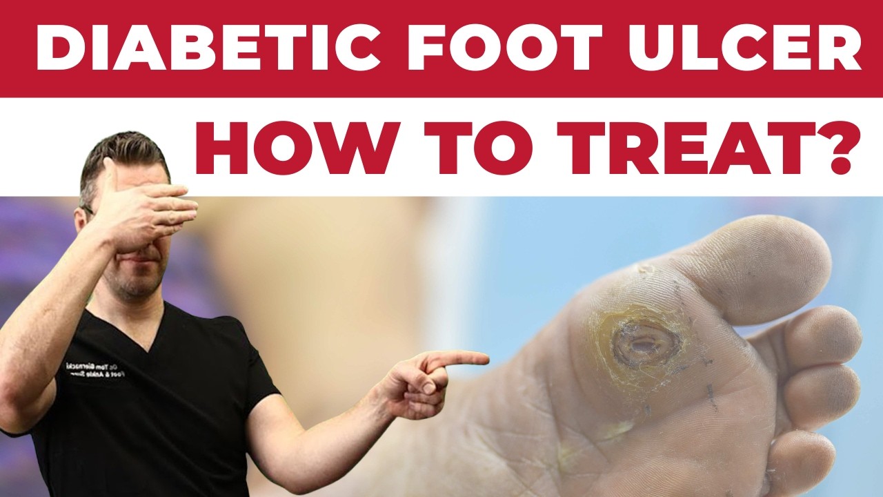
Diabetic Foot Ulcers
The most serious type of foot wound. Occur in areas of pressure or friction, often without pain due to neuropathy. Require immediate, aggressive wound care to prevent infection and amputation.
Venous & Arterial Ulcers
Wounds caused by poor circulation. Venous ulcers typically occur on the inner ankle; arterial ulcers on the tips of toes or outer ankle. Compression therapy and vascular referral often needed.
Post-Surgical Wounds
Following foot or ankle surgery, proper wound care is critical for optimal healing. We manage all post-operative wounds with careful attention to infection prevention and scar management.
Traumatic Wounds & Lacerations
Cuts, punctures, and abrasions to the foot and ankle. Proper cleaning, debridement, and closure prevents infection and promotes proper healing without complications.
Infected Wounds & Cellulitis
Infected foot wounds require urgent evaluation. We culture wounds, prescribe appropriate antibiotics, perform surgical debridement when needed, and coordinate hospitalization for serious infections.
Pressure Wounds (Bedsores)
Pressure ulcers on the feet in immobile or bedbound patients. Require specialized offloading, wound care, and nutritional support to promote healing.
Watch: Diabetic Foot Care Checklist: 10 Daily Habits
![Diabetic Foot Ulcer Treatment & Early Stages [Diabetic Neuropathy]](https://www.michiganfootdoctors.com/wp-content/cache/flying-press/74c9e9e0d518cbe6e669aab11d9fa7ea.jpg)
Dr. Tom Biernacki, DPM explains the clinical basics — then covers what we do differently in our Howell and Bloomfield Hills clinics. Subscribe on YouTube for weekly walkthroughs.
Our Advanced Wound Care Approach
- Wound debridement — Removal of dead, infected, or devitalized tissue to stimulate healthy healing.
- Advanced wound dressings — Foam, hydrocolloid, alginate, and silver-impregnated dressings matched to wound characteristics.
- Offloading — Total contact casting, removable cast walkers, and custom padding to eliminate pressure from healing wounds.
- Infection management — Wound cultures, targeted antibiotic therapy, and surgical debridement for infected wounds.
- Growth factor therapy — Becaplermin (Regranex) and other advanced biologics to stimulate wound healing in chronic ulcers.
- Hyperbaric oxygen referral — Coordination with hyperbaric oxygen therapy centers for complex chronic wounds when appropriate.
Expert Foot Wound Care in Michigan
Same-week appointments · Howell & Bloomfield Hills · Board-certified wound care specialists · Most insurance accepted
Book Urgent Wound EvaluationOr call (810) 206-1402
Foot & Ankle Wound Care — Michigan Podiatrist
Dr. Biernacki explains advanced wound care for diabetic foot ulcers, post-surgical wounds, and chronic non-healing wounds — including MLS laser, offloading devices, and tissue grafts.

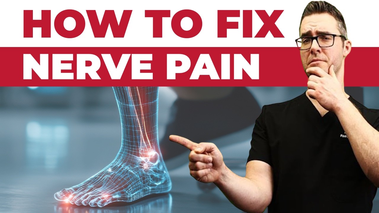
Why Diabetic Foot Wounds Are Different
A diabetic foot wound is not just a cut that won’t heal. Diabetes changes the biology of wound repair at the cellular level, so what looks like a minor blister, corn edge, or shoe-rub abrasion on one patient becomes a limb-threatening ulcer in another. The three drivers we see every week in clinic are neuropathy (so the foot does not feel the injury), peripheral arterial disease (so oxygen-rich blood cannot reach the tissue), and an altered inflammatory response (so the cleanup and rebuild phases of healing stall in place). Each one on its own is a problem. In combination, they are why a small scrape on a diabetic foot can become an amputation 90 days later if it is not managed aggressively from day one.
In our Howell and Bloomfield Hills clinics, we treat every new diabetic foot wound as a stacked triad: how much sensation remains, how well the foot perfuses, and how much glucose is running through the bloodstream at the moment of injury. Only after we quantify each of those three do we decide what dressing, what offloading, and what referral the patient needs. A wound that would close in 7 days on a healthy 35-year-old may need 12 weeks of structured care on a diabetic with an ankle-brachial index of 0.6, and the plan looks completely different the moment we see that number on the Doppler printout.
The most dangerous part of this biology is silent. Protective sensation drops before the patient notices. We see patients walk in with a quarter-sized ulcer on the plantar first metatarsal head who genuinely did not know it was there. That is not carelessness — that is small-fiber neuropathy doing exactly what small-fiber neuropathy does. If you have had diabetes for more than 10 years, you should assume that some of your foot’s protective sensation is gone until a podiatrist tests you with a 10-gram monofilament and proves otherwise.
Wagner Classification: What Your Wound Grade Actually Means
When you see us for a diabetic foot wound, the first number we assign is a Wagner grade. This 0–5 staging system tells every clinician on the care team how deep the wound goes, whether infection is involved, and whether tissue death has started. Grade is the single most important driver of where you are treated (office, outpatient, or admitted), how fast you need a vascular workup, and whether the conversation is about healing or about salvage.
Wagner 0 is a pre-ulcerative foot — callus, deformity, previous healed ulcer, but no break in the skin yet. This is the stage at which prevention pays the biggest dividend, and it is why we recommend a full diabetic foot screen every 3–6 months if you have any risk factor. Wagner 1 is a superficial ulcer through the epidermis and dermis. Most of these close with offloading plus dressings if we catch them early. Wagner 2 extends through subcutaneous tissue and may expose tendon or joint capsule. Wagner 3 reaches bone or involves abscess or osteomyelitis. Wagner 4 is localized gangrene, and Wagner 5 is extensive gangrene of the whole foot.
We photograph every wound on the first visit and every 14 days after. Progression up the Wagner scale is the fastest trigger for escalating treatment — MRI, hospital admission, IV antibiotics, or vascular consultation. Progression down the scale is how we measure whether our current plan is working. If a wound has not moved down at least half a grade in four weeks, we change the plan. Good wound care is measured — it does not rely on hope.
ABI, Toe Pressures, and Why Vascular Testing Comes First
Before we choose a dressing, before we prescribe an antibiotic, and before we consider whether to debride, we want to know how well blood is reaching the wound. The gold-standard bedside tools are the ankle-brachial index (ABI) and toe-brachial index (TBI). ABI is a Doppler-measured ratio of ankle systolic pressure to arm systolic pressure. A value of 0.9–1.3 is normal. Below 0.9 signals peripheral arterial disease. Below 0.5 means a wound is unlikely to close without revascularization. Above 1.3 usually means the vessels are calcified and non-compressible — in which case we switch to toe-brachial index, because the digital arteries stay compressible even when the ankle vessels do not.
In our clinic, we measure ABI on every diabetic wound patient at the first visit and again at the 30-day mark if the wound is not closing. We also order a transcutaneous oxygen measurement (TcPO2) when ABI is borderline — a TcPO2 below 30 mmHg at the wound edge predicts poor healing even when ABI looks acceptable. These numbers do not just drive our podiatric plan. They drive the decision to loop in a vascular surgeon for angiography, angioplasty, bypass, or stenting. Treating a wound without knowing perfusion is like pouring water into a glass without checking whether the glass has a hole in the bottom.
This is also why we strongly caution against grandma-remedy compression socks on any diabetic foot with an untested ABI. Compression on an arterially compromised foot accelerates tissue death. Doctor Hoy’s, topical anti-inflammatories, and all of the over-the-counter wound kits on the market assume normal perfusion. If your ABI is 0.6, the rules change — and we need to be the ones who write the plan.
Offloading Is the #1 Factor in Diabetic Wound Healing
Most patients think dressings are what close a diabetic foot wound. The data says otherwise. The single strongest predictor of whether a plantar ulcer heals is whether the patient successfully offloads the wound — meaning they stop walking on it while the tissue rebuilds. A wound cannot close under repetitive peak pressure, no matter how expensive the dressing. Every footstep re-injures the wound bed and restarts the clock. Offloading is the clinical lever with the biggest effect size in our entire toolkit, and it is the one patients resist the most because it changes their day-to-day life.
The gold standard is a total contact cast (TCC). Fiberglass, padded at bony prominences, molded to the foot, removed only in the office. Worn 24/7. A well-applied TCC drops peak plantar pressure on the wound by 80–90% and closes Wagner 1 ulcers in about 6 weeks on average in the published literature. We apply TCCs in both our Howell and Bloomfield Hills offices for appropriate patients — generally those with adequate perfusion (ABI ≥ 0.7), no active infection, and no active Charcot. For patients who cannot tolerate a cast, we step down to a removable cast walker (CROW boot), which is about 70% as effective when worn consistently and close to 0% effective when left on the shelf. For patients who cannot tolerate a boot, a healing shoe with a custom offloading insole fitted to the exact wound location is the minimum acceptable plan.
In the home, we add a knee scooter or crutches for any patient who has to cross a kitchen floor and two crucial rules: no barefoot walking ever, and no shortcut trips to the bathroom at night without the offloading device. The bathroom-at-2am shortcut is the single most common reason a wound that should have healed in 6 weeks takes 16. We would rather send a patient home with a knee scooter and a bed-side commode than have them lose toes to a trip they thought was too short to matter.
Advanced Dressings and the Debridement Ladder
The wound dressing world has exploded over the last 15 years. We now carry dressings that deliver silver, iodine, honey, collagen, hydrogel, hydrofiber, foam, alginate, PHMB, and cellular or tissue-based products (CTPs) like amniotic membrane and epidermal grafts. No single dressing fits every wound, which is why we match dressing to wound phase rather than prescribing by habit. The phases we manage are exudate control, bacterial load reduction, debridement, granulation support, and epithelialization — and each phase has a preferred dressing category.
A heavily draining wound gets an absorbent foam or alginate changed every 1–3 days. A colonized or biofilm-heavy wound gets a silver or PHMB antimicrobial dressing and often sharp debridement the same visit. A clean granulating wound gets a hydrogel or moisturizing dressing that protects the fragile new tissue. A stalled wound that has not shrunk in 4 weeks becomes a candidate for advanced CTPs (amniotic membrane products, dehydrated human amnion/chorion, collagen/ORC, or epidermal grafts) — each of which can restart a stuck wound in the right patient.
Debridement is the other half of the dressing equation. The goal of debridement is to convert a chronic wound (with senescent cells and biofilm) back into an acute wound that knows how to heal. We perform sharp debridement at the bedside every 1–2 weeks as long as the wound needs it — scalpel and curette, removing callused margins, non-viable slough, and visibly colonized tissue until we reach clean, bleeding edges. For patients who do not tolerate sharp debridement, we step down to enzymatic debridement (collagenase), biologic debridement (medical-grade larvae, which we can refer out), or autolytic debridement with hydrogel — in that order of effectiveness.
Recovery Timeline: What 12 Weeks of Diabetic Wound Care Actually Looks Like
Patients ask us on day one how long this will take. The honest answer is: a Wagner 1 ulcer with adequate perfusion and strict offloading averages 6–8 weeks to closure. A Wagner 2 ulcer averages 8–12 weeks. A Wagner 3 ulcer with osteomyelitis will usually need 6 weeks of IV antibiotics plus another 6–8 weeks after that before the wound is closed. These are averages. The patient who closes fastest is the patient who shows up every week, offloads every step, tests their blood sugar four times a day, and calls us the moment they see a worrisome change. The patient who takes longest is the one who skips visits because the wound seems smaller and removes the boot to go to their granddaughter’s graduation.
Weeks 1–2: Intake, ABI, wound photography, labs (HbA1c, CBC, basic metabolic panel, ESR and CRP if infection is suspected), baseline X-ray to rule out osteomyelitis, and first dressing plus offloading device fitted. Weeks 3–4: Measurable wound area reduction of 30–50% is the target. If we are not hitting it, we escalate — MRI, vascular referral, or advanced CTP. Weeks 5–8: Most Wagner 1 wounds close in this window. Wagner 2 wounds are filling in with granulation. Weeks 9–12: Epithelialization complete or nearly complete. Final dressing change. Transition to preventive footwear. Months 3–12: Diabetic shoe prescription, monthly nail and callus care, 3-month recheck schedule — because a patient who has had one ulcer has a 30–40% lifetime recurrence risk at the same site unless we intervene with proper footwear and pressure redistribution.
Differential Diagnosis: Not Every Foot Wound Is a Diabetic Ulcer
A break in the skin on a diabetic foot is not always a diabetic neuropathic ulcer, and treatment changes when the diagnosis changes. We work through the differential every first visit because mis-treating a venous ulcer as a neuropathic ulcer (or vice versa) wastes weeks. The conditions that most often masquerade as “diabetic wound” in our practice are venous stasis ulcers (weeping, irregular borders, medial malleolar location, hemosiderin staining), ischemic arterial ulcers (punched-out borders, distal toes or lateral foot, dry/black base, intensely painful unless neuropathy is also present), pressure injuries (heel or lateral malleolus, bedbound or wheelchair patients), and Charcot-related ulcers (midfoot collapse with plantar bone prominence under the cuboid or navicular).
Less common but life-changing diagnoses we have to actively rule out include squamous cell carcinoma arising in a chronic wound (Marjolin ulcer), pyoderma gangrenosum (rapidly expanding necrotic ulcer with violaceous borders — often misdiagnosed and made worse by debridement), and calciphylaxis in end-stage renal disease patients. Any wound that has failed standard care for more than 90 days, looks clinically atypical, or has been debrided multiple times without improvement earns a punch biopsy in our office. Missing a Marjolin ulcer costs lives. We would rather biopsy 10 ordinary wounds than miss one cancer.
Red Flags: When a Foot Wound Becomes an Emergency
Most diabetic foot wounds are managed safely in the office. A subset are surgical and hospital-level emergencies from the moment they walk in — and missing one of these can cost a limb or a life inside a weekend. If you have a diabetic foot wound and notice any of the following, do not wait for your next appointment. Call our office, go to an emergency room, or both.
- Sudden spreading redness, streaking, or warmth — early sign of cellulitis or necrotizing infection.
- Fever, chills, or confusion — systemic infection, especially dangerous in older diabetics.
- Foul odor or visible pus draining from the wound — abscess or deep space infection.
- Sudden pain increase in a previously painless neuropathic wound — usually means deep tissue infection, gas-forming organism, or ischemia.
- Black tissue at the wound edges or on the toes — ischemia or gangrene; vascular emergency.
- Cold, mottled, or pulseless foot — acute limb ischemia; time-critical.
- Blood sugar suddenly uncontrolled (readings over 300 with ketones) — infection pushing glucose up.
- Exposed bone or the ability to probe to bone with a cotton swab — osteomyelitis until proven otherwise.
Any two of the above in combination with diabetes plus a foot wound is an ER trip, not an office call. In Michigan, both McLaren Oakland and St. Joseph Mercy in Howell will admit a diabetic foot infection for IV antibiotics when appropriate, and we maintain good relationships with the hospitalist teams at both so we can continue wound management in-hospital and take patients back in clinic after discharge.
Three Real Patient Scenarios We See Every Week
Scenario 1 — The silent ulcer. A 64-year-old Type 2 diabetic comes in because his wife noticed blood on his sock. He denies pain. Plantar first metatarsal head has a 1.2 cm Wagner 1 ulcer with a thick callused rim. ABI is 0.95. HbA1c is 8.4. Plan: sharp debridement of the callused rim, silver foam dressing, total contact cast, weekly follow-up. Target closure: 6–8 weeks. Long-term plan: custom diabetic shoes with Plastazote insole, glucose optimization with primary care, quarterly recheck for life. We see some version of this patient every single week.
Scenario 2 — The ischemic forefoot. A 72-year-old with 20 years of diabetes, prior stent, and active smoking presents with a painful, dry, black-edged ulcer on the tip of the second toe. ABI is 0.4 on the affected side. Plan: same-day referral to vascular surgery for angiography, no wet dressing until we know perfusion status, prophylactic antibiotic if infection signs emerge, strict non-weightbearing, smoking cessation counseling. Outcome depends entirely on whether the vascular team can revascularize. We do not debride an ischemic ulcer until perfusion is restored — debriding ischemic tissue just enlarges the wound.
Scenario 3 — The Charcot emergency. A 58-year-old diabetic with known neuropathy presents with a warm, swollen, red midfoot and a plantar ulcer under the cuboid. X-ray shows midfoot collapse. This is active Charcot neuroarthropathy with a secondary ulcer — not cellulitis. Plan: TCC, complete immobilization, skin temperature monitoring (we want a 2°C or greater drop between affected and contralateral foot before advancing), vitamin D optimization, and — once the Charcot is inactive — custom CROW boot followed by custom accommodative footwear for life. Treating Charcot as cellulitis with antibiotics alone is the most common error we inherit from outside offices, and it destroys midfoot architecture that cannot be rebuilt.
Advanced Questions Patients Ask About Diabetic Wound Care
Do I need hyperbaric oxygen? HBOT is reserved for Wagner 3 ulcers with osteomyelitis or failed diabetic foot ulcers after 30 days of standard care with adequate perfusion — and only when insurance criteria are met. We refer to HBOT partners in Oakland County when the criteria are met. HBOT is not a first-line tool and is not magic.
Can I shower with a diabetic foot wound? Yes, but not a soak. Brief showers with a waterproof cover over the dressing are fine. No baths, no hot tubs, no pools, no lakes until the wound is fully closed and epithelialized. Waterborne pseudomonas infection of a diabetic wound is a disaster we have seen more than once in this lake-heavy state.
Why do you keep taking pictures of my foot? Because a photograph is the only objective way to measure healing between visits. We compare week-to-week photos to see if the wound is shrinking, stalling, or growing — and we do not trust memory on a 6-week timeline. If your wound is not at least 30% smaller in 4 weeks, we change the plan. The photos are what tell us that.
Is Medicare going to cover all of this? Medicare covers diabetic wound care, in-office debridement, total contact casts, most advanced dressings, and diabetic therapeutic shoes and inserts through the Medicare Therapeutic Shoe Program (one pair of shoes plus three pairs of inserts per calendar year if qualifying criteria are met). Our front desk runs the paperwork for you. Advanced CTPs, negative pressure wound therapy (wound vac), and HBOT each have their own coverage criteria — we handle the prior authorization when it applies.
Worried about a foot wound right now? Same-day appointments are almost always available for active wounds. Call (810) 206-1402 or book online. Both our Howell and Bloomfield Hills offices stock the full diabetic wound care toolkit — total contact casts, silver dressings, amniotic membrane products, and offloading shoes — so we can start treatment the same visit.
Products That Support Diabetic Wound Healing
These are the products we actually stock at our Howell and Bloomfield Hills clinics and hand to patients dealing with diabetic wound care. Each one has clinical evidence behind it — and each one has a patient it is not right for. We have included that, too, because we want you to pick well. Links below support the research behind this site; prices and availability vary.
- DASS 15–20 mmHg Compression Socks (after ABI confirmed) — Controls edema that slows wound healing — but only after we have screened your arterial flow in clinic (ABI ≥ 0.7). Not ideal for: anyone with PAD who has not had an ABI screening — compression over ischemic tissue is harmful. See what we stock.
- PowerStep Pinnacle + custom offloading insert — Offloads pressure from under the metatarsal heads where most diabetic wounds develop. We modify these in clinic based on wound location. Not ideal for: active forefoot ulcers — we use total-contact casting or a specialty wound shoe instead. See what we stock.
- Medical-grade collagen or alginate dressing (in-clinic application) — We apply and change these in our wound care program. Home dressings should match what we send you home with — do not substitute OTC gauze. Not ideal for: anyone trying to self-manage a non-healing wound at home — come see us. See what we stock.
Not sure which is right for your foot? We fit and demo these in clinic and can pair them with the right shoe. (810) 206-1402 · book online. Same-day appointments available.
Related Conditions We Treat
If you or a loved one is managing diabetic foot care, these related patient guides cover prevention, referral pathways, and day-to-day care:
- Diabetic Wound Prevention
- Wound Care Referral in Michigan
- Michigan Diabetic Care Team
- Medicare Diabetic Foot Care & Custom Shoes
- Topical Products for Diabetic Foot Pain & Neuropathy
Ready for in-office care? Call (810) 206-1402 or book online. Same-day appointments available at our Howell and Bloomfield Hills clinics.
Frequently Asked Questions
How long does a diabetic foot ulcer take to heal?
Uncomplicated diabetic foot ulcers heal in 4–12 weeks with proper offloading, debridement, and dressing protocols. Infected wounds may require antibiotics and hospitalization. Early aggressive treatment dramatically reduces amputation risk — contact us at the first sign of a wound.
What is total contact casting for wound care?
Total contact casting is the gold-standard offloading device for plantar diabetic foot ulcers. A custom plaster or fiberglass cast redistributes pressure completely away from the wound, allowing healing even in ambulatory patients. It is far more effective than removable boots for compliance.
When is a wound care emergency?
Seek emergency care for: spreading redness, red streaks up the leg (cellulitis), fever, foul odor, black or gray tissue (gangrene), or wounds that have not improved in 2 weeks. Diabetic foot infections can progress to limb-threatening conditions within hours.
Watch Dr. Tom on Foot Wound Care
Dr. Tom walks through the daily wound care protocol that prevents 80% of diabetic foot ulcers from progressing to amputation.
Foot Wound Home Care Essentials
Minor foot wounds in diabetic or neuropathic patients can spiral quickly. These four items, combined with same-day podiatry evaluation for any wound >48 hours, prevent the majority of ulcers:
Hibiclens Antimicrobial Skin Cleanser
Chlorhexidine wound cleanser — safer and more effective than hydrogen peroxide or iodine for repeated home use.
Check Amazon Price →Silver Antimicrobial Bandages
Silver inhibits staph and pseudomonas — the two bacteria responsible for most diabetic ulcer progression.
Check Amazon Price →Eucerin Advanced Repair Foot Cream
Ceramide + urea prevents the skin cracking that becomes the next wound — daily use on unaffected skin around wounds.
Check Amazon Price →Non-Binding Diabetic Socks
Seamless toe + non-binding band — critical for neuropathic feet with healing wounds.
Check Amazon Price →Affiliate disclosure: Amazon links are affiliate links — we earn a small commission if you buy through them, at no cost to you. We only recommend products we actually prescribe to patients at Balance Foot & Ankle.
Related from Balance Foot & Ankle
Foot pain — Frequently Asked Questions
When should I see a podiatrist for foot pain?
If symptoms persist beyond 2 weeks of self-care, interfere with daily activity, or worsen suddenly, schedule a podiatrist evaluation. Early intervention typically shortens recovery and prevents chronic compensation patterns.
Will I need imaging or surgery?
Most foot pain cases resolve with conservative care—custom orthotics, supportive shoe changes, anti-inflammatory protocols, and targeted physical therapy. Imaging (X-ray, ultrasound, MRI) is reserved for cases that fail conservative treatment or when structural pathology is suspected. Surgery is rarely the first option.
Does insurance cover foot pain treatment in Michigan?
Most major Michigan insurance plans (BCBS, BCN, Priority Health, HAP, Medicare, Medicaid HMOs, United, Aetna, Cigna) cover medically necessary podiatric care. Custom orthotics may have separate DME coverage rules. Our team verifies your specific benefits before your visit.
Related Treatments at Balance Foot & Ankle
Common conditions we treat in our Howell and Bloomfield Hills offices.