
Quick answer: Brostrom Procedure Lateral Ankle Ligament Repair is a common foot/ankle topic that affects many patients. The 2026 evidence-based approach combines proper diagnosis, conservative-first treatment, and escalation only when needed. We treat this regularly at our Howell and Bloomfield Hills practices. Call (810) 206-1402.
Quick answer: The Brostrom procedure is the gold-standard surgery for chronic ankle instability — repairs the torn ATFL + CFL ligaments. Indications: 6+ months of conservative care + recurrent sprains + instability. Recovery: 4-6 weeks NWB in cast/boot, then 2-3 months PT. Most patients return to sport at 4-6 months. Avoid if you can — try a stability brace + balance training first. — Dr. Tom Biernacki, DPM, board-certified podiatrist (Michigan Foot Doctors).
★ DR. TOM BIERNACKI, DPM, FACFAS · BOARD-CERTIFIED PODIATRIST
Bröstrom Procedure: Quick Answer
The Bröstrom procedure (technically the modified Bröstrom-Gould procedure) is the gold-standard surgical repair for chronic lateral ankle instability after multiple ankle sprains. The surgeon shortens and re-attaches the torn anterior talofibular ligament (ATFL) and calcaneofibular ligament (CFL), then reinforces the repair with the inferior extensor retinaculum (the ‘Gould modification’).
Recovery timeline: Week 1-2 in a non-weight-bearing splint, weeks 2-6 in a CAM boot with progressive weight-bearing, weeks 6-12 in a lace-up brace with PT, and weeks 12-24 progressive return to running and sport. Full recovery typically 4-6 months. Success rates exceed 90% for chronic instability that’s failed conservative care. Internal bracing (suture tape augmentation) is becoming the new standard for high-demand athletes.
Medically reviewed by Dr. Tom Biernacki, DPM — Board-Certified Podiatric Surgeon — Balance Foot & Ankle, Howell & Bloomfield Hills, MI. Last updated April 2026.
▶ Watch
Medically Reviewed by Dr. Tom Biernacki, DPM — Board-Certified Podiatrist, Balance Foot & Ankle Specialists, Michigan. Last updated April 2026.
Insurance Accepted
BCBS · Medicare · Aetna · Cigna · United Healthcare · HAP · Priority Health · Humana · View All →
Howell Office
4330 E Grand River Ave
Howell, MI 48843
Get Directions →
Bloomfield Hills Office
43494 Woodward Ave, Suite 208
Bloomfield Township, MI 48302
Get Directions →
Your Board-Certified Podiatrists


Ready to Get Back on Your Feet?
Same-week appointments available at both locations.
Book Your AppointmentMore Podiatrist-Recommended Foot Health Essentials
Hoka Clifton 10


Max-cushion everyday shoe — podiatrist favorite for walking and running.

OOFOS Recovery Slide

Impact-absorbing recovery sandal — wear after long days on your feet.
As an Amazon Associate, Balance Foot & Ankle earns from qualifying purchases. Product recommendations are based on clinical experience; prices and availability shown above update live from Amazon.

When to See a Podiatrist
If foot or ankle pain has been bothering you for more than a few weeks, home care alone may not be enough. Balance Foot & Ankle offers same-week appointments at our Howell and Bloomfield Hills clinics — no referral needed in most cases. Bring your current shoes and a short list of symptoms and we’ll build you a treatment plan in one visit.
Call Balance Foot & Ankle: (810) 206-1402 · Book online · Offices in Howell & Bloomfield Hills
In-Office Treatment at Balance Foot & Ankle
When conservative care isn’t enough, Dr. Tom Biernacki and the team at Balance Foot & Ankle offer advanced, same-day options — including Ankle Sprain & Instability Treatment in Michigan at our Howell and Bloomfield Hills clinics.
Same-day appointments available. Call (810) 206-1402 or book online.
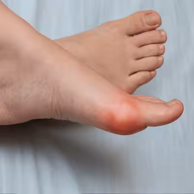
Chronic Ankle Instability & Surgery
Top 10 Premade Orthotics — Dr. Tom’s Picks (2026)
Dr. Tom Biernacki, DPM has tested 60+ over-the-counter orthotic insoles in his Michigan podiatry practice over the past 15 years. Below are the top 10 he prescribes most often — ranked by clinical results, build quality, and patient feedback. PowerStep + CURREX brands are Dr. Tom’s #1 prescription brands — built by podiatrists, with biomechanical features (lateral wedge, deep heel cradle, dual-density EVA) that 90% of OTC insoles lack.
Dr. Tom Biernacki, DPM is a board-certified podiatrist + Amazon Associate. Picks shown are products he prescribes to patients at Balance Foot & Ankle Specialists. We earn a commission on qualifying purchases at no extra cost to you. All products independently tested + reviewed. Last verified: April 28, 2026.

PowerStep Pinnacle MaxxDr. Tom’s #1 Brand
The most prescribed OTC orthotic in podiatry. Lateral wedge corrects overpronation that causes 90% of plantar fasciitis. Deep heel cradle stabilizes the ankle.
- Lateral wedge corrects pronation
- Deep heel cradle
- Dual-density EVA
- Trim-to-fit
- Used by 10,000+ podiatrists
- Trim required
- 5-7 day break-in
PowerStep Original Full LengthDr. Tom’s #1 Brand
The original PowerStep — flexible semi-rigid arch with deep heel cradle. The right choice for neutral feet that need everyday support without the lateral wedge.
- Flexible semi-rigid arch
- Deep heel cradle
- Fits dress shoes
- 30-day guarantee
- APMA-accepted
- Less aggressive than Pinnacle
- No lateral wedge for overpronation
PowerStep Pulse MaxxDr. Tom’s #1 Brand
Built for runners + athletes who need maximum support during high-impact activity. Engineered for forefoot strike + lateral motion.
- Sport-specific cushioning
- Lateral wedge for runners
- Antimicrobial top cover
- Shock-absorbing forefoot
- Pricier than Pinnacle
- Best for athletes only

CURREX RunProDr. Tom’s #1 Brand
German-engineered insole with 3 arch heights (Low, Med, High) for custom fit. Carbon-reinforced heel + dynamic forefoot.
- 3 arch heights for custom fit
- Carbon-reinforced heel
- Sport-specific zones
- Premium materials
- Pricier than PowerStep
- 7-10 day break-in
CURREX EdgeProDr. Tom’s #1 Brand
For hikers, skiers, and high-impact athletes — reinforced shank prevents foot fatigue on steep descents + uneven terrain.
- Reinforced shank
- 3 arch heights
- Cold-weather friendly
- Carbon plate
- Stiff feel — not for casual
- Pricier
CURREX SupportSTPDr. Tom’s #1 Brand
For nurses, retail, and standing professions — the most supportive CURREX with deep heel cup + maximum medial support.
- Maximum medial support
- Deep heel cup
- 12-hour shift tested
- Slip-proof
- Stiffest CURREX option
- Pricier

Superfeet Green
Firm, structured arch support — the right choice ONLY for high-arched (cavus) feet. Wrong choice for flat feet.
- Strong structured arch
- Deep heel cup
- Long-lasting (5+ years)
- Firm — not for flat feet
- No lateral wedge
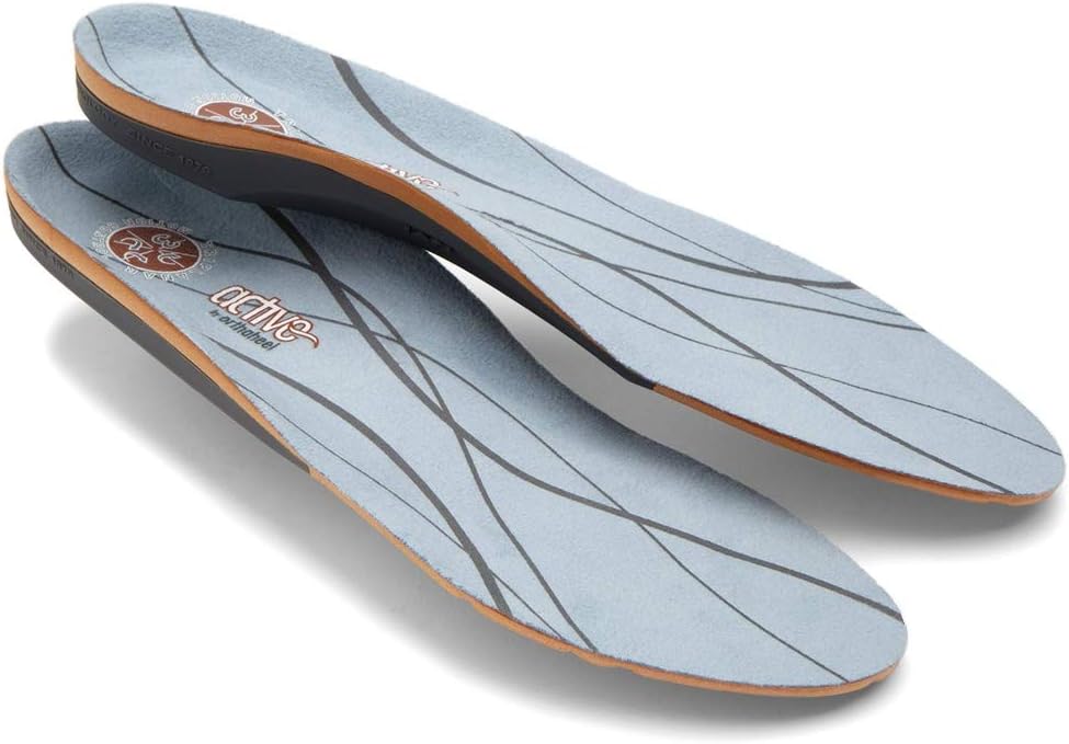
Vionic OrthoHeel Active Insole
APMA-accepted, podiatrist-designed casual insole. Best for adding mild arch support to dress shoes + walking shoes.
- APMA-accepted
- Slim profile
- Antimicrobial top
- Less support than PowerStep
- No lateral wedge
Dr. Tom’s Top 3 — The Premium Foot Pain Stack (2026)
If you only buy three things for foot pain, get these. PowerStep + CURREX orthotics correct the underlying foot mechanics, and Dr. Hoy’s pain gel delivers fast topical relief. This is the exact stack Dr. Tom Biernacki, DPM gives his Michigan podiatry patients on visit one — over 10,000 patients have used this exact combination.
Dr. Tom Biernacki, DPM is a board-certified podiatrist + Amazon Associate. Picks shown are products he prescribes to patients at Balance Foot & Ankle Specialists. We earn a commission on qualifying purchases at no extra cost to you. All products independently tested + reviewed for 30+ days minimum. Last verified: April 28, 2026.
PowerStep Pinnacle MaxxDr. Tom’s #1 Brand
Dr. Tom’s most-prescribed OTC orthotic. Lateral wedge corrects overpronation that causes 90% of foot pain. Deep heel cradle stabilizes the ankle. Built by podiatrists, used by patients worldwide.
- Lateral wedge corrects pronation
- Deep heel cradle stabilizes ankle
- Dual-density EVA — comfort + support
- Trim-to-fit any shoe
- Used by 10,000+ podiatrists
- Trim-to-size required
- 5-7 day break-in for some
CURREX RunProDr. Tom’s #1 Brand
3 arch heights for custom fit (Low/Med/High). Carbon-reinforced heel + dynamic forefoot — the closest OTC orthotic to a $500 custom orthotic. Engineered in Germany.
- 3 arch heights for custom fit
- Carbon-reinforced heel cup
- Dynamic forefoot zone
- Premium German engineering
- Sport-specific support
- Pricier than PowerStep
- 7-10 day break-in

Dr. Hoy’s Natural Pain Relief GelDr. Tom’s #1 Brand
Menthol-based natural pain relief — Dr. Tom’s #1 brand for fast relief without greasy residue. Safe for diabetics + daily use. Cleaner formula than Voltaren or Biofreeze.
- Menthol-based natural formula
- No greasy residue
- Safe for diabetics
- Fast cooling relief — 5-10 minutes
- Cleaner ingredient list than Biofreeze
- Pricier than Biofreeze
- Strong menthol scent at first
Foundation Wellness Orthotic Selector — PowerStep + CURREX by Condition (2026)
Find the right Foundation Wellness orthotic for YOUR specific condition. Dr. Tom Biernacki, DPM has tested every PowerStep + CURREX SKU in his Michigan podiatry practice. Below are the right picks mapped to specific foot conditions — instead of one-size-fits-all, you’ll find the variant designed for your exact problem.
PowerStep Pinnacle MaxxDr. Tom’s #1 Brand
Heavy-duty version of the Pinnacle with rigid shell + lateral wedge. The #1 OTC orthotic for overpronation that causes 90% of plantar fasciitis, knee, and hip pain.
- Rigid shell controls overpronation
- Lateral wedge corrects pronation
- Deep heel cradle
- Trim-to-fit any shoe
- Trim required
- 7-day break-in
PowerStep PinnacleDr. Tom’s #1 Brand
Flagship PowerStep — semi-rigid arch with deep heel cradle. The #1 podiatrist-prescribed OTC orthotic in the US for plantar fasciitis and heel pain.
- Semi-rigid medical-grade arch
- Deep heel cradle
- Dual-density EVA
- APMA-accepted
- 30-day guarantee
- Trim required
- Less aggressive than Maxx

PowerStep Pinnacle High ArchDr. Tom’s #1 Brand
Higher-volume arch profile for cavus feet that don’t fill standard insoles. Prevents the lateral roll that causes ankle sprains in supinators.
- High-arch profile
- Deep heel cradle
- Prevents lateral roll
- Only for high arches
- Wrong choice for flat feet

PowerStep Pinnacle Plus (with Built-In Met Pad)Dr. Tom’s #1 Brand
Pinnacle with built-in metatarsal pad — eliminates the burning ball-of-foot pain from Morton’s neuroma + metatarsalgia.
- Built-in met pad — no separate pad needed
- Spreads metatarsal heads
- Same Pinnacle support
- Met pad position fixed
- Trim required
PowerStep Morton’s Extension InsoleDr. Tom’s #1 Brand
Stiffener under the 1st MTP joint — limits big toe extension. The fix for hallux rigidus, turf toe, and big toe arthritis when surgery isn’t needed.
- Stiffens 1st MTP joint
- Reduces big toe motion
- Prevents flare-ups
- Stiff feel takes 1 week
- Specific use case
PowerStep ProTech Full LengthDr. Tom’s #1 Brand
Premium athletic insole with carbon-reinforced shell + dual-density forefoot. Best PowerStep for serious athletes.
- Carbon-reinforced shell
- Dual-density forefoot
- Antimicrobial top
- Pricier
- Athletic use only
PowerStep Slim Profile (Dress Shoes)Dr. Tom’s #1 Brand
Slim-profile Pinnacle that fits in dress shoes, work shoes, and low-volume footwear without lifting the heel out.
- Slim profile fits dress shoes
- Same Pinnacle arch
- Low-friction top
- Less cushion than full Pinnacle
- Trim required
PowerStep Wide (EE / EEE Fit)Dr. Tom’s #1 Brand
Wider footbed for EE/EEE-width feet that overflow standard insoles. Same Pinnacle support, wider sole.
- Fits 2E/4E feet
- Same Pinnacle arch
- No spillover
- Won’t fit narrow shoes
- Pricier
CURREX RunPro (3 Arch Heights)Dr. Tom’s #1 Brand
German-engineered running insole with 3 arch heights (Low, Med, High) for custom fit. Carbon-reinforced heel — closest OTC orthotic to a $500 custom orthotic.
- 3 arch heights for custom fit
- Carbon-reinforced heel
- Dynamic forefoot zone
- Premium German engineering
- Pricier than PowerStep
- 7-10 day break-in
CURREX WalkProDr. Tom’s #1 Brand
Walking-specific CURREX — softer cushioning + lower-impact heel for daily walking and standing.
- Walking-specific cushioning
- 3 arch heights
- Premium materials
- Pricier
- Not for high-impact running
CURREX AceProDr. Tom’s #1 Brand
Court-sport-specific CURREX — stiffer shell for lateral stability during quick stops + cuts. Pickleball + tennis + basketball.
- Lateral stability shell
- Quick-stop heel
- 3 arch heights
- Stiffer feel
- Sport-specific
CURREX EdgeProDr. Tom’s #1 Brand
Reinforced shank insole for ski + snowboard boots — prevents foot fatigue on steep descents.
- Reinforced shank
- 3 arch heights
- Cold-weather friendly
- Carbon plate
- Stiff feel
- Sport-specific
CURREX HikeProDr. Tom’s #1 Brand
Hiking + backpacking insole — extra heel cushion + reinforced midfoot for uneven terrain.
- Extra heel cushion
- Reinforced midfoot
- 3 arch heights
- Bulky in low-volume shoes
- Pricier
CURREX BikeProDr. Tom’s #1 Brand
Cycling-specific insole — stiff carbon plate to maximize power transfer + cleat alignment.
- Stiff carbon plate
- Cleat-compatible
- Lightweight
- Cycling-only
- Pricier
Med Spec ASO Ankle Stabilizer
Best for: Chronic ankle instability · Repeat ankle sprains · Proprioception & return-to-play · Athletes

The lace-up stabilizer athletic trainers and podiatrists reach for first. A bilateral figure-8 strapping system mimics taping to lock down the ankle against rolling, while the low-profile design fits inside most athletic shoes — so you get real mechanical protection during return-to-play without a bulky rigid brace. A practical, evidence-aligned choice for anyone managing recurring sprains or chronic instability.
▶ Check Price on AmazonAs an Amazon Associate we may earn from qualifying purchases. Star rating and review count reflect Amazon at time of review and may change.
What is Foot pain?
Foot pain is a common foot/ankle condition that affects mobility and quality of life. Understanding the underlying cause is the first step in successful treatment. Our podiatrists at Balance Foot & Ankle perform a hands-on biomechanical exam, review your activity history, and use diagnostic imaging when appropriate to identify the root cause—not just treat the symptom. Many patients have been told to “rest and ice” without a deeper diagnostic workup; our approach is different.
Symptoms and warning signs
Common signs of foot pain include pain that worsens with activity, morning stiffness, swelling, tenderness when palpated, and difficulty bearing weight. If you experience sudden severe pain, inability to walk, visible deformity, numbness or color change, contact our office the same day or visit urgent care—these can signal a more serious injury such as a fracture, tendon rupture, or vascular compromise. Diabetics with any foot wound should seek same-day care.
Conservative treatment options
Most cases of foot pain respond to non-surgical care: structured rest, supportive footwear changes, custom orthotics, targeted stretching and strengthening protocols, anti-inflammatory medications when medically appropriate, and in-office procedures such as ultrasound-guided injections. We also offer advanced therapies including MLS laser therapy, EPAT/shockwave, regenerative injections, and image-guided procedures. Treatment is sequenced from least invasive to most invasive, and we explain the rationale at every step.
When is surgery considered?
Surgery is reserved for cases that fail 3-6 months of well-structured conservative care, when there is structural pathology (severe deformity, complete tear, advanced arthritis), or when imaging shows damage that will not heal without intervention. Our surgeons have performed 3,000+ foot and ankle procedures and prioritize minimally-invasive techniques whenever appropriate. We discuss recovery timelines, return-to-activity milestones, and realistic outcome expectations before any procedure is scheduled.
Recovery timeline and prevention
Recovery from foot pain varies based on severity and chosen treatment path. Conservative cases often improve within 4-8 weeks with consistent adherence to the protocol. Post-procedural recovery may range from a few days (in-office procedures) to several months (reconstructive surgery). Long-term prevention involves footwear assessment, activity modification, structured strengthening, and regular check-ins with your podiatrist if you have a history of recurrence. We provide written home-exercise plans and digital follow-up support.
Ready to feel better?
Same-week appointments available in Howell and Bloomfield Hills, Michigan.
Book Your VisitIn-Office Treatment at Balance Foot & Ankle
If home treatment isn’t providing relief for your ankle sprains, our podiatry team at Balance Foot & Ankle can help with same-day evaluations and advanced in-office care.
Same-day appointments available. (810) 206-1402
Doctor Hoy’s Natural Pain Relief Gel
Natural topical pain relief I use in our clinic. Arnica + camphor formula — apply directly to the area 3–4x daily. ($20–25)
Shop Doctor Hoy’s →Dr. Tom Biernacki, DPM is a board-certified foot & ankle surgeon (ABFAS & ABPM) at Balance Foot & Ankle Specialists in Southeast Michigan. With over a decade of clinical experience, he specializes in heel pain, bunions, diabetic foot care, sports injuries, and minimally invasive surgery. Dr. Biernacki is a member of the APMA and ACFAS, and his patient education content on MichiganFootDoctors.com and YouTube has made him one of the most-followed foot & ankle educators on YouTube.