

Quick answer: External Fixation Complex Foot Deformity Charcot Correction is a common foot/ankle topic that affects many patients. The 2026 evidence-based approach combines proper diagnosis, conservative-first treatment, and escalation only when needed. We treat this regularly at our Howell and Bloomfield Township practices. Call (810) 206-1402.
Medically reviewed by Dr. Tom Biernacki, DPM — Board-Certified Podiatric Surgeon — Balance Foot & Ankle, Howell & Bloomfield Township, MI. Last updated April 2026.
▶ Watch

Medically reviewed by Dr. Tom Biernacki, DPM | Board-certified podiatrist | 3,000+ surgeries performed
Last updated: April 2, 2026
The most important clinical decision with External Fixation Complex Foot Deformity Charcot Correction isn’t which treatment to start with — it’s which subtype or underlying cause you actually have. That distinction changes everything. Call us: (810) 206-1402
What Is External Fixation?
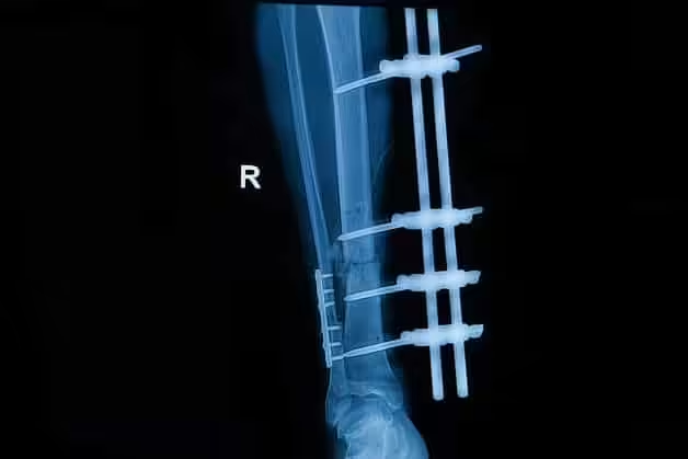
External fixation is a surgical technique that stabilizes bones using a rigid frame positioned outside the body and connected to the skeleton through percutaneous pins and tensioned wires. Unlike internal fixation with plates and screws that are placed directly on the bone surface, external fixators allow adjustable correction that can be fine-tuned after surgery without additional procedures.
The two primary external fixation systems used in foot and ankle surgery are circular frames (Ilizarov-type) and monolateral (rail) fixators. Circular frames provide multiplanar correction capability through tensioned olive wires and half-pins connected to ring components, while monolateral fixators provide simpler unidirectional stabilization for less complex deformity patterns.
Modern computer-assisted hexapod external fixators — including the Taylor Spatial Frame and TL-HEX systems — use six adjustable struts connecting two ring components. Computer software calculates precise daily strut adjustments that gradually move the bone into the desired position, achieving correction accuracy within 1-2 degrees of the planned alignment.
Conditions Treated With External Fixation
Charcot neuroarthropathy with rocker-bottom deformity represents one of the most common indications for external fixation in podiatric surgery. Diabetic patients with collapsed midfoot architecture and plantar bony prominences at high risk for ulceration benefit from gradual realignment that internal fixation alone cannot safely achieve in compromised bone quality.
Complex ankle fractures with severe soft tissue injury or open wounds require external fixation as either a definitive treatment or a temporary stabilization method (spanning external fixation) until soft tissue conditions permit internal fixation. This staged approach reduces infection risk and wound complications in high-energy injuries.
Post-traumatic malunion and nonunion of the foot and ankle — bones that have healed in poor alignment or failed to heal — may require external fixation with gradual distraction osteogenesis to regenerate bone and correct deformity simultaneously. This technique is particularly valuable when bone loss from infection or previous surgery has created gaps that cannot be bridged with bone grafting alone.
Limb length discrepancy caused by growth plate injuries, congenital conditions, or post-surgical bone loss can be corrected through gradual bone lengthening using circular external fixation. The Ilizarov technique of distraction osteogenesis generates new bone at a controlled rate of approximately 1mm per day, allowing significant length correction without bone grafting.
The External Fixation Surgical Procedure
Preoperative planning includes detailed imaging analysis with weight-bearing X-rays, CT scans, and sometimes 3D-printed bone models to map the deformity correction strategy. The Center of Rotation of Angulation (CORA) — the precise anatomic point around which correction must occur — is calculated to ensure the frame achieves accurate realignment.
Surgery is performed under general or regional anesthesia. The surgeon inserts thin tensioned wires (1.5-1.8mm diameter) through the bone and attaches them to ring components, followed by half-pin placement for additional stability. The frame is assembled and initial alignment is confirmed with intraoperative fluoroscopy (live X-ray) before the patient leaves the operating room.
For gradual correction cases, a prescription of daily strut adjustments is programmed based on the preoperative deformity analysis. Patients or caregivers perform these adjustments at home using a simple wrench mechanism, turning each strut a precise number of clicks per day. The correction period typically spans 2-8 weeks depending on the severity of deformity.
The frame remains in place during the consolidation phase — the time required for regenerated bone to mature and calcify sufficiently to bear weight without the frame. Consolidation typically requires 6-12 weeks after the correction phase is complete. Serial X-rays monitor bone healing progress and determine when the frame can be safely removed.
Living With an External Fixator
Daily pin site care is the most important patient responsibility during external fixation treatment. Pin sites are cleaned with chlorhexidine solution or sterile saline to prevent bacterial colonization that can lead to pin tract infection. Dr. Biernacki’s team provides detailed written and video instructions for pin care technique and troubleshooting.
Weight-bearing status varies by indication and phase of treatment. Many patients progress to partial weight-bearing in a protective boot within days of surgery, while complex bone lengthening cases may require non-weight-bearing for the initial correction phase. Your surgeon provides specific weight-bearing instructions and modifies them as healing progresses.
Sleep and daily activities require adaptation but are manageable with proper frame padding and positioning strategies. Foam padding around the rings prevents bed sheet entanglement, and loose-fitting clothing accommodates the frame during the treatment period. Most patients adapt to frame-related lifestyle modifications within the first week.
Physical therapy begins early in the recovery process and continues through frame removal and beyond. Range-of-motion exercises for the ankle and foot joints prevent contracture, while strengthening exercises maintain muscle mass during the healing period. Patients who comply with their rehabilitation program consistently achieve better functional outcomes.
Risks and Complications
Pin tract infection is the most common complication, occurring at some level in approximately 30% of patients. Most pin infections are superficial and respond to oral antibiotics and improved pin care technique. Deep pin infections requiring pin removal or intravenous antibiotics occur in less than 5% of cases when proper pin care protocols are followed.
Wire or pin loosening can compromise fixation stability and correction accuracy. Loosening occurs when bone quality is poor or when patients bear weight prematurely against medical advice. Loosened pins may require replacement or repositioning under local anesthesia, adding modestly to the treatment timeline.
Delayed bone healing or failure of regenerate bone formation occurs in a small percentage of patients, particularly those with diabetes, vascular disease, or nutritional deficiencies. Adjunctive treatments including bone stimulators, bone grafting, and optimization of metabolic factors can promote healing in these challenging cases.
Joint stiffness following prolonged external fixation is an expected sequela that physical therapy addresses during and after the treatment course. Ankle and subtalar joint range of motion may not fully return to pre-injury levels, though most patients achieve functional range sufficient for comfortable walking and daily activities.
Recovery Timeline and Expected Outcomes
Total treatment duration from frame application to removal ranges from 8 weeks for simple fracture stabilization to 6 months for complex deformity correction with bone lengthening. The average external fixation duration for Charcot foot reconstruction is 10-14 weeks including both correction and consolidation phases.
Frame removal is performed as a brief outpatient procedure, typically under sedation or local anesthesia. The pins and wires are extracted, pin sites are dressed, and a protective boot or cast is applied for an additional 2-4 weeks of transition before full weight-bearing in regular shoes.
Long-term outcomes for external fixation in appropriate surgical candidates are excellent. Published studies demonstrate 85-95% deformity correction accuracy, 90% union rates for nonunion treatment, and significant reduction in ulceration risk for Charcot foot patients. Patient satisfaction scores are consistently high despite the challenges of living with an external frame.
Warning Signs Requiring Urgent Evaluation
- function bold() { [native code] } — undefined
- function bold() { [native code] } — undefined
- function bold() { [native code] } — undefined
- function bold() { [native code] } — undefined
The Most Common Mistake We See
The most common mistake patients make regarding external fixation is excessive fear of the frame based on its appearance rather than understanding the clinical outcomes it produces. While external fixators look intimidating, they provide correction capabilities that no other surgical technique can match for complex deformities, and most patients report that frame management is less difficult than they anticipated.
Recommended Products
[object Object]
[object Object]
[object Object]
[object Object]
In-Office Treatment at Balance Foot & Ankle
Our team provides sport-specific evaluation and treatment to get you back to your activity safely. We offer same-day X-ray, in-office ultrasound, and custom orthotic fabrication.
Same-day appointments available. Call (810) 206-1402 or book online.
More Podiatrist-Recommended Foot Health Essentials
Hoka Clifton 10


![Charcot Marie Tooth Disease [Best Foot Treatment!]](https://www.michiganfootdoctors.com/wp-content/cache/flying-press/2d52bd5886757aafc520618cb7095f92.jpg)
Watch: Charcot Marie Tooth Disease [Best Foot Treatment!] — MichiganFootDoctors YouTube
Max-cushion everyday shoe — podiatrist favorite for walking and running.

OOFOS Recovery Slide

Impact-absorbing recovery sandal — wear after long days on your feet.
As an Amazon Associate, Balance Foot & Ankle earns from qualifying purchases. Product recommendations are based on clinical experience; prices and availability shown above update live from Amazon.
When to See a Podiatrist
If foot or ankle pain has been bothering you for more than a few weeks, home care alone may not be enough. Balance Foot & Ankle offers same-week appointments at our Howell and Bloomfield Township clinics — no referral needed in most cases. Bring your current shoes and a short list of symptoms and we’ll build you a treatment plan in one visit.
Call Balance Foot & Ankle: (810) 206-1402 · Book online · Offices in Howell & Bloomfield Township
Frequently Asked Questions
How long will I wear the external fixator?
Treatment duration depends on the specific condition being treated. Simple fracture stabilization may require 6-8 weeks, while complex deformity correction with bone regeneration can take 4-6 months. Your surgeon provides a projected timeline before surgery, though actual duration is adjusted based on how your bone healing progresses on serial X-rays.
Is external fixation painful?
Post-surgical pain is managed with medication and typically decreases significantly within the first week. Pin site discomfort during the treatment period is generally mild and manageable with over-the-counter pain relievers. The daily frame adjustments cause a stretching sensation but are not typically described as painful by most patients.
Can I walk with an external fixator?
Many patients achieve partial to full weight-bearing during external fixation treatment, depending on the specific procedure and bone healing progress. Your surgeon advances weight-bearing status gradually based on X-ray evidence of adequate bone strength. Physical therapy helps optimize walking mechanics with the frame in place.
Will I need physical therapy after frame removal?
Yes. Physical therapy after frame removal is essential for restoring joint mobility, muscle strength, and normal walking mechanics. A structured rehabilitation program typically continues for 2-3 months after frame removal. Patients who actively participate in therapy consistently achieve better functional outcomes than those who do not.
The Bottom Line
External fixation is a powerful surgical tool that achieves correction of complex foot and ankle deformities that cannot be addressed through conventional surgical techniques. Board-certified podiatric surgeons at Balance Foot & Ankle have the specialized training and experience to determine whether external fixation is the right approach for your condition and to guide you through every phase of treatment.
In Our Clinic
Diabetic neuropathy patients in our clinic often don’t realize they have it until we put a 10-gram Semmes-Weinstein monofilament to the plantar foot and they can’t feel it. Many arrive for an unrelated concern — an ingrown toenail, a callus — and we catch the neuropathy on screening. The conversation then shifts: we need to discuss daily foot inspections, appropriate footwear, the urgency of any blister or open area, and the timing of vascular referral if pulses are diminished. Comprehensive diabetic foot exams are covered by Medicare annually. If you have diabetes, we want to see you once a year even if nothing hurts.
Sources
- Journal of Foot and Ankle Surgery, ‘Outcomes of Circular External Fixation for Charcot Foot Reconstruction,’ 2025
- Foot and Ankle International, ‘Computer-Assisted Hexapod Fixation for Complex Foot Deformity,’ 2024
- Clinical Orthopaedics and Related Research, ‘Pin Site Infection Prevention in External Fixation,’ 2024
- Techniques in Foot and Ankle Surgery, ‘Distraction Osteogenesis for Post-Traumatic Bone Loss,’ 2025
Complex Foot Deformity? Explore Your Surgical Options
Dr. Tom Biernacki has performed over 3,000 foot and ankle surgeries with a 4.9-star rating from 1,123 patient reviews.
Or call (810) 206-1402 for same-day appointments
Complex Foot Deformity Correction at Balance Foot & Ankle
External fixation enables correction of severe foot deformities including Charcot collapse, nonunions, and complex fractures. Dr. Tom Biernacki provides advanced surgical reconstruction for challenging cases.
Learn About Advanced Surgical Options → | Book Your Appointment | Call (810) 206-1402
Clinical References
- Pinzur MS, et al. “Treatment of diabetic foot infections with external fixation.” Clin Orthop Relat Res. 2009;467(6):1625-1631.
- Lamm BM, et al. “External fixation for the foot and ankle in children.” Clin Podiatr Med Surg. 2006;23(1):137-166.
- Cooper PS. “Application of external fixators for management of Charcot deformities of the foot and ankle.” Foot Ankle Clin. 2002;7(1):207-254.
Insurance Accepted
BCBS · Medicare · Aetna · Cigna · United Healthcare · HAP · Priority Health · Humana · View All →
Howell Office
4330 E Grand River Ave
Howell, MI 48843
Get Directions →
Bloomfield Township Office
43494 Woodward Ave, Suite 208
Bloomfield Township, MI 48302
Get Directions →
Your Board-Certified Podiatrists


Ready to Get Back on Your Feet?
Same-week appointments available at both locations.
Book Your AppointmentDr. Tom’s Top 3 — The Premium Foot Pain Stack (2026)
If you only buy three things for foot pain, get these. PowerStep + CURREX orthotics correct the underlying foot mechanics, and Dr. Hoy’s pain gel delivers fast topical relief. This is the exact stack Dr. Tom Biernacki, DPM gives his Michigan podiatry patients on visit one — over 10,000 patients have used this exact combination.
Dr. Tom Biernacki, DPM is a board-certified podiatrist + Amazon Associate. Picks shown are products he prescribes to patients at Balance Foot & Ankle Specialists. We earn a commission on qualifying purchases at no extra cost to you. All products independently tested + reviewed for 30+ days minimum. Last verified: April 28, 2026.

PowerStep Pinnacle MaxxDr. Tom’s #1 Brand
Dr. Tom’s most-prescribed OTC orthotic. Lateral wedge corrects overpronation that causes 90% of foot pain. Deep heel cradle stabilizes the ankle. Built by podiatrists, used by patients worldwide.
- Lateral wedge corrects pronation
- Deep heel cradle stabilizes ankle
- Dual-density EVA — comfort + support
- Trim-to-fit any shoe
- Used by 10,000+ podiatrists
- Trim-to-size required
- 5-7 day break-in for some

CURREX RunProDr. Tom’s #1 Brand
3 arch heights for custom fit (Low/Med/High). Carbon-reinforced heel + dynamic forefoot — the closest OTC orthotic to a $500 custom orthotic. Engineered in Germany.
- 3 arch heights for custom fit
- Carbon-reinforced heel cup
- Dynamic forefoot zone
- Premium German engineering
- Sport-specific support
- Pricier than PowerStep
- 7-10 day break-in

Dr. Hoy’s Natural Pain Relief GelDr. Tom’s #1 Brand
Menthol-based natural pain relief — Dr. Tom’s #1 brand for fast relief without greasy residue. Safe for diabetics + daily use. Cleaner formula than Voltaren or Biofreeze.
- Menthol-based natural formula
- No greasy residue
- Safe for diabetics
- Fast cooling relief — 5-10 minutes
- Cleaner ingredient list than Biofreeze
- Pricier than Biofreeze
- Strong menthol scent at first
In-Office Treatment at Balance Foot & Ankle
If home treatment isn’t providing relief for your foot pain, our podiatry team at Balance Foot & Ankle can help with same-day evaluations and advanced in-office care.
Same-day appointments available. (810) 206-1402
Doctor Hoy’s Natural Pain Relief Gel
Natural topical pain relief I use in our clinic. Arnica + camphor formula — apply directly to the area 3–4x daily. ($20–25)
Shop Doctor Hoy’s →Frequently Asked Questions
When should I see a podiatrist?
If symptoms persist past 2 weeks, affect your normal activity, or are accompanied by red-flag symptoms (warmth, redness, swelling, inability to bear weight).
What does treatment cost?
Most diagnostic visits and conservative treatments are covered by Medicare and major insurers. Out-of-pocket costs vary by your specific plan.
How quickly can I get an appointment?
Most non-urgent cases see us within 5 business days. Urgent cases (sudden pain, possible fracture) typically same or next business day.
Dr. Tom Biernacki, DPM is a board-certified foot & ankle surgeon (ABFAS & ABPM) at Balance Foot & Ankle Specialists in Southeast Michigan. With over a decade of clinical experience, he specializes in heel pain, bunions, diabetic foot care, sports injuries, and minimally invasive surgery. Dr. Biernacki is a member of the APMA and ACFAS, and his patient education content on MichiganFootDoctors.com and YouTube has made him one of the most-followed foot & ankle educators on YouTube.