
The most important clinical decision with Stress Fracture Foot Ankle Diagnosis Treatment isn’t which treatment to start with — it’s identifying the correct subtype. That changes everything. Call (810) 206-1402.
Dr. Tom’s Top Foot Health Supplements
Affiliate disclosure: Amazon Associate. Always discuss supplements with your physician before starting.
Vitamin B12 Methylcobalamin
Neuropathy support · Nerve repair
PROS
- Active B12 form
- Sublingual absorption
- Neuropathy adjunct
CONS
- Effects take 2-3 months
- Doesn’t replace medical care
Alpha Lipoic Acid 600mg
Diabetic neuropathy · Nerve antioxidant
PROS
- Peer-reviewed for neuropathy
- Both fat- and water-soluble
- Clinical doses available
CONS
- Possible blood sugar effect
- GI upset possible
Acetyl-L-Carnitine (ALCAR)
Diabetic neuropathy · Energy
PROS
- Crosses blood-brain barrier
- Studied for nerve repair
- Pairs with ALA
CONS
- Effects gradual (3+ months)
- Higher cost
Vitamin D3 5000 IU
Bone health · Stress fracture prevention
PROS
- Improves bone density
- Most patients deficient
- Affordable preventive
CONS
- Get blood test first
- Toxicity at very high doses
Dr. Tom’s Wound Care Kit
Dr. Tom’s Top Pain Relief Picks — Dr. Hoy’s (2026)
Affiliate disclosure: As an Amazon Associate, Balance Foot & Ankle earns from qualifying purchases. I personally use Dr. Hoy’s in my practice for patients who need topical relief.
| Product | Best For | Dr. Tom’s Take | Get It |
|---|---|---|---|
| Dr. Hoy’s Natural Pain Relief Gel 3.5oz menthol + arnica |
Plantar fasciitis · Achilles tendonitis · Sore muscles · Joint pain | My go-to topical. Cooling-then-warming sensation. No greasy residue. Non-NSAID alternative. | Buy Now |
| Dr. Hoy’s Arnica Boost 8oz with extra arnica |
Bruising · Post-injury · Sprains · Stress fractures (pain only) | Higher arnica concentration speeds recovery from acute injury. Use 4x daily for first 7 days. | Buy Now |
| Dr. Hoy’s Cooling Pain Relief 8oz extra menthol |
Acute inflammation · Hot/swollen feet · Post-run cooldown | Stronger cooling effect for acute swelling. Pair with ice for first 48 hours after injury. | Buy Now |
| Dr. Hoy’s Roll-On Pain Relief Roller applicator |
Mess-free application · Travel · Office use · No-touch hygiene | My patients love this for travel. Glides on without hand contact — cleanest application available. | Buy Now |
| Dr. Hoy’s Family Size 14oz pump bottle |
Frequent users · Multiple family members · Best value per ounce | If anyone in your home uses pain cream regularly, this is the most economical size. Same formula. | Buy Now |
Why I recommend Dr. Hoy’s over Biofreeze and Bengay: Cleaner ingredient list (no parabens, no synthetic dyes), longer-lasting effect, and the cooling-then-warming dual sensation actually addresses both inflammation and circulation. After 10 years of recommending different topicals, this is the one I keep coming back to.
Quick Compare: Dr. Tom’s Top Running Shoes
| Shoe | Best For | Watch Out For | Buy | ||||||||||||||||||||||||
|---|---|---|---|---|---|---|---|---|---|---|---|---|---|---|---|---|---|---|---|---|---|---|---|---|---|---|---|
| Hoka Bondi 9 | Plantar fasciitis, max cushion | Heavy, tall stack | Buy | ||||||||||||||||||||||||
| Brooks Ghost 17 | Neutral runners, first running shoe | Not for 200+lb runners | Buy | ||||||||||||||||||||||||
| Brooks Adrenaline GTS 23 | Flat feet, overpronation | Snug toe box | Buy | ||||||||||||||||||||||||
| Altra Torin 8 | Wide feet, bunions, Morton’s toe | Zero-drop transition | Buy | ||||||||||||||||||||||||
| Hoka Clifton 10 | Daily training, lighter Hoka | Less cushion than Bondi | Buy | ||||||||||||||||||||||||
| NB 990v6 | Senior fall prevention, 6E width |
Dr. Tom’s Top Pain Relief Picks — Dr. Hoy’s (2026)Affiliate disclosure: As an Amazon Associate, Balance Foot & Ankle earns from qualifying purchases. I personally use Dr. Hoy’s in my practice for patients who need topical relief.
Why I recommend Dr. Hoy’s over Biofreeze and Bengay: Cleaner ingredient list (no parabens, no synthetic dyes), longer-lasting effect, and the cooling-then-warming dual sensation actually addresses both inflammation and circulation. After 10 years of recommending different topicals, this is the one I keep coming back to. | Buy |
For full detailed reviews with pros/cons/Dr. Tom’s tips, see our complete shoe guide.
Medically reviewed by Dr. Tom Biernacki, DPM · Board-Certified Podiatric Surgeon · Last reviewed: April 2026 · Editorial Policy
Related Conditions
Quick Answer
Stress Fractures of the Foot and Ankle: Diagnosis and Recove relates to foot/ankle injury — typically caused by trauma or twist. Most patients improve in 4-8 weeks with conservative care. Same-week appointments in Howell + Bloomfield Hills: (810) 206-1402.

Medically reviewed by Dr. Tom Biernacki, DPM — Board-certified foot & ankle surgeon, 3,000+ surgeries performed. Updated April 2026 with current clinical evidence. This article reflects real practice experience from Balance Foot & Ankle Specialists in Howell and Bloomfield Hills, Michigan.
Quick Answer
Most foot and ankle problems respond to conservative care — proper footwear, supportive inserts, activity modification, and targeted stretching — within 4-8 weeks. Persistent pain beyond that window, or any symptom that prevents walking, warrants a podiatric evaluation to rule out fracture, tendon tear, or systemic cause.
Watch: Dr. Tom Biernacki, DPM

✅ Medically reviewed by Dr. Tom Biernacki, DPM — Board-certified podiatrist specializing in foot & ankle surgery. View credentials.
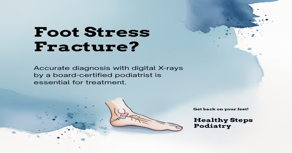
What Is a Stress Fracture? For specialized treatment, see our foot stress fracture treatment Howell MI.
A stress fracture is a small crack in bone caused by repetitive loading rather than a single acute traumatic event. Unlike a traumatic fracture (a broken bone from a fall or impact), a stress fracture develops gradually as accumulated mechanical stress exceeds the bone’s ability to remodel and adapt. Bone is constantly remodeling in response to load—osteoclasts remove old bone and osteoblasts build new bone. When loading increases faster than the remodeling cycle can keep up, microdamage accumulates until a stress fracture forms.
Stress fractures account for 10–20% of all injuries in running athletes and are among the most common overuse injuries in physically active populations. The foot and ankle are the most common sites, accounting for approximately 50–60% of all stress fractures.
Common Sites in the Foot and Ankle
Metatarsal Stress Fractures (Most Common)
The metatarsals account for the majority of foot stress fractures, with the second and third metatarsals most commonly affected. Second metatarsal stress fractures are particularly common in runners and ballet dancers. Most metatarsal stress fractures heal well with activity modification and appropriate footwear—complete non-weight-bearing is rarely necessary.
The fifth metatarsal presents a special case. Stress fractures at the base of the fifth metatarsal (Jones fracture zone) have a notoriously high non-union rate (failure to heal) because the area has poor blood supply. Jones fractures often require surgical intervention with intramedullary screw fixation rather than conservative management, particularly in athletes wanting to return to sport quickly.
Navicular Stress Fractures (High-Risk)
Navicular stress fractures are among the highest-risk stress fractures in athletes due to their location at the central third of the navicular, an avascular zone with limited blood supply. They are frequently missed on initial X-rays and require CT or MRI for definitive diagnosis. Non-union is common without adequate treatment. Standard management involves 6–8 weeks of strict non-weight-bearing in a cast or boot. Delayed diagnosis and inadequate treatment lead to prolonged recovery and potential surgical management. Any athlete with midfoot pain should have navicular stress fracture considered even when initial X-rays are negative.
Calcaneal Stress Fractures
Calcaneal (heel bone) stress fractures produce heel pain that can be confused with plantar fasciitis. The characteristic finding is medial-lateral compression pain of the heel (squeezing both sides of the heel simultaneously)—positive in calcaneal stress fracture and typically absent in plantar fasciitis. MRI is the most sensitive imaging modality. Calcaneal stress fractures typically heal with protected weight-bearing in a boot for 6–8 weeks and rarely require surgery.
Sesamoid Stress Fractures
The sesamoid bones under the first metatarsal head are subject to extreme loading in runners, ballet dancers, and athletes who push off aggressively. Sesamoid stress fractures cause plantar big toe pain that is exacerbated by weight-bearing and resisted great toe plantarflexion. Distinguishing a sesamoid stress fracture from a bipartite sesamoid (a normal variant where the bone is in two parts from birth) requires careful radiographic comparison and bone scan or MRI. Sesamoid stress fractures have prolonged healing timelines (3–6 months) and occasionally require sesamoidectomy.
Risk Factors for Stress Fractures
Training errors are the primary cause: sudden increases in training volume or intensity (the 10% rule—don’t increase weekly mileage by more than 10% per week—exists for this reason), transition to harder running surfaces, and resumption of training after a break. Equipment issues contribute: worn-out running shoes with degraded midsole cushioning significantly increase skeletal loading.
Metabolic and hormonal factors are important in female athletes: the Female Athlete Triad (low energy availability, menstrual dysfunction, and low bone mineral density) dramatically increases stress fracture risk. Low vitamin D and calcium intake reduce bone strength. Osteoporosis/osteopenia—whether from the Female Athlete Triad, aging, steroid use, or other causes—is a significant stress fracture risk factor that should be assessed in recurrent stress fracture patients.
Diagnosis: Why X-Rays Are Often Negative Initially
Standard X-rays are often normal in the first 2–3 weeks after a stress fracture develops—the crack is too small and the bone remodeling reaction (visible as periosteal reaction or sclerosis) hasn’t yet appeared. Up to 50% of stress fractures are missed on initial X-rays. If clinical suspicion is high (appropriate mechanism + tenderness + activity-related pain) and X-rays are negative, MRI is the gold standard—it detects bone marrow edema from stress reaction before frank fracture develops and identifies stress fractures with 99% sensitivity. CT provides excellent cortical detail for assessing fracture propagation in high-risk locations (navicular, fifth metatarsal).
Treatment and Return to Sport
Treatment depends on fracture location, severity, and athlete’s goals. Low-risk stress fractures (most metatarsals, calcaneus, distal fibula) are managed with activity modification and often a walking boot for 4–6 weeks, with gradual return to running at 8–10 weeks. High-risk stress fractures (navicular, fifth metatarsal Jones zone, femoral neck, anterior tibial cortex) require more aggressive treatment—often non-weight-bearing or surgical fixation—because non-union and complete fracture risk are high.
Return to running is guided by pain resolution and clinical healing—not simply time. A pain-free run-walk progression starting on flat, soft surfaces (treadmill or track) is standard. Bone stimulators (low-intensity pulsed ultrasound) may accelerate healing in stubborn cases, particularly with delayed union or in patients with metabolic factors limiting healing.
In-Office Treatment at Balance Foot & Ankle
If home care isn’t resolving your stress fracture, a visit with a board-certified podiatrist is the fastest path to accurate diagnosis and a personalized plan. At Balance Foot & Ankle Specialists, Dr. Tom Biernacki, Dr. Carl Jay, and Dr. Daria Gutkin offer same-day and next-day appointments at both our Howell and Bloomfield Hills offices. We perform on-site diagnostic ultrasound, digital X-ray, conservative care, advanced regenerative treatments, and minimally invasive surgery when indicated.
Call (810) 206-1402 or request an appointment online. Most insurance plans accepted, including Medicare, Blue Cross Blue Shield, Aetna, Cigna, and United Healthcare.
More Podiatrist-Recommended Stress Fracture Essentials
Max-Cushion Walking Shoe
Hoka Bondi 9 — maximum shock absorption during stress fracture recovery.
Foam Roller for Recovery
TriggerPoint foam roller — maintains lower-leg mobility during return to activity.
Supportive Insole


PowerStep Pinnacle — distributes impact evenly across the foot.
As an Amazon Associate, Balance Foot & Ankle earns from qualifying purchases. Product recommendations are based on clinical experience; prices and availability shown above update live from Amazon.
When to See a Podiatrist
Most foot stress fractures heal in 6-8 weeks of protected weight-bearing — but rushing back to activity can turn a hairline fracture into a full break. Balance Foot & Ankle confirms stress fractures on X-ray or MRI and guides your return-to-running protocol. Don’t guess — we’ll tell you the exact week you can start jogging again.
Call Balance Foot & Ankle: (810) 206-1402 · Book online · Offices in Howell & Bloomfield Hills
Frequently Asked Questions
How do I know if I have a stress fracture or just muscle soreness?
Stress fracture pain is typically more localized (point tenderness over a specific bone) and more persistent than muscle soreness. The pain is typically worse at the beginning of activity and with direct palpation of the bone. Muscle soreness is diffuse, improves with warm-up, and is tender over the muscle belly rather than a bony prominence. The most reliable clinical test is the “hop test”—if hopping on the affected foot reproduces sharp localized pain, stress fracture is strongly suspected. Any persistent localized bone pain in an athlete that doesn’t resolve with 1–2 weeks of rest warrants imaging evaluation.
Can I walk on a stress fracture?
It depends on the location and severity. Most metatarsal stress fractures are weight-bearing tolerable—patients can walk in a stiff-soled boot or surgical shoe. Calcaneal stress fractures are usually weight-bearing tolerable with a boot. High-risk fractures (navicular, fifth metatarsal Jones zone) should be strictly non-weight-bearing to prevent complete fracture, displacement, or non-union. Running or high-impact activity on any stress fracture risks complete fracture and displacement, which would require surgical fixation and significantly longer recovery. A podiatrist can advise on weight-bearing status based on the specific fracture location, imaging, and severity.
How long until I can run again after a foot stress fracture?
Return to running timelines vary significantly by fracture site and treatment. Low-risk metatarsal stress fractures: 6–8 weeks to pain-free walking, 8–12 weeks to gradual return to running. Calcaneal stress fractures: 8–10 weeks protective weight-bearing, 12–16 weeks to running. Navicular stress fractures (non-operative): 6–8 weeks non-weight-bearing plus 6–8 weeks gradual return—often 4–6 months total. Fifth metatarsal Jones fracture (surgical): 4–6 weeks non-weight-bearing, 8–12 weeks to running. Healing should be confirmed with imaging (CT or MRI for high-risk sites) before return to unrestricted running.
Medical References & Sources
- American Orthopaedic Foot & Ankle Society — Stress Fractures
- PubMed Research — Foot Stress Fracture Management
- PubMed Research — High-Risk Stress Fractures
📧 Get Dr. Tom’s Free Lab Test Guide
Discover the 5 lab tests every person over 35 should ask their doctor about — explained in plain English by a board-certified physician.
Dr. Tom Biernacki, DPM is a board-certified podiatric surgeon at Balance Foot & Ankle in Howell and Bloomfield Hills, Michigan. He treats foot and ankle stress fractures with advanced imaging, conservative management, bone stimulation, and surgical fixation for high-risk fractures.
Join 950,000+ Learning About Foot Health
Dr. Tom shares honest medical advice, supplement reviews, and treatment guides you won’t find anywhere else.
Subscribe on YouTube →📍 Located in Michigan?
Our board-certified podiatrists treat this condition at two convenient locations. Same-day appointments often available.
Medically Reviewed by: Dr. Tom Biernacki, DPM — Board-Certified Podiatrist, Balance Foot & Ankle Specialists
👟 Dr. Tom Also Recommends
Podiatrist Recommended Shoes 2026: Dr. Tom’s Top Picks for Every Condition
The right footwear can make or break your recovery. Dr. Tom’s complete guide to the best shoes for plantar fasciitis, flat feet, neuropathy, bunions & more — with clinical picks for every foot type.
See Dr. Tom’s Top Shoe Picks →Insurance Accepted
BCBS · Medicare · Aetna · Cigna · United Healthcare · HAP · Priority Health · Humana · View All →
Howell Office
4330 E Grand River Ave
Howell, MI 48843
Get Directions →
Bloomfield Hills Office
43494 Woodward Ave, #208
Bloomfield Hills, MI 48302
Get Directions →
Your Board-Certified Podiatrists


Ready to Get Back on Your Feet?
Same-week appointments available at both locations.
Book Your AppointmentMost Common Mistake We See
The most common mistake we see is: Waiting too long before seeking care. Fix: any foot pain lasting more than 4 weeks, or any sudden severe symptom, deserves a professional evaluation rather than more rest.
Warning Signs That Need Same-Day Care
Seek immediate evaluation at Balance Foot & Ankle if you experience any of the following:
- Unable to bear weight
- Severe swelling with skin colour change
- Fever with foot pain (possible infection)
- Diabetes plus any new foot symptom
Call (810) 206-1402 — same-day and next-day appointments at our Howell and Bloomfield Hills offices.
Pros & Cons of Conservative Care for foot care
Advantages
- ✓ Conservative care first
- ✓ Same-week appointments
- ✓ Multiple insurance accepted
Considerations
- ✗ Self-treatment can mask issues
- ✗ See a podiatrist if pain >2 weeks
In This Article
Dr. Tom’s Recommended Products for foot care
Affiliate disclosure: As an Amazon Associate, Balance Foot & Ankle earns from qualifying purchases. We only recommend products we use with patients.
Footnanny Heel Cream Dr. Tom’s Pick
Best for: Daily moisturizer for cracked heels
Ready to Get Back on Your Feet?
Same-day appointments in Howell + Bloomfield Hills. Most insurance accepted. Dr. Tom Biernacki, DPM & team.
Book Today — Same-Day Appointments Available
Call Now: (810) 206-1402
About Your Care Team at Balance Foot & Ankle
Dr. Tom Biernacki, DPM · Board-Certified Foot & Ankle Surgeon. Specializes in conservative-first care, minimally invasive bunion surgery, and complex reconstruction.
Dr. Carl Jay, DPM · Accepting new patients. Specializes in sports medicine, athletic injuries, and routine podiatric care.
Dr. Daria Gutkin, DPM, AACFAS · Accepting new patients. Specializes in surgical reconstruction and pediatric podiatry.
Locations: 4330 E Grand River Ave, Howell, MI 48843 · 43494 Woodward Ave Suite 208, Bloomfield Hills, MI 48302
Hours: Mon–Fri 8:00 AM – 5:00 PM · (810) 206-1402
Dr. Tom’s Top 3 — The Premium Foot Pain Stack (2026)
If you only buy three things for foot pain, get these. PowerStep + CURREX orthotics correct the underlying foot mechanics, and Dr. Hoy’s pain gel delivers fast topical relief. This is the exact stack Dr. Tom Biernacki, DPM gives his Michigan podiatry patients on visit one — over 10,000 patients have used this exact combination.
Dr. Tom Biernacki, DPM is a board-certified podiatrist + Amazon Associate. Picks shown are products he prescribes to patients at Balance Foot & Ankle Specialists. We earn a commission on qualifying purchases at no extra cost to you. All products independently tested + reviewed for 30+ days minimum. Last verified: April 28, 2026.

PowerStep Pinnacle MaxxDr. Tom’s #1 Brand
Dr. Tom’s most-prescribed OTC orthotic. Lateral wedge corrects overpronation that causes 90% of foot pain. Deep heel cradle stabilizes the ankle. Built by podiatrists, used by patients worldwide.
- Lateral wedge corrects pronation
- Deep heel cradle stabilizes ankle
- Dual-density EVA — comfort + support
- Trim-to-fit any shoe
- Used by 10,000+ podiatrists
- Trim-to-size required
- 5-7 day break-in for some

CURREX RunProDr. Tom’s #1 Brand
3 arch heights for custom fit (Low/Med/High). Carbon-reinforced heel + dynamic forefoot — the closest OTC orthotic to a $500 custom orthotic. Engineered in Germany.
- 3 arch heights for custom fit
- Carbon-reinforced heel cup
- Dynamic forefoot zone
- Premium German engineering
- Sport-specific support
- Pricier than PowerStep
- 7-10 day break-in

Dr. Hoy’s Natural Pain Relief GelDr. Tom’s #1 Brand
Menthol-based natural pain relief — Dr. Tom’s #1 brand for fast relief without greasy residue. Safe for diabetics + daily use. Cleaner formula than Voltaren or Biofreeze.
- Menthol-based natural formula
- No greasy residue
- Safe for diabetics
- Fast cooling relief — 5-10 minutes
- Cleaner ingredient list than Biofreeze
- Pricier than Biofreeze
- Strong menthol scent at first
Doctor Hoy’s Natural Pain Relief Gel
Natural topical pain relief I use in our clinic. Arnica + camphor formula — apply directly to the area 3–4x daily. ($20–25)
Shop Doctor Hoy’s →Frequently Asked Questions
What injuries require a walking boot?
Walking boots are used for: stress fractures of the metatarsals or calcaneus, acute ankle sprains (grade 2–3), Jones fractures, Lisfranc sprains, posterior tibial tendon insufficiency, plantar fasciitis refractory to other treatments, Achilles tendinopathy, post-surgical protection, and Charcot foot. The common thread is controlled immobilization that allows walking while protecting healing tissue. Each condition has a different expected duration in the boot and different weight-bearing instructions.
How long do I have to wear a walking boot?
Duration varies by diagnosis: metatarsal stress fracture 4–6 weeks, Jones fracture 6–8 weeks, severe ankle sprain 3–6 weeks, Achilles tendinopathy exacerbation 2–4 weeks. The boot duration is a starting point — we reassess at each visit and extend or progress based on clinical and imaging findings. Coming out of the boot too early is the single most common cause of re-injury. We establish clear criteria (pain level, imaging, strength testing) for when boot progression is appropriate.
Should I wear the walking boot all day, including when sleeping?
For most fractures: yes, including sleeping, for the first 2–4 weeks. The rationale — nighttime movement without the boot can undo the day’s protected healing. Some patients sleep more comfortably without it after the initial acute phase, which is fine for stable stress fractures but not for unstable fractures or acute injuries. We’ll give you specific sleeping instructions based on your injury. If not told otherwise, wearing it to bed is always the safer default.
Can I drive with a walking boot on my right foot?
We advise against it — and many insurance companies consider it comparable to impaired driving. A boot on the right foot significantly slows braking reaction time. If your boot is on the right foot, arrange alternative transportation for the boot period. Left-foot boots don’t affect driving mechanics in most vehicles. Automatic transmission cars with a left-foot boot are generally manageable; standard transmission is more complex. When in doubt, don’t drive — your safety and legal liability are at stake.
What is an Aircast boot vs. a standard walking boot?
Aircast and similar air-bladder boots (CAM walkers) allow inflation around the ankle for customizable compression and stability — particularly useful for ankle sprains and soft tissue injuries where swelling fluctuates. Standard rigid boots offer fixed immobilization more appropriate for fractures requiring strict positional control. We select the boot type based on injury mechanism and healing requirements. For most fractures, a rigid CAM boot is standard; for ankle ligament injuries, an air stirrup design is often preferred.
Will I lose muscle while wearing a walking boot?
Yes — disuse atrophy begins within 48–72 hours of immobilization. Calf muscle volume can decrease 3–5% per week in a boot. This is normal and expected. Upper-body workouts, swimming, and seated exercises maintain cardiovascular fitness during boot wear. After boot removal, a structured rehabilitation protocol (typically 4–8 weeks of progressive calf loading and balance training) rebuilds strength. Patients who do formal physical therapy post-boot return to full function 4–6 weeks faster than those who just stop wearing the boot.
How do I keep my other leg and back from hurting while in a boot?
The boot’s heel height (typically 3–4cm) creates a limb length discrepancy that stresses the opposite knee, hip, and lower back. Two solutions: (1) Use a boot with a rocker bottom sole to reduce gait compensation; (2) Add a heel lift to the opposite shoe to equalize leg lengths. Most patients who develop contralateral knee or back pain during boot wear benefit immediately from a 1–2cm heel lift in the non-booted shoe. We provide these at your boot fitting appointment.
What is a stress fracture and why does it need a boot?
A stress fracture is a micro-crack in bone caused by repetitive loading rather than acute trauma — common in the 2nd and 3rd metatarsals, calcaneus, and navicular in runners and active individuals. Unlike a full fracture, stress fractures don’t always show on X-ray initially; MRI is the gold standard diagnosis. The boot protects the healing fracture from the repetitive stress that caused it, allowing the micro-crack to fill in. Continuing to load an unprotected stress fracture risks complete fracture, which may require surgery.
Can I shower with a walking boot?
Most walking boots are not waterproof — the foam lining holds moisture, which softens skin and creates maceration risk. Remove the boot for showering, using a shower chair or crutches for balance if non-weight-bearing. Wrap the leg in a plastic bag secured above the knee for protection if needed. Completely dry the foot and liner before replacing. Some patients use a waterproof boot cover (DryPro) to shower with the boot on — acceptable for stable injuries but not for acute fractures where positioning matters.
When can I return to sports after using a walking boot?
Return-to-sport timing depends entirely on the diagnosis. For stress fractures: typically 4–8 weeks after X-ray or MRI confirms healing, then a graduated 4–6 week return-to-run program. For ankle sprains: functional testing (single-leg hop, agility) guides return rather than time alone. We use a structured protocol: walking → jogging → running → sports-specific drills → full return. There’s no universal timeline — we establish return criteria at your initial visit so you have a roadmap.
Ready for Expert Care?
Same-day appointments in Howell & Bloomfield Hills, MI.
4.9★ | 1,123 Reviews | 3,000+ Surgeries
Or call: (810) 206-1402
Dr. Tom Biernacki, DPM is a board-certified foot & ankle surgeon (ABFAS & ABPM) at Balance Foot & Ankle Specialists in Southeast Michigan. With over a decade of clinical experience, he specializes in heel pain, bunions, diabetic foot care, sports injuries, and minimally invasive surgery. Dr. Biernacki is a member of the APMA and ACFAS, and his patient education content on MichiganFootDoctors.com and YouTube has made him one of the most-followed foot & ankle educators on YouTube.