
Medically reviewed by Dr. Tom Biernacki, DPM
Board-certified podiatric surgeon | Balance Foot & Ankle, Howell & Bloomfield Township, MI
Last reviewed: May 2026

| Location | Risk Level | Blood Supply | Treatment | WB Status | Return to Sport |
|---|---|---|---|---|---|
| 2nd–4th Metatarsal Shaft | Low | Good | Stiff-soled shoe or boot 4–6 wk | Protected WB in stiff shoe | 6–8 weeks |
| Calcaneus | Low | Good | Boot 4–6 weeks | Protected WB | 6–10 weeks |
| Fibula (distal shaft) | Low | Good | Boot 4–6 weeks | Weight-bearing tolerated | 6–8 weeks |
| 1st Metatarsal | Moderate | Moderate | Boot 6 weeks — avoids hallux stress | Minimal WB | 8–12 weeks |
| 5th Metatarsal Zone I (avulsion) | Low | Good | Boot or hard-sole shoe 4–6 wk | Protected WB | 6–8 weeks |
| 5th Metatarsal Zone II (Jones) | HIGH ⚠️ | Watershed zone — poor | Surgical fixation (IMN screw) for athletes | NWB 6–8 wk post-op | 12–16 weeks |
| Navicular (N-zone) | HIGH ⚠️ | Avascular central zone | NWB cast 6–8 wk; surgery if displaced | Strict NWB | 3–6 months |
| Sesamoid | Moderate–High | Limited | J-pad, NWB boot 6–10 wk | NWB to protected WB | 3–6 months |
| Medial Malleolus | HIGH ⚠️ | Good but high stress | Surgery if displaced; NWB cast if stable | NWB 6–8 weeks | 3–5 months |
| Risk Factor | Mechanism | Magnitude of Risk Increase | Modifiable? | Intervention |
|---|---|---|---|---|
| Rapid training load increase (>10%/week) | Bone remodeling lags demand | 3–4x increased risk | Yes | 10% rule — increase mileage by max 10%/week |
| Low bone mineral density (osteopenia/OP) | Reduced bone strength at baseline | 2–5x increased risk | Partially | DEXA scan, calcium/D3, bisphosphonates if needed |
| Female Athlete Triad / RED-S | Low energy availability → low estrogen → bone loss | 4–7x increased risk | Yes — nutrition | Caloric sufficiency, sports dietitian, endocrinology |
| Vitamin D deficiency | Impaired bone mineralization | 2–3x increased risk | Yes | 25-OH-D level; supplement to >40 ng/mL |
| High arch (pes cavus) | Reduced shock absorption → increased bone stress | 2–3x vs. neutral foot | Partially | Cushioned orthotics, cushioned shoes |
| Running on hard surfaces (asphalt/concrete) | Higher ground reaction forces | 1.5–2x vs. soft surface | Yes | Mix surface training — trails, track, grass |
| Worn-out running shoes (>500 miles) | Midsole compression loss → less shock absorption | 1.5–2x risk | Yes | Replace shoes every 300–500 miles regardless of appearance |
A foot or ankle stress fracture often gets dismissed as just sore feet for weeks before imaging finally catches it. The right boot for 4-6 weeks usually fully heals it without surgery.
You’re in the right place. Dr. Tom Biernacki, DPM, FACFAS — board-certified foot & ankle surgeon with 3,000+ surgeries — explains exactly what stress fracture treatment in Michigan means and what works. Call (810) 206-1402 for same-day appointment at Howell or Bloomfield Township.
Quick answer:Stress fracture of the foot (most common: 2nd metatarsal, navicular, 5th metatarsal base) causes point tenderness and diffuse aching that worsens with activity. Treatment: non-weight-bearing in a boot for 4-8 weeks. Navicular and Jones fractures heal poorly without strict non-weight-bearing. MRI is more sensitive than X-ray for early detection. Call (810) 206-1402.ll (810) 206-1402.
Medically Reviewed | Dr. Tom Biernacki, DPM | Board-Certified Podiatric Surgeon | Balance Foot & Ankle, Michigan
Related Conditions
In This Article
- What is a foot stress fracture?
- Stress Fractures in the Foot: Michigan Podiatrist’s Clinical Guide
- Mechanism and Pathophysiology
- High-Risk vs. Low-Risk Stress Fractures: The Critical Classification
- Navicular Stress Fractures: Treat Aggressively
- Metatarsal Stress Fractures in Michigan Runners
- The Female Athlete Triad and Relative Energy Deficiency (RED-S)
- Training Error Identification
- Return to Sport After Stress Fracture
- Dr. Tom's Product Recommendations
- Frequently Asked Questions
- Frequently Asked Questions
- What is Stress fracture?
- Symptoms and warning signs
- Conservative treatment options
- When is surgery considered?
- Recovery timeline and prevention


![Calcaneus Stress Fracture Treatment [Heel Stress Fracture RECOVERY!]](https://www.michiganfootdoctors.com/wp-content/cache/flying-press/cdfca81fb25b3ea56c6af7d70a9e3fb0.jpg)
Watch: Calcaneus Stress Fracture Treatment [Heel Stress Fracture RECOVERY!] — MichiganFootDoctors YouTube
Stress Fractures in the Foot: Michigan Podiatrist’s Clinical Guide
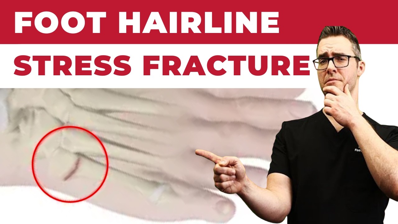
Stress fractures are among the most commonly missed diagnoses in active Michigan patients — frequently misidentified as shin splints, plantar fasciitis, or generalized foot pain until the fracture progresses to a complete break. Understanding the clinical presentation, risk stratification, and imaging sequence for stress fractures prevents the catastrophic outcomes (Jones fracture non-union, navicular complete fracture) that result from treating high-risk stress fractures as low-risk.
Mechanism and Pathophysiology
Bone undergoes constant remodeling — osteoclastic resorption followed by osteoblastic new bone formation — in response to mechanical loading. When loading increases faster than remodeling can adapt, microscopic damage accumulates. The progression: trabecular microdamage → periosteal stress reaction → cortical stress reaction → complete fracture. Early stages (stress reaction) are invisible on plain radiographs; MRI detects marrow edema within days. Plain X-ray may not show periosteal reaction for 2–4 weeks, explaining why “X-rays were negative” doesn’t rule out a stress fracture.
High-Risk vs. Low-Risk Stress Fractures: The Critical Classification
Low-risk stress fractures (compressive side, good blood supply, low non-union rate): 2nd, 3rd, and 4th metatarsal shafts; calcaneal body; fibula. Treatment: weight-bearing as tolerated in a protective boot, activity restriction, return to sport in 4–8 weeks with appropriate progression.
High-risk stress fractures (tension side, watershed vascularity, or biomechanically unfavorable): Navicular central third (15–30% non-union without strict NWB), Jones fracture Zone 2 (proximal 5th metatarsal metaphyseal-diaphyseal junction, 15–20% non-union, surgical consideration for athletes), sesamoids (medial tibial sesamoid most common, poor healing with ambulation), great toe proximal phalanx (tension stress), medial malleolus. These require immediate non-weight-bearing, orthopedic or podiatric surgery consultation, and often intramedullary screw fixation for competitive athletes who cannot accept 8–12 weeks NWB.
Navicular Stress Fractures: Treat Aggressively
The navicular central third is the highest-risk stress fracture in sport. Its watershed vascular supply — the central third receives end-arterial flow from dorsal and plantar navicular arteries with a true avascular zone in the middle — means that stress reaction in this zone progresses to complete fracture with remarkable speed under continued loading. Athletes presenting with dorsal midfoot aching that worsens with running and is precisely tender over the “N-spot” (direct digital pressure over the navicular dorsal surface) have a navicular stress fracture until proven otherwise.
Management: MRI (or CT if MRI equivocal) → confirmed navicular stress fracture → strict NWB in cast for 6 weeks → CT to confirm healing → protected weight-bearing and graduated return to sport. Complete navicular fractures in competitive athletes often receive intramedullary screw fixation to allow earlier rehabilitation — surgery is time faster than NWB casting for high-performance athletes with tight competitive calendars.
Metatarsal Stress Fractures in Michigan Runners
The 2nd metatarsal is the most common stress fracture in runners — its relative rigidity compared to the mobile 1st ray causes disproportionate loading under forefoot strike patterns. The fracture occurs most commonly at the metatarsal neck (distal third) in dancers (dancer’s fracture of the 2nd MT) and mid-shaft in runners. Management: rigid-soled boot for 4–6 weeks, activity modification, return to run program with 10% weekly mileage escalation.
The 5th metatarsal has three distinct stress zones with dramatically different prognoses (Zone 1 avulsion, Zone 2 Jones fracture, Zone 3 diaphyseal stress fracture) — detailed in the fracture classification framework Dr. Biernacki applies at every foot fracture evaluation.
The Female Athlete Triad and Relative Energy Deficiency (RED-S)
Female athletes presenting with stress fractures should be screened for the female athlete triad: low energy availability (chronic caloric restriction relative to training load), menstrual dysfunction (oligomenorrhea, amenorrhea), and low bone mineral density (assessed with DEXA scan). The triad creates a hormonal and metabolic environment where bone resorption exceeds formation — the fracture is the consequence, not the cause. Without addressing energy availability and hormonal status, stress fractures recur despite optimal mechanical treatment.
The expanded concept — Relative Energy Deficiency in Sport (RED-S) — includes male athletes and broader health consequences including cardiovascular, endocrine, immunologic, and psychological domains. Michigan cross-country, distance running, gymnastics, and figure skating athletes are the highest-risk populations.
Training Error Identification
The 10% rule (never increase weekly mileage more than 10% per week) is the most widely cited training guideline for injury prevention. Violations are the most common identifiable precipitant for metatarsal stress fractures in recreational runners. Other training errors: sudden surface change (treadmill to road, road to track), transition to minimalist footwear without gradual adaptation, and return-to-sport after a detraining period at previous volume.
Gait analysis identifies biomechanical contributors: excessive stride length (longer stride = higher ground reaction force per step), rear-foot strike with vertical oscillation (higher impact loading), and crossover gait (medially directed impact vector increasing 2nd metatarsal loading). Gait retraining to increase cadence by 5–10% and reduce overstriding significantly reduces metatarsal stress fracture recurrence.
Return to Sport After Stress Fracture
Return-to-run progression begins when imaging confirms healing (absence of edema on MRI, cortical bridging on CT) and the patient is pain-free with daily walking. A graduated program — walk/run intervals progressing over 4–6 weeks to full running volume — is followed by sport-specific loading before competitive return. For high-risk fractures (navicular, Jones Zone 2), CT confirmation of bony bridging before any impact activity is mandatory.
Dr. Tom's Product Recommendations
Ossur Rebound Air Walker Boot
⭐ Highly Rated
The clinical-standard removable cast walker for metatarsal and calcaneal stress fractures requiring protected weight-bearing. Pneumatic air bladder provides circumferential compression and secure fit. Rigid rocker sole reduces metatarsal head loading during push-off. The most prescribed boot for low-risk foot stress fractures.
Dr. Tom says: “Wore this for 6 weeks for a 2nd metatarsal stress fracture. Rigid sole kept me mobile at work while protecting the fracture completely.”
Low-risk metatarsal and calcaneal stress fractures, protected weight-bearing phase
Not appropriate for navicular or high-risk fractures where strict NWB is required
Disclosure: We earn a commission at no extra cost to you.
CURREX RunPro Insoles
⭐ Highly Rated
Biomechanically engineered running insoles that optimize load distribution across the metatarsals during return-to-run. Research shows CURREX dynamic arch technology reduces peak metatarsal stress by modulating arch stiffness through the gait cycle. Profile-matched to arch height for personalized stress distribution.
Dr. Tom says: “After my 2nd metatarsal stress fracture, my podiatrist prescribed CURREX for my return to running. Completed my half marathon training block without any recurrence.”
Return-to-run after metatarsal stress fracture, stress fracture prevention in runners
Profile selection critical — arch height match required for optimal benefit
Disclosure: We earn a commission at no extra cost to you.
Brooks Ghost 16 Running Shoe
⭐ Highly Rated
Neutral cushioning road running shoe with consistent, predictable ride and adequate metatarsal cushioning for return-to-sport after stress fracture. The Ghost 16’s balanced heel-to-toe drop (12mm) and DNA LOFT foam midsole provide impact absorption without sacrificing road feedback. Available in wide widths.
Dr. Tom says: “My podiatrist cleared me to run in these after my metatarsal stress fracture healed. The cushioning gave me confidence to rebuild my mileage safely.”
Return-to-run after stress fracture, neutral gait, mileage rebuilding phase
High-arch runners may need more medial support — consider Brooks Adrenaline
Disclosure: We earn a commission at no extra cost to you.
✅ Pros / Benefits
- High-risk vs. low-risk stress fracture classification — navicular and Jones Zone 2 treated aggressively
- MRI ordering (not just X-ray) when clinical suspicion is high — early detection prevents progression
- Female athlete triad screening — bone health and energy availability assessed
- Gait analysis identifies training error contributors to prevent recurrence
- Return-to-run protocol with CT confirmation for high-risk fractures
❌ Cons / Risks
- Navicular stress fractures require 6–12 weeks NWB — significant time loss from sport
- DEXA scan for bone density assessment requires separate facility scheduling
- Surgical fixation for Jones Zone 2 and navicular fractures in competitive athletes requires OR scheduling
Dr. Tom Biernacki’s Recommendation
The stress fracture mistake I see most often is treating a navicular stress fracture like a second metatarsal stress fracture. Navicular gets a boot and told to rest for two weeks. Two weeks later they’re back, still in pain, and now we’ve lost three weeks of healing. Navicular is a high-risk fracture. It goes non-weight-bearing in a cast, full stop, until CT shows healing. That’s not conservative — that’s correct.
— Dr. Tom Biernacki, DPM | Board-Certified Podiatric Surgeon | Balance Foot & Ankle
Frequently Asked Questions
Why doesn’t an X-ray show my stress fracture?
Plain X-rays can’t detect the early stages of stress fracture — the periosteal reaction visible on X-ray takes 2–4 weeks to develop, and small cortical cracks are often invisible even on high-quality films. MRI shows bone marrow edema within days of injury onset, making it the gold standard for early stress fracture detection. If your X-ray is negative but your pain and clinical presentation are consistent with a stress fracture, MRI is the next step.
How long am I out of running with a metatarsal stress fracture?
Low-risk metatarsal stress fractures (2nd, 3rd, 4th shaft) typically require 4–6 weeks in a protective boot followed by a 4–6 week graduated return-to-run program — total time from injury to full training is 8–12 weeks. High-risk fractures (navicular, Jones Zone 2) require strict non-weight-bearing for 6–8 weeks plus a longer graduated return — total timeline 12–20 weeks, sometimes longer if healing is delayed.
Can I still exercise with a foot stress fracture?
Low-impact, non-weight-bearing exercise (pool running, swimming, cycling on a stationary bike with the boot) is typically possible during stress fracture healing and significantly reduces deconditioning. This should be cleared with your podiatrist based on fracture location and severity. High-risk fractures (navicular especially) may require complete rest from all impact activity until imaging confirms adequate healing.
How do I prevent my stress fractures from coming back?
Stress fracture recurrence is preventable with the right interventions: strict 10% weekly mileage rule during any rebuilding phase, bone health assessment (Vitamin D, calcium, DEXA if indicated), correction of training errors (cadence, footwear, surface), biomechanical evaluation for structural contributors (hyperpronation, cavovarus, leg length discrepancy), and adequate nutrition for bone remodeling. Most recurrences reflect inadequate attention to one of these factors.
Michigan Foot Pain? See Dr. Biernacki In Person
4.9★ rated | 1,123 Reviews | 3,000+ Surgeries
Same-week appointments · Howell & Bloomfield Township
📞 (810) 206-1402 Book Online →Frequently Asked Questions
When should I see a podiatrist?
If symptoms persist past 2 weeks, affect your normal activity, or are accompanied by red-flag symptoms (warmth, redness, swelling, inability to bear weight).
What does treatment cost?
Most diagnostic visits and conservative treatments are covered by Medicare and major insurers. Out-of-pocket costs vary by your specific plan.
How quickly can I get an appointment?
Most non-urgent cases see us within 5 business days. Urgent cases (sudden pain, possible fracture) typically same or next business day.
What is Stress fracture?
Stress fracture is a common foot/ankle condition that affects mobility and quality of life. Understanding the underlying cause is the first step in successful treatment. Our podiatrists at Balance Foot & Ankle perform a hands-on biomechanical exam, review your activity history, and use diagnostic imaging when appropriate to identify the root cause—not just treat the symptom. Many patients have been told to “rest and ice” without a deeper diagnostic workup; our approach is different.
Symptoms and warning signs
Common signs of stress fracture include pain that worsens with activity, morning stiffness, swelling, tenderness when palpated, and difficulty bearing weight. If you experience sudden severe pain, inability to walk, visible deformity, numbness or color change, contact our office the same day or visit urgent care—these can signal a more serious injury such as a fracture, tendon rupture, or vascular compromise. Diabetics with any foot wound should seek same-day care.
Conservative treatment options
Most cases of stress fracture respond to non-surgical care: structured rest, supportive footwear changes, custom orthotics, targeted stretching and strengthening protocols, anti-inflammatory medications when medically appropriate, and in-office procedures such as ultrasound-guided injections. We also offer advanced therapies including MLS laser therapy, EPAT/shockwave, regenerative injections, and image-guided procedures. Treatment is sequenced from least invasive to most invasive, and we explain the rationale at every step.
When is surgery considered?
Surgery is reserved for cases that fail 3-6 months of well-structured conservative care, when there is structural pathology (severe deformity, complete tear, advanced arthritis), or when imaging shows damage that will not heal without intervention. Our surgeons have performed 3,000+ foot and ankle procedures and prioritize minimally-invasive techniques whenever appropriate. We discuss recovery timelines, return-to-activity milestones, and realistic outcome expectations before any procedure is scheduled.
Recovery timeline and prevention
Recovery from stress fracture varies based on severity and chosen treatment path. Conservative cases often improve within 4-8 weeks with consistent adherence to the protocol. Post-procedural recovery may range from a few days (in-office procedures) to several months (reconstructive surgery). Long-term prevention involves footwear assessment, activity modification, structured strengthening, and regular check-ins with your podiatrist if you have a history of recurrence. We provide written home-exercise plans and digital follow-up support.
Ready to feel better?
Same-week appointments available in Howell and Bloomfield Township, Michigan.
Book Your VisitFrequently Asked Questions
What injuries require a walking boot?
Walking boots are used for: stress fractures of the metatarsals or calcaneus, acute ankle sprains (grade 2–3), Jones fractures, Lisfranc sprains, posterior tibial tendon insufficiency, plantar fasciitis refractory to other treatments, Achilles tendinopathy, post-surgical protection, and Charcot foot. The common thread is controlled immobilization that allows walking while protecting healing tissue. Each condition has a different expected duration in the boot and different weight-bearing instructions.
How long do I have to wear a walking boot?
Duration varies by diagnosis: metatarsal stress fracture 4–6 weeks, Jones fracture 6–8 weeks, severe ankle sprain 3–6 weeks, Achilles tendinopathy exacerbation 2–4 weeks. The boot duration is a starting point — we reassess at each visit and extend or progress based on clinical and imaging findings. Coming out of the boot too early is the single most common cause of re-injury. We establish clear criteria (pain level, imaging, strength testing) for when boot progression is appropriate.
Should I wear the walking boot all day, including when sleeping?
For most fractures: yes, including sleeping, for the first 2–4 weeks. The rationale — nighttime movement without the boot can undo the day’s protected healing. Some patients sleep more comfortably without it after the initial acute phase, which is fine for stable stress fractures but not for unstable fractures or acute injuries. We’ll give you specific sleeping instructions based on your injury. If not told otherwise, wearing it to bed is always the safer default.
Can I drive with a walking boot on my right foot?
We advise against it — and many insurance companies consider it comparable to impaired driving. A boot on the right foot significantly slows braking reaction time. If your boot is on the right foot, arrange alternative transportation for the boot period. Left-foot boots don’t affect driving mechanics in most vehicles. Automatic transmission cars with a left-foot boot are generally manageable; standard transmission is more complex. When in doubt, don’t drive — your safety and legal liability are at stake.
What is an Aircast boot vs. a standard walking boot?
Aircast and similar air-bladder boots (CAM walkers) allow inflation around the ankle for customizable compression and stability — particularly useful for ankle sprains and soft tissue injuries where swelling fluctuates. Standard rigid boots offer fixed immobilization more appropriate for fractures requiring strict positional control. We select the boot type based on injury mechanism and healing requirements. For most fractures, a rigid CAM boot is standard; for ankle ligament injuries, an air stirrup design is often preferred.
Will I lose muscle while wearing a walking boot?
Yes — disuse atrophy begins within 48–72 hours of immobilization. Calf muscle volume can decrease 3–5% per week in a boot. This is normal and expected. Upper-body workouts, swimming, and seated exercises maintain cardiovascular fitness during boot wear. After boot removal, a structured rehabilitation protocol (typically 4–8 weeks of progressive calf loading and balance training) rebuilds strength. Patients who do formal physical therapy post-boot return to full function 4–6 weeks faster than those who just stop wearing the boot.
How do I keep my other leg and back from hurting while in a boot?
The boot’s heel height (typically 3–4cm) creates a limb length discrepancy that stresses the opposite knee, hip, and lower back. Two solutions: (1) Use a boot with a rocker bottom sole to reduce gait compensation; (2) Add a heel lift to the opposite shoe to equalize leg lengths. Most patients who develop contralateral knee or back pain during boot wear benefit immediately from a 1–2cm heel lift in the non-booted shoe. We provide these at your boot fitting appointment.
What is a stress fracture and why does it need a boot?
A stress fracture is a micro-crack in bone caused by repetitive loading rather than acute trauma — common in the 2nd and 3rd metatarsals, calcaneus, and navicular in runners and active individuals. Unlike a full fracture, stress fractures don’t always show on X-ray initially; MRI is the gold standard diagnosis. The boot protects the healing fracture from the repetitive stress that caused it, allowing the micro-crack to fill in. Continuing to load an unprotected stress fracture risks complete fracture, which may require surgery.
Can I shower with a walking boot?
Most walking boots are not waterproof — the foam lining holds moisture, which softens skin and creates maceration risk. Remove the boot for showering, using a shower chair or crutches for balance if non-weight-bearing. Wrap the leg in a plastic bag secured above the knee for protection if needed. Completely dry the foot and liner before replacing. Some patients use a waterproof boot cover (DryPro) to shower with the boot on — acceptable for stable injuries but not for acute fractures where positioning matters.
When can I return to sports after using a walking boot?
Return-to-sport timing depends entirely on the diagnosis. For stress fractures: typically 4–8 weeks after X-ray or MRI confirms healing, then a graduated 4–6 week return-to-run program. For ankle sprains: functional testing (single-leg hop, agility) guides return rather than time alone. We use a structured protocol: walking → jogging → running → sports-specific drills → full return. There’s no universal timeline — we establish return criteria at your initial visit so you have a roadmap.
In-Office Treatment at Balance Foot & Ankle
If home treatment isn’t providing relief for your foot and ankle issues, our podiatry team at Balance Foot & Ankle can help with same-day evaluations and advanced in-office care.
Ready to Get Relief?
Same-day appointments available in Howell & Bloomfield Township, MI
4.9★ | 1,123 Reviews | 3,000+ Surgeries
Or call: (810) 206-1402
Seen at both offices — Howell and Bloomfield Township.

Dr. Tom Biernacki, DPM is a board-certified foot & ankle surgeon (ABFAS & ABPM) at Balance Foot & Ankle Specialists in Southeast Michigan. With over a decade of clinical experience, he specializes in heel pain, bunions, diabetic foot care, sports injuries, and minimally invasive surgery. Dr. Biernacki is a member of the APMA and ACFAS, and his patient education content on MichiganFootDoctors.com and YouTube has made him one of the most-followed foot & ankle educators on YouTube.