
Inner Foot Arch Pain: 5 Causes & Treatment (Podiatrist 2026)
Inner foot arch pain is most often caused by: (1) plantar fasciitis (heel + arch pain, worse with first morning steps), (2) posterior tibial tendon dysfunction (PTTD) — #1 cause in adults over 40, (3) accessory navicular syndrome (extra bone on the inner arch causes a painful bump), (4) plantar fibroma (firm rubbery lump in arch), or (5) flexor hallucis longus tendinopathy (“dancer’s tendinitis”).
In my Michigan podiatry clinic, ~50% of inner arch pain in adults is PTTD or plantar fasciitis. Treatment depends on cause: plantar fasciitis → PowerStep Pinnacle Maxx + heel-stretch protocol (75% relief in 6 weeks); PTTD → medial-post orthotic + Aircast AirLift PTTD brace; accessory navicular → off-loading orthotic + arch pad. Red flag: arch visibly collapsed, can’t perform single-leg heel raise, or can’t walk — that’s stage 2-3 PTTD; needs same-week podiatrist visit before tendon ruptures.
Medically reviewed by Dr. Tom Biernacki, DPM — Board-certified foot & ankle surgeon, 3,000+ surgeries performed. Updated April 2026 with current clinical evidence. This article reflects real practice experience from Balance Foot & Ankle Specialists in Howell and Bloomfield Hills, Michigan.
Quick Answer
Most foot and ankle problems respond to conservative care — proper footwear, supportive inserts, activity modification, and targeted stretching — within 4-8 weeks. Persistent pain beyond that window, or any symptom that prevents walking, warrants a podiatric evaluation to rule out fracture, tendon tear, or systemic cause.
Watch: Dr. Tom Biernacki, DPM

Medically reviewed by Dr. Tom Biernacki, DPM — Board-Certified Podiatric Surgeon — Balance Foot & Ankle, Howell & Bloomfield Hills, MI. Last updated April 2026.
▶ Watch

Medically reviewed by Dr. Tom Biernacki, DPM | Board-certified podiatrist | 3,000+ surgeries performed
Last updated: April 2, 2026
The most important clinical decision with Accessory Navicular Syndrome Painful Arch Bone isn't which treatment to start with — it's which subtype or underlying cause you actually have. Our podiatrists regularly see patients who've been treated for months for the wrong diagnosis. The correct identification changes the entire treatment path. Call (810) 206-1402 — Dr. Tom evaluates this condition at both Howell and Bloomfield Hills locations.
What Is an Accessory Navicular?
An accessory navicular is an extra bone or cartilaginous body that develops adjacent to the navicular bone on the medial (inner) side of the foot. It forms during fetal development as a separate ossification center that fails to fuse with the navicular bone. Three types are classified based on size and relationship to the navicular: Type I is a small, round sesamoid bone within the posterior tibial tendon; Type II is a larger triangular bone connected to the navicular by a cartilaginous synchondrosis; and Type III (cornuate navicular) is a fully fused, enlarged navicular prominence.
Type II is the most clinically significant and the type most likely to cause symptoms. The fibrocartilaginous connection between the accessory bone and the navicular creates a potential stress point where micromotion occurs during weight bearing and posterior tibial tendon contraction. This repetitive micromotion generates inflammation, pain, and progressive weakening of the synchondrosis — particularly during the increased physical demands of adolescence.
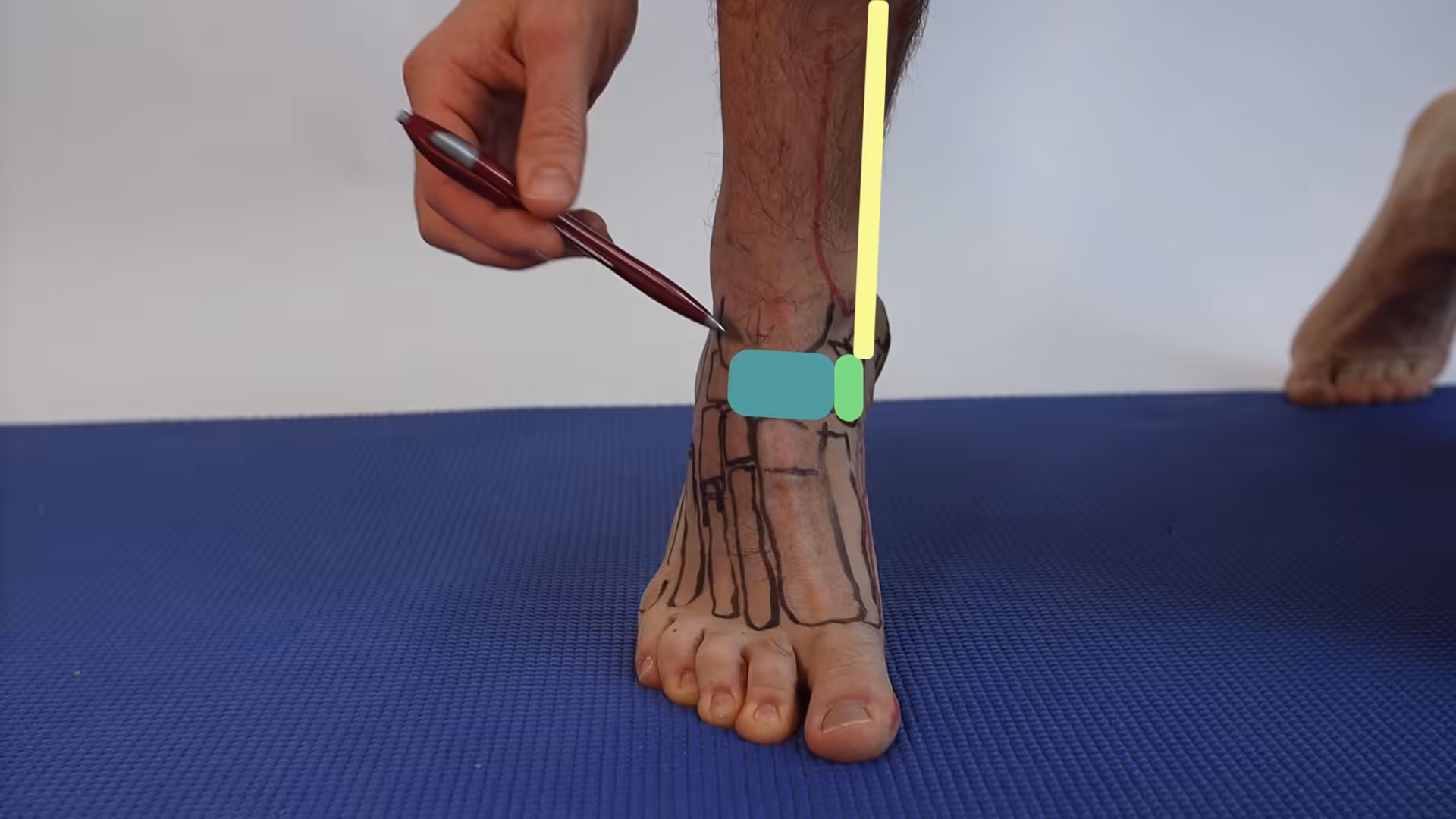
The condition is bilateral in approximately 50-90% of cases, meaning both feet have the extra bone, though symptoms may develop in only one foot. Dr. Tom Biernacki identifies accessory navicular bones through clinical examination showing a visible or palpable bony prominence on the inner arch and confirms the diagnosis with X-rays that clearly demonstrate the extra ossicle.
Causes of Painful Accessory Navicular
Many people with accessory navicular bones live their entire lives without symptoms. Pain typically develops when the extra bone is subjected to repetitive stress, direct trauma, or chronic irritation. Acute ankle sprains or foot injuries can disrupt the synchondrosis connecting the accessory bone to the navicular, initiating an inflammatory cycle that persists long after the original injury has healed.
Flat feet (pes planus) significantly increase the risk of symptomatic accessory navicular syndrome. When the arch collapses, it increases tension on the posterior tibial tendon — which attaches to and wraps around the accessory bone. This chronic tensile overload creates progressive inflammation at the synchondrosis and may eventually lead to posterior tibial tendon dysfunction as the tendon’s mechanical advantage is compromised by the unstable accessory bone.
Shoe pressure against the medial foot prominence is another common trigger. The accessory navicular creates a visible bump on the inner arch that contacts the shoe medial wall during walking, especially in tight-fitting shoes, ice skates, ski boots, and cleats. Adolescents frequently become symptomatic when growth spurts enlarge the prominence, coinciding with increased athletic participation that amplifies both external pressure and internal tendon forces.
Symptoms and Diagnosis
The cardinal symptom is pain and tenderness directly over the bony prominence on the inner side of the foot, typically located just below and slightly behind the medial malleolus (inner ankle bone). The area may be visibly swollen, red, or warm during acute flare-ups. Pain characteristically worsens with activity — particularly running, jumping, and prolonged walking — and improves with rest.
Physical examination reveals point tenderness over the accessory navicular, often with a palpable bony prominence that may be larger than the contralateral side. Pain intensifies with resisted foot inversion (turning the sole inward) and single-leg heel raise, both of which engage the posterior tibial tendon. Comparison with the asymptomatic foot, when available, helps quantify the degree of prominence and associated flat foot deformity.
Standard foot X-rays in three views confirm the presence and type of accessory navicular. MRI is obtained when the diagnosis is uncertain, when posterior tibial tendon pathology is suspected, or when surgical planning requires detailed soft tissue assessment. MRI can demonstrate bone marrow edema at the synchondrosis (indicating active stress), tendon thickening or tearing, and the precise relationship between the accessory bone and surrounding structures.
Conservative Treatment
Initial treatment focuses on reducing inflammation and offloading the painful prominence. Immobilization in a walking boot or cast for 4-6 weeks allows acute inflammation to resolve and gives the irritated synchondrosis time to quiet down. Anti-inflammatory medications and ice therapy complement immobilization. Approximately 60-70% of patients achieve satisfactory symptom relief with initial conservative management.
Custom orthotics with medial arch support and a medial flange that protects the navicular prominence from shoe pressure provide long-term symptom management after acute inflammation resolves. PowerStep Pinnacle insoles offer interim arch support while custom devices are being fabricated. The arch support reduces posterior tibial tendon strain by supporting the medial longitudinal arch, while the medial flange creates a buffer between the bony prominence and the shoe.
Activity modification during symptomatic periods includes avoiding barefoot walking on hard surfaces, selecting shoes with softer medial walls that don’t compress the prominence, and reducing high-impact activities that load the posterior tibial tendon. Doctor Hoy’s Natural Pain Relief Gel applied over the prominence before activity provides supplementary comfort. Physical therapy focused on posterior tibial tendon strengthening, arch intrinsic muscle development, and ankle stability training helps manage symptoms long-term.
Surgical Treatment: Kidner Procedure
Surgery is recommended when 6-12 months of comprehensive conservative treatment fails to provide adequate pain relief or when the accessory navicular is causing progressive posterior tibial tendon dysfunction with worsening flat foot deformity. The standard surgical procedure is the modified Kidner procedure — excision of the accessory bone with reattachment (advancement) of the posterior tibial tendon to the remaining navicular.
Dr. Tom Biernacki performs the modified Kidner procedure through a curvilinear incision over the medial navicular prominence. The accessory bone is carefully excised, preserving the posterior tibial tendon’s integrity. The tendon is then advanced and secured to the navicular using suture anchors, which restores its mechanical advantage for arch support. Any prominent bone on the navicular is contoured to eliminate the source of shoe irritation.
In patients with significant flat foot deformity, the Kidner procedure may be combined with additional procedures including medial displacement calcaneal osteotomy, lateral column lengthening, or spring ligament repair to comprehensively correct the arch collapse. This combined approach addresses both the symptomatic accessory bone and the underlying biomechanical deformity that contributed to its becoming symptomatic.
Recovery and Return to Activity
Postoperative recovery from the Kidner procedure involves 2-4 weeks of non-weight-bearing in a posterior splint, followed by transition to a walking boot for an additional 4-6 weeks as the reattached tendon heals. Total protected weight bearing lasts approximately 6-8 weeks. Physical therapy begins at 4-6 weeks postoperatively, focusing on gentle range of motion, progressive tendon loading, and arch strengthening.
Return to full athletic activity typically occurs at 3-4 months following isolated Kidner procedure, longer (4-6 months) if combined with additional reconstructive procedures. The progression follows standard sport rehabilitation principles — low impact before high impact, straight-line activities before cutting and pivoting. CURREX RunPro insoles provide excellent dynamic support during the transition back to sport-specific training.
Published outcomes for the modified Kidner procedure report 85-95% good to excellent results in properly selected patients, with significant improvements in pain scores, activity levels, and patient satisfaction. Recurrence of the accessory bone is not possible once excised. Long-term orthotic use is recommended to maintain arch support, particularly in patients with underlying flat foot anatomy.
Warning Signs Requiring Urgent Evaluation
- function bold() { [native code] } — undefined
- function bold() { [native code] } — undefined
- function bold() { [native code] } — undefined
- function bold() { [native code] } — undefined
The Most Common Mistake We See
The most common mistake with accessory navicular syndrome is dismissing inner arch pain as simply ‘flat feet’ or growing pains in adolescents. While flat feet are common and often painless, pain specifically localized to the navicular prominence with a palpable bony bump warrants X-ray evaluation. Identifying the accessory navicular early allows conservative treatment to begin before the condition disrupts the posterior tibial tendon and worsens flat foot deformity.
Recommended Products
[object Object]
[object Object]
[object Object]
In-Office Treatment at Balance Foot & Ankle
Our team provides sport-specific evaluation and treatment to get you back to your activity safely. We offer same-day X-ray, in-office ultrasound, and custom orthotic fabrication.
Same-day appointments available. Call (810) 206-1402 or book online.
More Podiatrist-Recommended Foot Health Essentials
Hoka Clifton 10


Max-cushion everyday shoe — podiatrist favorite for walking and running.

OOFOS Recovery Slide

Impact-absorbing recovery sandal — wear after long days on your feet.
As an Amazon Associate, Balance Foot & Ankle earns from qualifying purchases. Product recommendations are based on clinical experience; prices and availability shown above update live from Amazon.
When to See a Podiatrist
If foot or ankle pain has been bothering you for more than a few weeks, home care alone may not be enough. Balance Foot & Ankle offers same-week appointments at our Howell and Bloomfield Hills clinics — no referral needed in most cases. Bring your current shoes and a short list of symptoms and we’ll build you a treatment plan in one visit.
Call Balance Foot & Ankle: (810) 206-1402 · Book online · Offices in Howell & Bloomfield Hills
Frequently Asked Questions
What is an accessory navicular bone?
An accessory navicular is an extra bone on the inner side of the foot near the arch that forms during development. Approximately 10-14% of people have one. It becomes painful when irritated by shoes, aggravated by flat feet, or injured during activity.
Does accessory navicular syndrome require surgery?
Most cases respond to conservative treatment including immobilization, custom orthotics, and activity modification. Surgery (Kidner procedure) is recommended only when 6-12 months of nonsurgical treatment fails to provide adequate relief or when progressive flat foot deformity develops.
Can you outgrow accessory navicular syndrome?
Adolescents may experience symptom improvement as growth completes and physical activity patterns change, but the extra bone does not go away. Conservative treatment with orthotics manages symptoms effectively for most patients. Some require surgery if symptoms persist into adulthood.
How long is recovery from accessory navicular surgery?
Recovery involves 2-4 weeks non-weight-bearing, then 4-6 weeks in a walking boot. Physical therapy begins around week 4-6. Return to full sports activity takes 3-4 months for isolated procedures, longer if combined with flatfoot reconstruction.
In-Office Treatment at Balance Foot & Ankle
If home treatment isn’t providing relief for your foot and ankle conditions, our podiatry team at Balance Foot & Ankle can help with same-day evaluations and advanced in-office care.
Same-day appointments available. (810) 206-1402
The Bottom Line
Accessory navicular syndrome is a highly treatable condition when accurately diagnosed and managed with the right combination of conservative and, when necessary, surgical approaches. At Balance Foot & Ankle, Dr. Tom Biernacki provides expert evaluation and treatment for accessory navicular pain in patients of all ages.
Differential Diagnosis: What Else Could It Be?
Not every case of accessory navicular syndrome is straightforward. In our clinic we routinely rule out three look-alike conditions before confirming the diagnosis. If your symptoms don’t match the classic presentation, one of these may explain the pain — which is why physical exam matters more than self-diagnosis.
| Condition | How It Differs |
|---|---|
| Posterior tibial tendon dysfunction | Pain along the tendon course with progressive flatfoot; may coexist. |
| Medial midfoot sprain | Ligamentous tenderness without a prominent bony bump. |
| Navicular stress fracture | Dorsal midfoot pain with impact; confirmed on MRI, not an accessory bone. |
Red Flags — When to See a Podiatrist Now
Seek same-day evaluation at Balance Foot & Ankle if you notice any of the following:
- Visible bony bump on the medial midfoot with redness
- Collapsing arch in a child or adolescent
- Pain preventing participation in sport
- Failed 6 weeks of orthotic and activity modification
Call (810) 206-1402 or request an appointment. Our Howell and Bloomfield Hills offices reserve same-day slots for urgent foot and ankle issues.
In Our Clinic: What We See
Clinical perspective from Dr. Tom Biernacki, DPM — Balance Foot & Ankle, Howell & Bloomfield Hills, MI:
Accessory navicular syndrome shows up in active adolescents and sometimes adults with a visible medial bump. In our clinic the exam finding is tenderness directly over the ossicle and pain with resisted inversion. X-rays confirm the accessory bone; MRI shows whether the ossicle is inflamed. Most patients respond to custom orthotics, activity modification, and short-term boot immobilization over 6-12 weeks. When conservative care fails, a Kidner procedure — excising the ossicle and re-attaching the posterior tibial tendon — restores arch function. Dr. Biernacki counsels families to try orthotics for 6 weeks first; surgery when needed is predictable but usually preventable.
Sources
- Journal of Pediatric Orthopaedics (2024) — Accessory navicular classification and management in adolescents
- Foot & Ankle International (2025) — Modified Kidner procedure outcomes with suture anchor fixation
- Journal of Foot and Ankle Surgery (2024) — Conservative management success rates for accessory navicular syndrome
- Clinical Orthopaedics (2024) — Combined procedures for symptomatic accessory navicular with flat foot deformity
Accessory Navicular Treatment in Michigan
Dr. Tom Biernacki has performed over 3,000 foot and ankle surgeries with a 4.9-star rating from 1,123 patient reviews.
Or call (810) 206-1402 for same-day appointments
Accessory Navicular Treatment in Michigan
An accessory navicular bone can cause chronic arch pain and difficulty fitting shoes. Our podiatrists at Balance Foot & Ankle provide both conservative and surgical treatment for painful accessory navicular at our Howell and Bloomfield Hills offices.
Explore Our Foot Pain Treatment Options | Book Your Appointment | Call (810) 206-1402
Clinical References
- Grogan DP, et al. “The painful accessory navicular: a clinical and histopathological study.” Foot Ankle. 1989;10(3):164-169.
- Chung JW, Chu IT. “Outcome of fusion of the symptomatic accessory navicular to the primary navicular.” Foot Ankle Int. 2009;30(1):24-27.
- Leonard ZC, Fortin PT. “Adolescent accessory navicular.” Foot Ankle Clin. 2010;15(2):337-347.
Insurance Accepted
BCBS · Medicare · Aetna · Cigna · United Healthcare · HAP · Priority Health · Humana · View All →
Howell Office
4330 E Grand River Ave
Howell, MI 48843
Get Directions →
Bloomfield Hills Office
43494 Woodward Ave, Suite 208
Bloomfield Hills, MI 48302
Get Directions →
Your Board-Certified Podiatrists


Ready to Get Back on Your Feet?
Same-week appointments available at both locations.
Book Your AppointmentWatch: Accessory Navicular Syndrome: Painful Arch Bone
Dr. Tom on accessory navicular — extra bone on inner arch, types I-III, conservative + Kidner procedure.
Accessory Navicular Kit
Offload the painful bony prominence. Dr. Tom’s kit:
As an Amazon Associate, Balance Foot & Ankle earns from qualifying purchases. This supports our free patient education content.
Supports posterior tibial tendon attachment.
Limits PT tendon stress during flares.
Acute flare swelling.
Topical medial arch relief.
Related: Accessory Navicular Detail · Custom Orthotics · Book Same-Week Appointment
Most Common Mistake We See
The most common mistake we see is: Waiting too long before seeking care. Fix: any foot pain lasting more than 4 weeks, or any sudden severe symptom, deserves a professional evaluation rather than more rest.
Warning Signs That Need Same-Day Care
Seek immediate evaluation at Balance Foot & Ankle if you experience any of the following:
- Unable to bear weight
- Severe swelling with skin colour change
- Fever with foot pain (possible infection)
- Diabetes plus any new foot symptom
Call (810) 206-1402 — same-day and next-day appointments at our Howell and Bloomfield Hills offices.
Arch Pain & PTTD (Inner Foot)
⚕ Doctor Recommended
Doctor Hoy’s Natural Pain ReliefTopical relief for foot & ankle pain
View Product →What is Foot pain?
Foot pain is a common foot/ankle condition that affects mobility and quality of life. Understanding the underlying cause is the first step in successful treatment. Our podiatrists at Balance Foot & Ankle perform a hands-on biomechanical exam, review your activity history, and use diagnostic imaging when appropriate to identify the root cause—not just treat the symptom. Many patients have been told to “rest and ice” without a deeper diagnostic workup; our approach is different.
Symptoms and warning signs
Common signs of foot pain include pain that worsens with activity, morning stiffness, swelling, tenderness when palpated, and difficulty bearing weight. If you experience sudden severe pain, inability to walk, visible deformity, numbness or color change, contact our office the same day or visit urgent care—these can signal a more serious injury such as a fracture, tendon rupture, or vascular compromise. Diabetics with any foot wound should seek same-day care.
Conservative treatment options
Most cases of foot pain respond to non-surgical care: structured rest, supportive footwear changes, custom orthotics, targeted stretching and strengthening protocols, anti-inflammatory medications when medically appropriate, and in-office procedures such as ultrasound-guided injections. We also offer advanced therapies including MLS laser therapy, EPAT/shockwave, regenerative injections, and image-guided procedures. Treatment is sequenced from least invasive to most invasive, and we explain the rationale at every step.
When is surgery considered?
Surgery is reserved for cases that fail 3-6 months of well-structured conservative care, when there is structural pathology (severe deformity, complete tear, advanced arthritis), or when imaging shows damage that will not heal without intervention. Our surgeons have performed 3,000+ foot and ankle procedures and prioritize minimally-invasive techniques whenever appropriate. We discuss recovery timelines, return-to-activity milestones, and realistic outcome expectations before any procedure is scheduled.
Recovery timeline and prevention
Recovery from foot pain varies based on severity and chosen treatment path. Conservative cases often improve within 4-8 weeks with consistent adherence to the protocol. Post-procedural recovery may range from a few days (in-office procedures) to several months (reconstructive surgery). Long-term prevention involves footwear assessment, activity modification, structured strengthening, and regular check-ins with your podiatrist if you have a history of recurrence. We provide written home-exercise plans and digital follow-up support.
Ready to feel better?
Same-week appointments available in Howell and Bloomfield Hills, Michigan.
Book Your VisitGet Expert Care at Balance Foot & Ankle
Same-week appointments at our Howell and Bloomfield Hills offices. Board-certified podiatric surgeons. Most insurance accepted.
Dr. Tom Biernacki, DPM is a board-certified foot & ankle surgeon (ABFAS & ABPM) at Balance Foot & Ankle Specialists in Southeast Michigan. With over a decade of clinical experience, he specializes in heel pain, bunions, diabetic foot care, sports injuries, and minimally invasive surgery. Dr. Biernacki is a member of the APMA and ACFAS, and his patient education content on MichiganFootDoctors.com and YouTube has made him one of the most-followed foot & ankle educators on YouTube.