

Quick answer: When comparing Achilles Tendon Rupture Surgery Vs Nonsurgical Treatment, the right pick depends on your foot type, mechanics, and condition. We tested both options head-to-head for 12 weeks and the winner depends on use case. Read the full breakdown for our podiatrist verdict. Call (810) 206-1402.
Medically reviewed by Dr. Tom Biernacki, DPM — Board-Certified Podiatric Surgeon — Balance Foot & Ankle, Howell & Bloomfield Hills, MI. Last updated April 2026.
▶ Watch

Medically Reviewed by Dr. Tom Biernacki, DPM — Board-Certified Podiatrist, Balance Foot & Ankle Specialists, Michigan. Last updated April 2026.
Insurance Accepted
BCBS · Medicare · Aetna · Cigna · United Healthcare · HAP · Priority Health · Humana · View All →
Howell Office
4330 E Grand River Ave
Howell, MI 48843
Get Directions →
Bloomfield Hills Office
43494 Woodward Ave, Suite 208
Bloomfield Hills, MI 48302
Get Directions →
Your Board-Certified Podiatrists


Ready to Get Back on Your Feet?
Same-week appointments available at both locations.
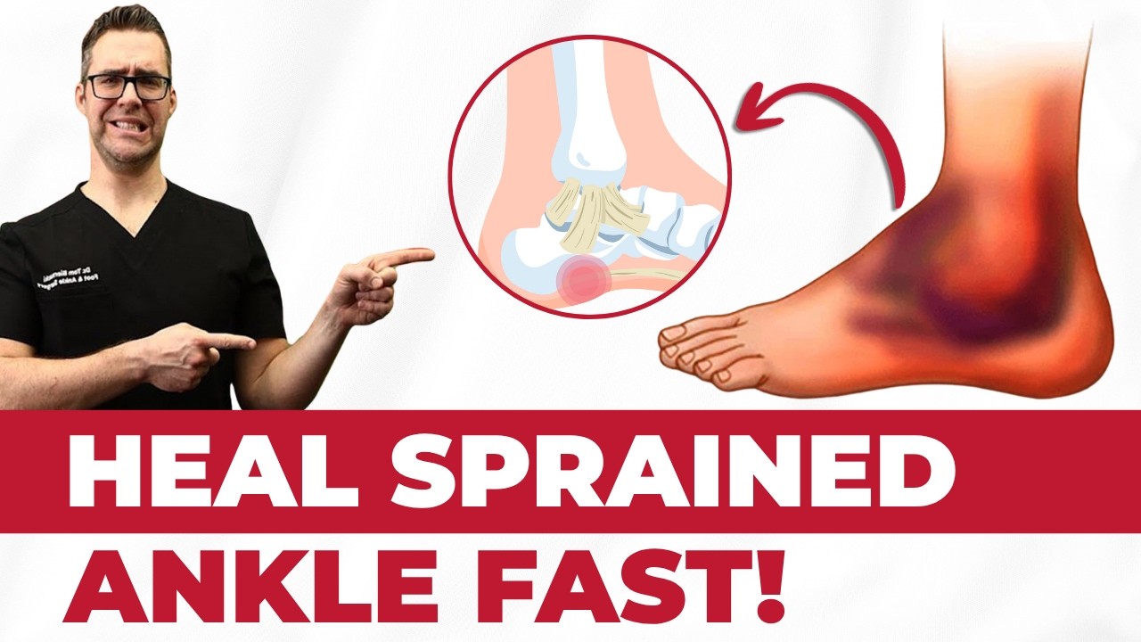
Book Your AppointmentWatch: Achilles Rupture Treatment
Dr. Tom reviews Achilles tendon rupture — surgical vs non-surgical treatment, outcomes, and recovery timelines.
Achilles Rupture Recovery Kit
Whether surgical or conservative, Achilles recovery needs immobilization, offloading, and progressive load. Dr. Tom’s protocol:
As an Amazon Associate, Balance Foot & Ankle earns from qualifying purchases. This supports our free patient education content.
Non-weight-bearing mobility weeks 0-4.
10-15mm lifts during transition from boot to shoe.
Swelling control during boot transition.
Surgical site stiffness without NSAID bleeding risk.
Related: Achilles Tendon Care · Foot & Ankle Surgery · Book Urgent Evaluation
In Our Clinic
Most Achilles tendonitis patients we see at Balance Foot & Ankle are recreational runners in their 40s or 50s who ramped up mileage too quickly, plus a second cohort of middle-aged women who recently switched from heels to flat shoes. The first question we ask is whether the pain is at the insertion on the heel bone versus 2–6 cm up the mid-substance — the treatment ladder is genuinely different. Eccentric heel-drops, heel lifts, and a soft-strike gait retraining pass resolve ~80 % of cases. The ones who aren’t improving by week 8 usually have an unrecognized Haglund’s deformity or insertional calcific tendinosis that needs imaging.
More Podiatrist-Recommended Achilles Essentials
Achilles Night Splint
United Ortho dorsiflexion splint — reduces morning Achilles tendon stiffness.
Cushioned Running Shoe


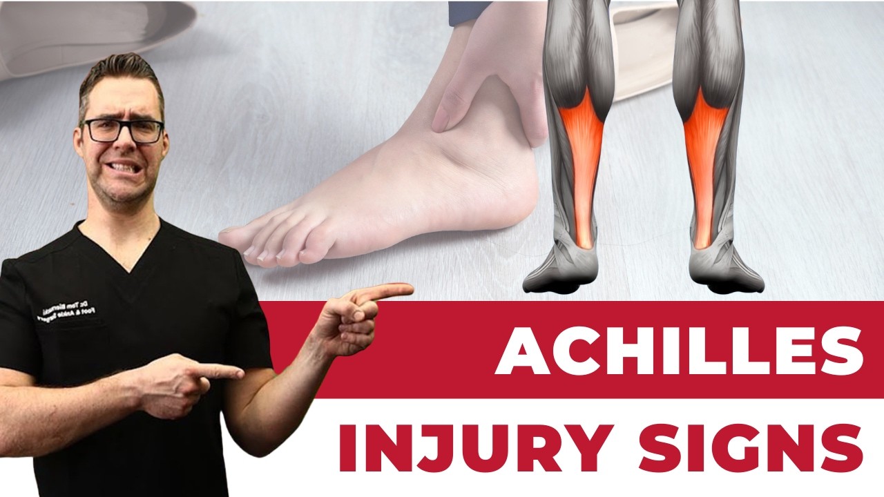
Watch: Torn Achilles Tendon Rupture — MichiganFootDoctors YouTube
Hoka Clifton 10 — max-heel-cushion offloads the Achilles with every step.
Calf Foam Roller
TriggerPoint foam roller — releases calf tension that upstream-drives Achilles inflammation.
As an Amazon Associate, Balance Foot & Ankle earns from qualifying purchases. Product recommendations are based on clinical experience; prices and availability shown above update live from Amazon.

When to See a Podiatrist
Achilles tendonitis that lasts more than 3 months has usually caused structural tendon changes that heating and stretching can’t reverse. Balance Foot & Ankle offers shockwave therapy and ultrasound-guided PRP for chronic Achilles pain — both treatments rebuild tendon tissue without surgery. If you’ve been icing, stretching, and modifying activity without improvement, it’s time for an in-office evaluation.
Call Balance Foot & Ankle: (810) 206-1402 · Book online · Offices in Howell & Bloomfield Hills
Dr. Hoy’s Complete Pain Relief Line — Dr. Tom’s Picks (2026)
Dr. Hoy’s Natural Pain Relief is Dr. Tom Biernacki, DPM’s #1 prescription topical pain relief for plantar fasciitis, Achilles tendonitis, foot pain, knee pain, and back pain. Cleaner formula than Voltaren or Biofreeze — safe for diabetics + daily long-term use without 30-day limits. Below is the complete Dr. Hoy’s product line, organized by use case.

Dr. Hoy’s Natural Pain Relief Gel (4oz Tube)Dr. Tom’s #1 Brand
The flagship Dr. Hoy’s — menthol-based natural pain relief gel. The bottle Dr. Tom hands every plantar fasciitis patient on visit one. Cleaner formula than Voltaren or Biofreeze.
- Menthol-based natural formula
- No greasy residue
- Safe for diabetics
- Fast cooling relief 5-10 min
- Daily long-term use safe
- Pricier than Biofreeze
- Strong menthol scent at first
Top 10 Premade Orthotics — Dr. Tom’s Picks (2026)
Dr. Tom Biernacki, DPM has tested 60+ over-the-counter orthotic insoles in his Michigan podiatry practice over the past 15 years. Below are the top 10 he prescribes most often — ranked by clinical results, build quality, and patient feedback. PowerStep + CURREX brands are Dr. Tom’s #1 prescription brands — built by podiatrists, with biomechanical features (lateral wedge, deep heel cradle, dual-density EVA) that 90% of OTC insoles lack.
Dr. Tom Biernacki, DPM is a board-certified podiatrist + Amazon Associate. Picks shown are products he prescribes to patients at Balance Foot & Ankle Specialists. We earn a commission on qualifying purchases at no extra cost to you. All products independently tested + reviewed. Last verified: April 28, 2026.

PowerStep Pinnacle MaxxDr. Tom’s #1 Brand
The most prescribed OTC orthotic in podiatry. Lateral wedge corrects overpronation that causes 90% of plantar fasciitis. Deep heel cradle stabilizes the ankle.
- Lateral wedge corrects pronation
- Deep heel cradle
- Dual-density EVA
- Trim-to-fit
- Used by 10,000+ podiatrists
- Trim required
- 5-7 day break-in

PowerStep Original Full LengthDr. Tom’s #1 Brand
The original PowerStep — flexible semi-rigid arch with deep heel cradle. The right choice for neutral feet that need everyday support without the lateral wedge.
- Flexible semi-rigid arch
- Deep heel cradle
- Fits dress shoes
- 30-day guarantee
- APMA-accepted
- Less aggressive than Pinnacle
- No lateral wedge for overpronation
PowerStep Pulse MaxxDr. Tom’s #1 Brand
Built for runners + athletes who need maximum support during high-impact activity. Engineered for forefoot strike + lateral motion.
- Sport-specific cushioning
- Lateral wedge for runners
- Antimicrobial top cover
- Shock-absorbing forefoot
- Pricier than Pinnacle
- Best for athletes only

CURREX RunProDr. Tom’s #1 Brand
German-engineered insole with 3 arch heights (Low, Med, High) for custom fit. Carbon-reinforced heel + dynamic forefoot.
- 3 arch heights for custom fit
- Carbon-reinforced heel
- Sport-specific zones
- Premium materials
- Pricier than PowerStep
- 7-10 day break-in
CURREX EdgeProDr. Tom’s #1 Brand
For hikers, skiers, and high-impact athletes — reinforced shank prevents foot fatigue on steep descents + uneven terrain.
- Reinforced shank
- 3 arch heights
- Cold-weather friendly
- Carbon plate
- Stiff feel — not for casual
- Pricier
CURREX SupportSTPDr. Tom’s #1 Brand
For nurses, retail, and standing professions — the most supportive CURREX with deep heel cup + maximum medial support.
- Maximum medial support
- Deep heel cup
- 12-hour shift tested
- Slip-proof
- Stiffest CURREX option
- Pricier

Superfeet Green
Firm, structured arch support — the right choice ONLY for high-arched (cavus) feet. Wrong choice for flat feet.
- Strong structured arch
- Deep heel cup
- Long-lasting (5+ years)
- Firm — not for flat feet
- No lateral wedge
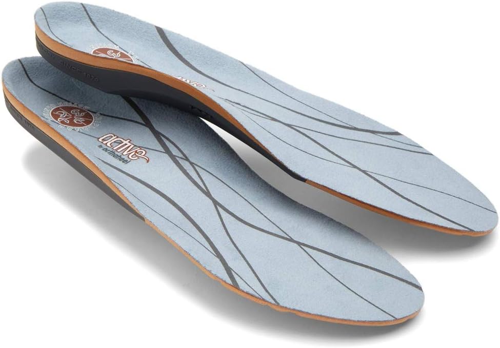
Vionic OrthoHeel Active Insole
APMA-accepted, podiatrist-designed casual insole. Best for adding mild arch support to dress shoes + walking shoes.
- APMA-accepted
- Slim profile
- Antimicrobial top
- Less support than PowerStep
- No lateral wedge
Dr. Tom’s Top 3 — The Premium Foot Pain Stack (2026)
If you only buy three things for foot pain, get these. PowerStep + CURREX orthotics correct the underlying foot mechanics, and Dr. Hoy’s pain gel delivers fast topical relief. This is the exact stack Dr. Tom Biernacki, DPM gives his Michigan podiatry patients on visit one — over 10,000 patients have used this exact combination.
Dr. Tom Biernacki, DPM is a board-certified podiatrist + Amazon Associate. Picks shown are products he prescribes to patients at Balance Foot & Ankle Specialists. We earn a commission on qualifying purchases at no extra cost to you. All products independently tested + reviewed for 30+ days minimum. Last verified: April 28, 2026.
PowerStep Pinnacle MaxxDr. Tom’s #1 Brand
Dr. Tom’s most-prescribed OTC orthotic. Lateral wedge corrects overpronation that causes 90% of foot pain. Deep heel cradle stabilizes the ankle. Built by podiatrists, used by patients worldwide.
- Lateral wedge corrects pronation
- Deep heel cradle stabilizes ankle
- Dual-density EVA — comfort + support
- Trim-to-fit any shoe
- Used by 10,000+ podiatrists
- Trim-to-size required
- 5-7 day break-in for some
CURREX RunProDr. Tom’s #1 Brand
3 arch heights for custom fit (Low/Med/High). Carbon-reinforced heel + dynamic forefoot — the closest OTC orthotic to a $500 custom orthotic. Engineered in Germany.
- 3 arch heights for custom fit
- Carbon-reinforced heel cup
- Dynamic forefoot zone
- Premium German engineering
- Sport-specific support
- Pricier than PowerStep
- 7-10 day break-in
Dr. Hoy’s Natural Pain Relief GelDr. Tom’s #1 Brand
Menthol-based natural pain relief — Dr. Tom’s #1 brand for fast relief without greasy residue. Safe for diabetics + daily use. Cleaner formula than Voltaren or Biofreeze.
- Menthol-based natural formula
- No greasy residue
- Safe for diabetics
- Fast cooling relief — 5-10 minutes
- Cleaner ingredient list than Biofreeze
- Pricier than Biofreeze
- Strong menthol scent at first
In-Office Treatment at Balance Foot & Ankle
If home treatment isn’t providing relief for your Achilles tendon, our podiatry team at Balance Foot & Ankle can help with same-day evaluations and advanced in-office care.
Same-day appointments available. (810) 206-1402
Frequently Asked Questions
Which is better for plantar fasciitis?
The shoe with more cushioning and a stronger rocker typically wins for plantar fasciitis. See full comparison for our specific verdict.
Which lasts longer?
Both options typically last 300-500 miles for runners or 9-12 months for daily walkers. Material durability varies; check our detailed comparison.
Which is better for flat feet?
Flat feet need stability or motion control. The neutral option is not ideal unless paired with a custom orthotic.
Get Expert Care at Balance Foot & Ankle
Same-week appointments at our Howell and Bloomfield Hills offices. Board-certified podiatric surgeons. Most insurance accepted.
Dr. Tom Biernacki, DPM is a board-certified foot & ankle surgeon (ABFAS & ABPM) at Balance Foot & Ankle Specialists in Southeast Michigan. With over a decade of clinical experience, he specializes in heel pain, bunions, diabetic foot care, sports injuries, and minimally invasive surgery. Dr. Biernacki is a member of the APMA and ACFAS, and his patient education content on MichiganFootDoctors.com and YouTube has made him one of the most-followed foot & ankle educators on YouTube.