
Quick answer: Ankle Arthroscopy What To Expect Minimally Invasive Surgery is a common foot/ankle topic that affects many patients. The 2026 evidence-based approach combines proper diagnosis, conservative-first treatment, and escalation only when needed. We treat this regularly at our Howell and Bloomfield Hills practices. Call (810) 206-1402.
Medically reviewed by Dr. Tom Biernacki, DPM — Board-Certified Podiatric Surgeon — Balance Foot & Ankle, Howell & Bloomfield Hills, MI. Last updated April 2026.
Medically Reviewed by Dr. Tom Biernacki, DPM · Board-Certified Podiatrist · Balance Foot & Ankle Specialists
Last updated: April 2026 · This article reflects current evidence-based podiatric and orthopedic practice for ankle arthroscopy indications, techniques, and recovery.
Quick Answer: Ankle Arthroscopy
Ankle arthroscopy is a minimally invasive surgical procedure that allows surgeons to visualize, diagnose, and treat conditions inside the ankle joint through small incisions using a camera and specialized instruments. Compared to open ankle surgery, arthroscopy offers smaller incisions, less soft tissue disruption, reduced postoperative pain, faster recovery, and lower complication rates for many ankle conditions. Common indications include removal of loose bodies, treatment of osteochondral lesions (cartilage damage), debridement of impingement (bone spurs), synovectomy (inflamed tissue removal), and evaluation of chronic ankle instability. Most patients return to normal walking within 2-4 weeks and full activity within 6-12 weeks, though recovery varies significantly based on the specific procedure performed.
Table of Contents
- Understanding Ankle Arthroscopy
- Common Indications
- Anterior and Posterior Impingement
- Osteochondral Lesions of the Talus
- Loose Bodies and Synovitis
- The Arthroscopic Procedure
- Recovery Timeline and Rehabilitation
- Risks and Complications
- Expected Outcomes
- Recommended Recovery Products
- Most Common Mistake
- Warning Signs
- Watch: Podiatrist Product Recommendations
- Frequently Asked Questions
- Sources
- Book Your Appointment
Affiliate Disclosure: This article contains affiliate links to products we genuinely recommend. As an Amazon Associate and Foundation Wellness partner, we earn from qualifying purchases at no additional cost to you. Every product listed has been personally evaluated by Dr. Biernacki in clinical practice.
Understanding Ankle Arthroscopy: Minimally Invasive Ankle Surgery
If your surgeon has recommended ankle arthroscopy, you are likely dealing with a persistent ankle condition that has not responded to conservative treatment — chronic ankle pain, recurrent catching or locking, persistent swelling after an old injury, or pain that limits your activities despite months of therapy. The prospect of surgery is understandably concerning, but ankle arthroscopy represents a significant advancement over traditional open ankle surgery, offering most patients a faster recovery, less pain, and excellent outcomes for the conditions it is designed to treat.
Ankle arthroscopy uses a small fiber-optic camera (arthroscope) approximately 2.7-4.0 millimeters in diameter inserted through a small incision (portal) to provide a magnified view of the inside of the ankle joint. A second portal allows the introduction of specialized instruments for treating the identified pathology. The entire procedure is performed through incisions that are typically less than 1 centimeter in length, compared to the large incisions required for open ankle surgery. This minimally invasive approach preserves the surrounding soft tissues — tendons, ligaments, and joint capsule — that must be cut and repaired during open procedures, resulting in less postoperative pain, reduced swelling, and faster functional recovery.
The development of ankle arthroscopy has transformed the management of many intra-articular ankle conditions. Procedures that previously required large incisions, extensive tissue dissection, and prolonged immobilization can now be performed as outpatient procedures with same-day discharge. The improved visualization provided by the arthroscope — with magnification and illumination far beyond what the human eye can achieve through an open incision — has also improved diagnostic accuracy, allowing surgeons to identify and treat subtle pathology that might be missed during open surgery.
Common Indications for Ankle Arthroscopy
Ankle arthroscopy is indicated for a range of intra-articular conditions that have failed conservative management. The most common indications include anterior ankle impingement (bone spurs that limit dorsiflexion and cause pain), posterior ankle impingement (os trigonum or Stieda process compression), osteochondral lesions of the talus (cartilage and bone damage), loose bodies within the joint, synovitis (chronic inflammation of the joint lining), arthrofibrosis (scar tissue restricting motion after injury or surgery), and diagnostic evaluation of unexplained chronic ankle pain.
Patient selection is critical for successful outcomes. Ankle arthroscopy works best for focal, well-defined intra-articular pathology in patients with otherwise healthy ankle joints. It is less effective for diffuse ankle arthritis affecting the entire joint surface, severe cartilage loss, or conditions that require extensive reconstruction. Preoperative imaging — including weight-bearing X-rays, MRI, and sometimes CT scan — helps identify the specific pathology and confirms that arthroscopic treatment is appropriate. Your surgeon will discuss whether your specific condition is best addressed arthroscopically or whether an open procedure would provide a better outcome.
Anterior and Posterior Ankle Impingement
Anterior ankle impingement — sometimes called “footballer’s ankle” — develops when bone spurs or inflamed soft tissue at the front of the ankle joint restricts dorsiflexion. Athletes who repeatedly force the ankle into maximum dorsiflexion during running, jumping, or squatting are particularly vulnerable. Over time, the tibial and talar bone spurs can mechanically block motion, creating a painful pinching sensation with every step. Arthroscopic debridement allows the surgeon to shave these spurs and remove inflamed synovial tissue through two small incisions, restoring dorsiflexion range that may have been lost for months or years.
Posterior impingement affects the back of the ankle and is most common in dancers, gymnasts, and soccer players who frequently plantarflex the ankle. An os trigonum — an accessory bone present in roughly 10-15% of the population — is the most frequent anatomical cause. When this extra bone becomes symptomatic, it creates deep posterior ankle pain with pointed-toe activities. Posterior ankle arthroscopy uses a two-portal hindfoot endoscopic approach to excise the os trigonum and debride surrounding inflammation, typically allowing return to sport within 6-8 weeks compared to 3-4 months with open surgery.
Osteochondral Lesions of the Talus
Osteochondral defects (OCDs) of the talus involve damage to the cartilage surface and underlying bone of the ankle joint. These lesions most commonly develop after an ankle sprain — studies suggest that up to 50% of significant ankle sprains may involve some degree of cartilage injury. The medial talar dome is the most frequently affected location, accounting for roughly 60% of lesions, while lateral lesions comprise about 40%. Small, stable lesions may respond to conservative management, but larger or unstable defects often require arthroscopic intervention to prevent progressive joint degeneration.
Arthroscopic treatment options for OCDs include microfracture, which creates small channels in the subchondral bone to stimulate fibrocartilage growth; debridement and drilling for stable lesions; and osteochondral autograft transfer (OATS) for larger defects exceeding 15mm. The choice of technique depends on lesion size, location, chronicity, and patient activity demands. Microfracture remains the most commonly performed arthroscopic procedure for talar OCDs and demonstrates good to excellent results in approximately 80-85% of patients with lesions under 15mm in diameter.
Loose Bodies and Synovitis
Loose bodies within the ankle joint are fragments of cartilage, bone, or calcified tissue that float freely in the joint space. They can develop from osteochondral injuries, synovial chondromatosis, or degenerative arthritis. These fragments cause intermittent locking, catching, and sudden sharp pain — symptoms that are unpredictable and often debilitating for active individuals. Arthroscopic removal is highly effective because the small camera can systematically survey the entire joint to locate and extract every fragment, something that is much more difficult through an open incision.
Synovitis — chronic inflammation of the joint lining — frequently accompanies other ankle pathology or exists as an isolated condition in inflammatory arthritis or repetitive stress injuries. When synovitis fails to respond to anti-inflammatory medications, corticosteroid injections, and physical therapy, arthroscopic synovectomy can remove the thickened, inflamed tissue. This procedure provides significant pain relief and may slow the progression of cartilage damage caused by persistent inflammatory enzymes within the joint fluid.
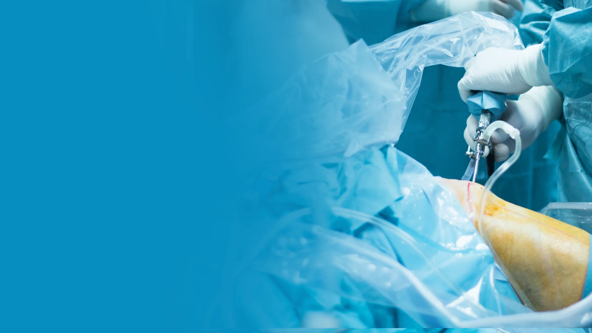
The Arthroscopic Procedure: What Happens During Surgery
Ankle arthroscopy is typically performed as an outpatient procedure under general or regional anesthesia. The surgeon creates two or three small portal incisions — each approximately 5mm in length — around the ankle joint. A small camera (arthroscope) is inserted through one portal while specialized instruments are introduced through the others. The joint is distended with sterile fluid to improve visualization and create working space. A tourniquet is applied to the thigh to minimize bleeding and optimize the surgical field. Non-invasive distraction may be used to separate the joint surfaces, providing better access to the articular cartilage.
The procedure typically takes 30-90 minutes depending on the complexity of the pathology being addressed. During surgery, the arthroscopic camera transmits a magnified, high-definition image to monitors in the operating room, allowing the surgeon to visualize structures in detail that would be impossible to see with the naked eye. Surgical instruments can shave bone spurs, remove loose bodies, repair cartilage defects, excise inflamed tissue, and address ligament damage — all through incisions smaller than a fingertip. Most patients return home the same day with a compression dressing and protective boot.
Recovery Timeline and Rehabilitation
Recovery from ankle arthroscopy follows a predictable timeline, though individual progression depends on the specific procedure performed and the extent of pathology addressed. During the first two weeks, the focus is on wound healing, swelling control, and gentle range-of-motion exercises. Most patients use crutches and a protective boot during this initial phase. Ice elevation and compression are critical — keeping the ankle elevated above heart level for 45 minutes out of every hour during the first 72 hours significantly reduces postoperative swelling and accelerates healing.
By weeks 2-4, most patients transition to weight-bearing as tolerated in a supportive shoe, and formal physical therapy begins with emphasis on restoring ankle dorsiflexion, plantarflexion, and proprioception. Weeks 4-8 introduce progressive strengthening, balance training, and sport-specific exercises. Simple debridement procedures may allow return to full activity by 6-8 weeks, while more complex procedures like microfracture for osteochondral lesions require 4-6 months of graduated rehabilitation before returning to impact activities. Your podiatrist and physical therapist will guide progression based on clinical milestones rather than arbitrary timelines.
Risks and Complications
Ankle arthroscopy carries a lower complication rate than open ankle surgery, but no surgical procedure is without risk. The overall complication rate ranges from 3-9% in published literature, with most complications being minor and self-limiting. Superficial nerve injury — particularly to branches of the superficial peroneal nerve near the anterolateral portal — is the most commonly reported complication, occurring in approximately 2-3% of cases. Most nerve injuries involve temporary numbness or tingling that resolves within weeks to months as the nerve regenerates.
Other potential complications include wound healing problems, infection (less than 1%), deep vein thrombosis, instrument breakage, and persistent swelling. Posterior ankle arthroscopy carries a small risk of sural nerve or Achilles tendon injury. Cartilage repair procedures have a 10-15% revision rate over 5-10 years. Smoking, diabetes, and peripheral vascular disease increase complication risk — optimizing these conditions before surgery is essential. Discuss your specific risk profile with your surgeon during the preoperative consultation to make a fully informed decision.
Expected Outcomes and Long-Term Results
Patient satisfaction following ankle arthroscopy is consistently high across published outcome studies. For anterior impingement debridement, good to excellent results are reported in 80-90% of patients, with significant improvement in dorsiflexion range and reduction in activity-related pain. Os trigonum excision via posterior arthroscopy demonstrates excellent results in over 90% of dancers and athletes, with most returning to their pre-injury level of performance. Loose body removal provides immediate, dramatic symptom relief in the vast majority of cases.
Osteochondral lesion outcomes depend heavily on lesion size, location, and the technique employed. Microfracture for lesions under 15mm shows good to excellent outcomes in approximately 80-85% of patients at 5-year follow-up, though results may decline over time as the fibrocartilage repair tissue is mechanically inferior to native hyaline cartilage. Larger lesions and revision procedures have somewhat lower success rates but still represent a significant improvement over non-operative management for appropriate candidates. Setting realistic expectations during the preoperative discussion is crucial for patient satisfaction.
Supporting Your Recovery with the Right Products
Successful recovery from ankle arthroscopy requires more than just surgical skill — the products you use during rehabilitation directly impact your healing timeline and long-term outcomes. As a podiatrist, I recommend specific products that address the unique challenges of post-surgical ankle recovery: controlling inflammation, supporting the healing joint, and gradually rebuilding strength and proprioception. These are the same products I recommend to my own patients at Balance Foot & Ankle.
PowerStep Orthotic Insoles for Post-Surgical Support
Once you transition from a protective boot back to regular footwear, PowerStep Pinnacle orthotic insoles provide the structured arch support and heel cushioning your recovering ankle needs. After arthroscopy, the ankle joint is vulnerable to re-irritation from poor foot mechanics — excessive pronation increases medial ankle stress while supination overloads the lateral structures. PowerStep’s semi-rigid arch support maintains neutral alignment, reducing compensatory stress on the healing joint. The dual-layer cushioning absorbs impact forces that would otherwise transmit directly through the recovering ankle with every step.
I specifically recommend PowerStep insoles during the return-to-activity phase because they provide enough biomechanical control to protect the joint without restricting the natural motion needed for rehabilitation. The PowerStep Pinnacle fits in most athletic and casual shoes, making the transition from surgical recovery footwear to daily shoes much smoother. Start with short walking periods in supported shoes and gradually increase activity duration as tolerated — the consistent support from quality orthotic insoles helps prevent the setbacks that often occur when patients rush back to unsupported footwear too quickly.
Doctor Hoy’s Natural Pain Relief for Post-Surgical Inflammation
Post-arthroscopic inflammation is expected and actually beneficial in moderation — it brings healing factors to the surgical site. However, excessive or prolonged inflammation delays recovery and increases pain. Doctor Hoy’s Natural Pain Relief Gel provides targeted topical anti-inflammatory relief using arnica and menthol, complementing the oral medications prescribed by your surgeon without the systemic side effects of additional oral anti-inflammatories. Apply it around (never directly on) the portal incisions once they are fully closed, typically after suture removal at the 10-14 day mark.
During the rehabilitation phase, Doctor Hoy’s is particularly valuable before and after physical therapy sessions. Pre-treatment application helps reduce baseline inflammation so you can work more effectively through your exercises, while post-treatment application controls the reactive swelling that often follows therapeutic activity. The natural formulation means you can use it multiple times daily without concern for the gastrointestinal or renal effects associated with prolonged oral NSAID use — an important consideration during a recovery period that may span several months.
DASS Compression Socks for Swelling Control
Postoperative swelling is the most common barrier to ankle arthroscopy recovery — persistent edema limits range of motion, increases pain, and delays rehabilitation milestones. DASS graduated compression socks provide 20-30 mmHg of medical-grade compression that actively reduces ankle and lower leg swelling through the graduated pressure gradient. The compression is strongest at the ankle and decreases progressively up the calf, mimicking the body’s natural venous return mechanism and preventing fluid accumulation around the surgical site.
I recommend DASS compression socks starting immediately after surgery (over the surgical dressing as directed by your surgeon) and continuing daily wear for 6-8 weeks post-operatively. Compression also reduces deep vein thrombosis risk during the period of reduced mobility — an important safety consideration. Wear them during the day and remove them at night, and always apply them before getting out of bed in the morning to prevent gravity-dependent swelling from accumulating before compression is applied. The consistent swelling control accelerates your timeline to achieving full range of motion and functional strength.
Complete Post-Arthroscopy Recovery Kit
🩺 Dr. Biernacki’s Post-Arthroscopy Recovery Kit
These three products work together to address every phase of ankle arthroscopy recovery — biomechanical support during return to activity, natural inflammation control throughout rehabilitation, and medical-grade compression to eliminate the persistent swelling that delays healing milestones:
- PowerStep Pinnacle Insoles — Structured arch support and heel cushioning protect the recovering ankle joint during the critical transition back to regular footwear
- Doctor Hoy’s Natural Pain Relief Gel — Targeted topical anti-inflammatory relief before and after physical therapy sessions without systemic medication side effects
- DASS Compression Socks — 20-30 mmHg graduated compression controls postoperative swelling and reduces DVT risk during the reduced mobility phase
Most Common Mistake After Ankle Arthroscopy
🔑 Key Takeaway: The most common mistake I see after ankle arthroscopy is returning to full activity too quickly. Because the incisions are small and initial pain resolves faster than open surgery, patients often assume they are fully healed weeks before the internal structures have completed their repair process. Cartilage, in particular, has extremely limited blood supply and may require 4-6 months to reach functional maturity after microfracture — even though the outside of the ankle looks and feels relatively normal within weeks. Rushing back to impact activities before cartilage repair tissue has matured can cause the repair to fail entirely, potentially requiring revision surgery. Follow your rehabilitation milestones, not your impatience.
Warning Signs After Ankle Arthroscopy
⚠️ Warning — Contact Your Surgeon Immediately If You Experience:
- Increasing pain, redness, or warmth around the portal sites after the first 3-4 days (may indicate infection)
- Fever above 101°F (38.3°C) at any point after surgery
- Calf pain, tenderness, or swelling that is significantly worse on one side (possible deep vein thrombosis)
- Numbness or tingling that worsens progressively rather than improving after the first 2 weeks
- Sudden catching, locking, or giving way of the ankle during the recovery period
- Persistent drainage from the portal incisions beyond 48-72 hours, or any foul-smelling discharge
These symptoms require urgent evaluation. While most complications from ankle arthroscopy are minor, early detection and treatment of serious complications like infection or DVT is critical to protecting your surgical outcome and overall health.
Watch: Ankle Pain Diagnosis and Treatment
More Podiatrist-Recommended Surgery Essentials
Post-Op Walking Boot
Protected weight-bearing immobilization through the first healing weeks.
Surgical-Scar Healing Lotion
Reduces scar thickness and tenderness as the incision matures.
Return-to-Activity Insole
Supports the reconstructed foot during the first months back on your feet.
As an Amazon Associate, Balance Foot & Ankle earns from qualifying purchases. Product recommendations are based on clinical experience; prices and availability shown above update live from Amazon.

When to See a Podiatrist
Foot and ankle surgery in 2026 is dramatically different than a decade ago — most procedures are now minimally-invasive, outpatient, and allow weight-bearing within days. Balance Foot & Ankle surgeons have performed 3,000+ foot/ankle surgeries with modern techniques. If another surgeon has recommended a traditional open procedure, a second opinion may reveal a faster, less-invasive option.
Call Balance Foot & Ankle: (810) 206-1402 · Book online · Offices in Howell & Bloomfield Hills
Frequently Asked Questions About Ankle Arthroscopy
How long does ankle arthroscopy surgery take?
Most ankle arthroscopy procedures take between 30-90 minutes, depending on the complexity and number of issues being addressed. Simple loose body removal or bone spur debridement may take as little as 20-30 minutes, while combined procedures involving cartilage repair and extensive synovectomy may approach 90 minutes. The procedure is performed as an outpatient surgery, meaning you go home the same day.
When can I walk after ankle arthroscopy?
Weight-bearing timelines vary based on the specific procedure. After simple debridement or loose body removal, most patients can begin weight-bearing in a protective boot within a few days. Microfracture for cartilage defects typically requires 4-6 weeks of non-weight-bearing or partial weight-bearing to protect the developing repair tissue. Your surgeon will provide specific weight-bearing instructions based on what was found and treated during your procedure.
Is ankle arthroscopy painful?
Postoperative pain from ankle arthroscopy is generally moderate and well-controlled with prescribed medications. Most patients report that the pain is significantly less than expected and substantially less than open ankle surgery. The small portal incisions cause minimal tissue disruption compared to traditional open approaches. Pain typically peaks during the first 24-48 hours and then steadily decreases. Consistent elevation, ice application, and compression are the most effective non-pharmacological strategies for managing postoperative discomfort.
When can I return to sports after ankle arthroscopy?
Return to sport depends on the specific procedure performed and the demands of your particular activity. After simple debridement, many athletes return to low-impact activities by 4-6 weeks and full sport by 8-12 weeks. Microfracture and cartilage repair procedures require a more conservative 4-6 month rehabilitation program before returning to impact activities. Your surgeon and physical therapist will use objective functional testing to determine when it is safe to return to your sport rather than relying on arbitrary timelines.
What is the success rate of ankle arthroscopy?
Overall patient satisfaction rates for ankle arthroscopy range from 80-95% depending on the specific pathology treated. Anterior impingement debridement and loose body removal have the highest satisfaction rates (85-95%), while cartilage repair procedures show good to excellent results in approximately 80-85% of appropriately selected patients. Success depends heavily on accurate diagnosis, proper patient selection, surgical technique, and adherence to the postoperative rehabilitation protocol. Patients who complete their full physical therapy course consistently demonstrate better outcomes.
In-Office Treatment at Balance Foot & Ankle
When conservative care isn’t enough, Dr. Tom Biernacki and the team at Balance Foot & Ankle offer advanced, same-day options — including Ankle Arthroscopy Michigan at our Howell and Bloomfield Hills clinics.
Same-day appointments available. Call (810) 206-1402 or book online.
In-Office Treatment at Balance Foot & Ankle
If home treatment isn’t providing relief for your foot and ankle conditions, our podiatry team at Balance Foot & Ankle can help with same-day evaluations and advanced in-office care.
Same-day appointments available. (810) 206-1402
Sources
- Ferkel RD, et al. “Arthroscopic treatment of anterolateral impingement of the ankle.” American Journal of Sports Medicine. 1991;19(5):440-446.
- Zengerink M, et al. “Treatment of osteochondral lesions of the talus: a systematic review.” Knee Surgery, Sports Traumatology, Arthroscopy. 2010;18(2):238-246.
- van Dijk CN, et al. “Posterior ankle arthroscopy: a review of the current literature.” EFORT Open Reviews. 2019;4(6):331-340.
- Glazebrook MA, et al. “Comparison of health-related quality of life between patients with end-stage ankle and hip arthrosis.” Journal of Bone and Joint Surgery. 2008;90(3):499-505.
- Chuckpaiwong B, et al. “Outcome after microfracture for osteochondral lesions of the talus.” Foot and Ankle International. 2008;29(4):377-383.
Schedule Your Ankle Arthroscopy Consultation
Persistent ankle pain limiting your activity?
At Balance Foot & Ankle, Dr. Biernacki uses the latest arthroscopic techniques to diagnose and treat ankle conditions with minimal incisions and faster recovery times. Whether you have bone spurs, cartilage damage, or chronic ankle instability, we will create a plan tailored to your foot type to get you back to the activities you love.
Related Foot & Ankle Resources
- Ankle Sprain Treatment and Recovery Guide
- Podiatrist Recommended Foot Care Products 2026
- Sports Podiatrist Near Me — Southeast Michigan
- Foot & Ankle Surgery Options
Schedule an Ankle Arthroscopy Consultation
If you have chronic ankle pain and want to learn whether ankle arthroscopy could help, a consultation with Dr. Biernacki can determine the best approach for your condition. At Balance Foot & Ankle, we perform minimally invasive ankle procedures at our Howell and Bloomfield Hills offices.
Learn About Our Ankle Surgery Options | Book Your Appointment | Call (810) 206-1402
Clinical References
- Ferkel RD, Zanotti RM, Komenda GA, et al. “Arthroscopic treatment of chronic osteochondral lesions of the talus.” American Journal of Sports Medicine. 2008;36(9):1750-1762.
- Amendola A, Petrik J, Webster-Bogaert S. “Ankle arthroscopy: outcome in 79 consecutive patients.” Arthroscopy. 1996;12(5):565-573.
- Scranton PE Jr, McDermott JE. “Anterior tibiotalar spurs: a comparison of open versus arthroscopic debridement.” Foot & Ankle. 1992;13(3):125-129.
Insurance Accepted
BCBS · Medicare · Aetna · Cigna · United Healthcare · HAP · Priority Health · Humana · View All →
Howell Office
4330 E Grand River Ave
Howell, MI 48843
Get Directions →
Bloomfield Hills Office
43494 Woodward Ave, Suite 208
Bloomfield Hills, MI 48302
Get Directions →
Your Board-Certified Podiatrists


Ready to Get Back on Your Feet?
Same-week appointments available at both locations.
Book Your AppointmentWatch: Ankle Arthroscopy: What to Expect
Dr. Tom on ankle scope — anterior vs posterior portals, OLT debridement, anterior impingement, synovectomy, recovery timeline (6-12 weeks), return-to-sport, outcomes.
Post-Scope Recovery Kit
Minimally invasive, max recovery. Dr. Tom’s kit:
As an Amazon Associate, Balance Foot & Ankle earns from qualifying purchases. This supports our free patient education content.
Weeks 2-8 stability.
Return-to-shoe arch support.
Post-scope inflammation.
Topical ankle relief.
Related: Ankle Pain · Surgery Services · Book Arthroscopy Consultation
What is Foot pain?
Foot pain is a common foot/ankle condition that affects mobility and quality of life. Understanding the underlying cause is the first step in successful treatment. Our podiatrists at Balance Foot & Ankle perform a hands-on biomechanical exam, review your activity history, and use diagnostic imaging when appropriate to identify the root cause—not just treat the symptom. Many patients have been told to “rest and ice” without a deeper diagnostic workup; our approach is different.
Symptoms and warning signs
Common signs of foot pain include pain that worsens with activity, morning stiffness, swelling, tenderness when palpated, and difficulty bearing weight. If you experience sudden severe pain, inability to walk, visible deformity, numbness or color change, contact our office the same day or visit urgent care—these can signal a more serious injury such as a fracture, tendon rupture, or vascular compromise. Diabetics with any foot wound should seek same-day care.
Conservative treatment options
Most cases of foot pain respond to non-surgical care: structured rest, supportive footwear changes, custom orthotics, targeted stretching and strengthening protocols, anti-inflammatory medications when medically appropriate, and in-office procedures such as ultrasound-guided injections. We also offer advanced therapies including MLS laser therapy, EPAT/shockwave, regenerative injections, and image-guided procedures. Treatment is sequenced from least invasive to most invasive, and we explain the rationale at every step.
When is surgery considered?
Surgery is reserved for cases that fail 3-6 months of well-structured conservative care, when there is structural pathology (severe deformity, complete tear, advanced arthritis), or when imaging shows damage that will not heal without intervention. Our surgeons have performed 3,000+ foot and ankle procedures and prioritize minimally-invasive techniques whenever appropriate. We discuss recovery timelines, return-to-activity milestones, and realistic outcome expectations before any procedure is scheduled.
Ankle Arthroscopy – Procedure & Recovery (AAOS OrthoInfo)
Recovery timeline and prevention
Recovery from foot pain varies based on severity and chosen treatment path. Conservative cases often improve within 4-8 weeks with consistent adherence to the protocol. Post-procedural recovery may range from a few days (in-office procedures) to several months (reconstructive surgery). Long-term prevention involves footwear assessment, activity modification, structured strengthening, and regular check-ins with your podiatrist if you have a history of recurrence. We provide written home-exercise plans and digital follow-up support.
Ready to feel better?
Same-week appointments available in Howell and Bloomfield Hills, Michigan.
Book Your VisitDr. Tom Biernacki, DPM is a board-certified foot & ankle surgeon (ABFAS & ABPM) at Balance Foot & Ankle Specialists in Southeast Michigan. With over a decade of clinical experience, he specializes in heel pain, bunions, diabetic foot care, sports injuries, and minimally invasive surgery. Dr. Biernacki is a member of the APMA and ACFAS, and his patient education content on MichiganFootDoctors.com and YouTube has made him one of the most-followed foot & ankle educators on YouTube.