
Medically reviewed by Dr. Tom Biernacki, DPM · Board-Certified Podiatric Surgeon · Last reviewed: April 2026 · Editorial Policy
The most important clinical decision with Arthroscopic Ankle Surgery Indications Portals Procedures isn’t which treatment to start with — it’s identifying the correct subtype. That changes everything. Call (810) 206-1402.
Quick Answer
Arthroscopic Ankle Surgery: Indications, Portal Anatomy, and relates to foot pain — typically caused by overuse, footwear, or biomechanics. Most patients improve in 6-12 weeks with conservative care. Same-week appointments in Howell + Bloomfield Township: (810) 206-1402.

Medically reviewed by Dr. Tom Biernacki, DPM — Board-certified foot & ankle surgeon, 3,000+ surgeries performed. Updated April 2026 with current clinical evidence. This article reflects real practice experience from Balance Foot & Ankle Specialists in Howell and Bloomfield Township, Michigan.
Quick Answer
Most foot and ankle problems respond to conservative care — proper footwear, supportive inserts, activity modification, and targeted stretching — within 4-8 weeks. Persistent pain beyond that window, or any symptom that prevents walking, warrants a podiatric evaluation to rule out fracture, tendon tear, or systemic cause.
Watch: Dr. Tom Biernacki, DPM

Medically Reviewed by Dr. Tom Biernacki, DPM — Board-Certified Podiatrist, Balance Foot & Ankle Specialists, Michigan. Last updated April 2026.
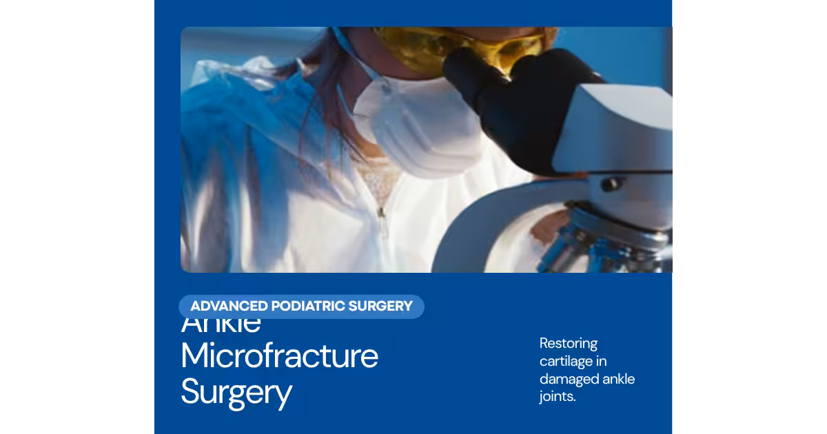
Ankle arthroscopy — insertion of a small camera into the ankle joint through two or three small portals to diagnose and treat intra-articular pathology — has transformed the management of ankle pain that was previously attributed to ‘chronic ankle sprain’ or treated with open surgery requiring weeks of non-weight-bearing recovery. Modern arthroscopic techniques allow treatment of osteochondral lesions, anterolateral impingement, synovitis, loose bodies, and ankle arthrofibrosis through portals as small as 4mm, with return to weight-bearing in days rather than weeks.
▶ Watch

Indications and Portal Anatomy
Common arthroscopic ankle diagnoses and treatments: anterolateral impingement syndrome — the scar tissue (Bassett’s ligament or anterolateral mass) formed after lateral ankle sprain that impinges against the talar dome with dorsiflexion; arthroscopic resection provides 80–90% pain relief; osteochondral lesions of the talus (OLT) — arthroscopic debridement and microfracture for small lesions; intra-articular loose bodies — removal of calcified fragments causing locking and pain; synovectomy for inflammatory arthritis; ankle arthrofibrosis — lysis of adhesions after prior surgery or prolonged immobilization. Standard portals: anteromedial portal (medial to the extensor hallucis longus, just medial to the saphenous neurovascular bundle) and anterolateral portal (just lateral to the peroneus tertius tendon, at risk for the superficial peroneal nerve) — these two portals allow visualization and instrumentation of the anterior ankle and most of the talar dome. Posterolateral portal: added for posterior ankle pathology (os trigonum, FHL tendinopathy, posterior impingement). Noninvasive ankle distraction: a strap around the heel provides 5–10mm of joint distraction to improve visualization of the talar dome and tibial plafond — allows inspection and treatment of central and posterior talar lesions without a trans-malleolar approach.
Recovery and Outcomes
Recovery: diagnostic arthroscopy — weight-bearing as tolerated immediately; anterolateral impingement resection — return to sport 6–8 weeks; OLT microfracture — non-weight-bearing 4–6 weeks, return to sport 4–6 months; arthrofibrosis lysis — aggressive physical therapy immediately after surgery is essential to prevent re-formation of adhesions. Dr. Biernacki at Balance Foot & Ankle performs ankle arthroscopy for anterolateral impingement, osteochondral lesions, and intra-articular ankle pathology. Call (810) 206-1402 at our Bloomfield Township or Howell office.
📧 Get Dr. Tom’s Free Lab Test Guide
Discover the 5 lab tests every person over 35 should ask their doctor about — explained in plain English by a board-certified physician.
📍 Located in Michigan?
Our board-certified podiatrists treat this condition at two convenient locations. Same-day appointments often available.
More Podiatrist-Recommended Surgery Essentials
OOFOS Recovery Slide


Post-op approved — impact-absorbing slide for early recovery.
HOKA Ora 3 Recovery Slide
Max-cushion recovery sandal — comfort for post-surgical swelling.
Hoka Bondi 9
Max-cushion walking shoe — ease into return-to-walking post-surgery.
As an Amazon Associate, Balance Foot & Ankle earns from qualifying purchases. Product recommendations are based on clinical experience; prices and availability shown above update live from Amazon.

When to See a Podiatrist
Foot and ankle surgery in 2026 is dramatically different than a decade ago — most procedures are now minimally-invasive, outpatient, and allow weight-bearing within days. Balance Foot & Ankle surgeons have performed 3,000+ foot/ankle surgeries with modern techniques. If another surgeon has recommended a traditional open procedure, a second opinion may reveal a faster, less-invasive option.
Call Balance Foot & Ankle: (810) 206-1402 · Book online · Offices in Howell & Bloomfield Township
Frequently Asked Questions
How do I know if I sprained or broke my ankle?
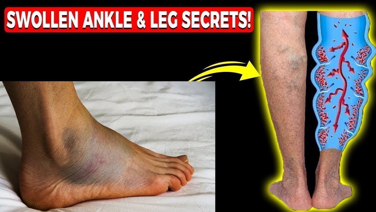
Both cause pain, swelling, and difficulty walking. Key differences: fractures often cause more immediate severe pain, tenderness directly over bone (not just ligament), and inability to bear any weight. X-rays and the Ottawa Ankle Rules help determine if imaging is needed.
How long does an ankle sprain take to heal?
Grade I (mild): 1–2 weeks. Grade II (moderate): 3–6 weeks. Grade III (complete tear): 2–3 months. Chronic instability from improperly treated sprains can persist and may require surgery.
What is the best treatment for a sprained ankle?
RICE protocol (Rest, Ice, Compression, Elevation) for the first 48–72 hours, followed by protected weight-bearing as tolerated. Physical therapy rehabilitation is critical for high-grade sprains to restore strength and proprioception and prevent chronic instability.
Need Treatment at Balance Foot & Ankle?
Dr. Tom Biernacki, Dr. Carl Jay, and Dr. Daria Gutkin see patients at our Howell and Bloomfield Township offices.
Book Online or call (810) 206-1402
Most Common Mistake We See
The most common mistake we see is: Waiting too long before seeking care. Fix: any foot pain lasting more than 4 weeks, or any sudden severe symptom, deserves a professional evaluation rather than more rest.
Warning Signs That Need Same-Day Care
Seek immediate evaluation at Balance Foot & Ankle if you experience any of the following:
- Unable to bear weight
- Severe swelling with skin colour change
- Fever with foot pain (possible infection)
- Diabetes plus any new foot symptom
Call (810) 206-1402 — same-day and next-day appointments at our Howell and Bloomfield Township offices.
Watch: Dr. Tom explains
Podiatrist-recommended products
As an Amazon Associate, Dr. Tom earns from qualifying purchases.
Post-arthroscopy support.
View on Amazon →Post-op cold therapy.
View on Amazon →Recovery arch support.
View on Amazon →Topical relief.
View on Amazon →Related resources
Ready to solve this? Book today.
Same-week appointments · Howell & Bloomfield Township · 4.9★ (1,123+ reviews)
☎ (810) 206-1402Book Online →Pros & Cons of Conservative Care for foot care
Advantages
- ✓ Conservative care first
- ✓ Same-week appointments
- ✓ Multiple insurance accepted
Considerations
- ✗ Self-treatment can mask issues
- ✗ See a podiatrist if pain >2 weeks
Dr. Tom’s Recommended Products for foot care
Affiliate disclosure: As an Amazon Associate, Balance Foot & Ankle earns from qualifying purchases. We only recommend products we use with patients.
Footnanny Heel Cream Dr. Tom’s Pick
Best for: Daily moisturizer for cracked heels
Ready to Get Back on Your Feet?
Same-day appointments in Howell + Bloomfield Township. Most insurance accepted. Dr. Tom Biernacki, DPM & team.
Book Today — Same-Day Appointments Available
Call Now: (810) 206-1402
About Your Care Team at Balance Foot & Ankle
Dr. Tom Biernacki, DPM · Board-Certified Foot & Ankle Surgeon. Specializes in conservative-first care, minimally invasive bunion surgery, and complex reconstruction.
Dr. Carl Jay, DPM · Accepting new patients. Specializes in sports medicine, athletic injuries, and routine podiatric care.
Dr. Daria Gutkin, DPM, AACFAS · Accepting new patients. Specializes in surgical reconstruction and pediatric podiatry.
Locations: 4330 E Grand River Ave, Howell, MI 48843 · 43494 Woodward Ave Suite 208, Bloomfield Township, MI 48302
Hours: Mon–Fri 8:00 AM – 5:00 PM · (810) 206-1402
In-Office Treatment at Balance Foot & Ankle
If home treatment isn’t providing relief for your foot and ankle conditions, our podiatry team at Balance Foot & Ankle can help with same-day evaluations and advanced in-office care.
Same-day appointments available. (810) 206-1402
Frequently Asked Questions
When should I see a podiatrist?
If symptoms persist past 2 weeks, affect your normal activity, or are accompanied by red-flag symptoms (warmth, redness, swelling, inability to bear weight).
What does treatment cost?
Most diagnostic visits and conservative treatments are covered by Medicare and major insurers. Out-of-pocket costs vary by your specific plan.
How quickly can I get an appointment?
Most non-urgent cases see us within 5 business days. Urgent cases (sudden pain, possible fracture) typically same or next business day.
Ready for Expert Care?
Same-day appointments in Howell & Bloomfield Township, MI.
4.9★ | 1,123 Reviews | 3,000+ Surgeries
Or call: (810) 206-1402
Dr. Tom Biernacki, DPM is a board-certified foot & ankle surgeon (ABFAS & ABPM) at Balance Foot & Ankle Specialists in Southeast Michigan. With over a decade of clinical experience, he specializes in heel pain, bunions, diabetic foot care, sports injuries, and minimally invasive surgery. Dr. Biernacki is a member of the APMA and ACFAS, and his patient education content on MichiganFootDoctors.com and YouTube has made him one of the most-followed foot & ankle educators on YouTube.