


Medically reviewed by Dr. Tom Biernacki, DPM · Board-Certified Podiatric Surgeon · Last reviewed: April 2026 · Editorial Policy
The most important clinical decision with Cavus Foot High Arch Causes Symptoms Treatment isn’t which treatment to start with — it’s identifying the correct subtype. That changes everything. Call (810) 206-1402.
Quick Answer
Cavus Foot (High Arch): Causes, Symptoms & Treatment B relates to arch concerns — typically caused by foot structure or fatigue. Most patients improve in 6-12 weeks with intervention with conservative care. Same-week appointments in Howell + Bloomfield Hills: (810) 206-1402.

Medically reviewed by Dr. Tom Biernacki, DPM — Board-certified foot & ankle surgeon, 3,000+ surgeries performed. Updated April 2026 with current clinical evidence. This article reflects real practice experience from Balance Foot & Ankle Specialists in Howell and Bloomfield Hills, Michigan.
Quick Answer
Most foot and ankle problems respond to conservative care — proper footwear, supportive inserts, activity modification, and targeted stretching — within 4-8 weeks. Persistent pain beyond that window, or any symptom that prevents walking, warrants a podiatric evaluation to rule out fracture, tendon tear, or systemic cause.
Watch: Dr. Tom Biernacki, DPM

Medically reviewed by Dr. Tom Biernacki, DPM — Board-Certified Podiatric Surgeon — Balance Foot & Ankle, Howell & Bloomfield Hills, MI. Last updated April 2026.
▶ Watch

📋 Dr. Tom Also Recommends
Podiatrist Recommended Orthotics 2026: Dr. Tom’s Top 10 Insoles & Arch Supports
A podiatrist’s complete clinical guide to the best insoles — custom orthotics, OTC picks, and what actually works for plantar fasciitis, flat feet, neuropathy & more.
Read the Full Guide →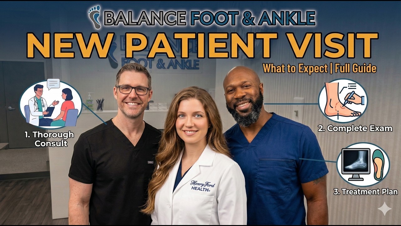
Medically Reviewed by Dr. Tom Biernacki, DPM — Board-Certified Podiatrist, Balance Foot & Ankle Specialists, Michigan. Last updated April 2026.
Cavus foot — a foot with an abnormally high arch — is less commonly discussed than flatfoot but can be equally symptomatic and functionally limiting. Unlike the mobile flatfoot that’s common in the general population, a high-arched foot is rigid and unable to absorb shock effectively, placing excessive pressure on the heel and ball of the foot, destabilizing the ankle, and predisposing patients to a characteristic cluster of painful conditions. Critically, cavus foot is frequently neurological in origin, making accurate diagnosis essential before treatment is planned.
What Defines Cavus Foot?
Cavus foot is defined by an elevated longitudinal arch on weight-bearing. Structurally, it involves plantarflexion of the first ray (the first metatarsal drops toward the ground), equinus of the hindfoot, claw toe deformities, and frequently a varus (inward-tilted) heel. The foot is rigid — it does not flatten when weight is applied — and absorbs shock poorly.
Causes of Cavus Foot
Neurological Causes (Most Important to Identify)
A significant proportion of cavus foot cases have an underlying neurological cause, particularly when the deformity is bilateral and progressive. The most common neurological associations include:
- Charcot-Marie-Tooth disease (CMT) — the most common hereditary peripheral neuropathy; produces progressive cavus deformity, peroneal muscle weakness, and intrinsic muscle atrophy over decades
- Friedreich’s ataxia — a spinocerebellar degenerative disease producing cavus foot, balance impairment, and cardiac involvement
- Spinal cord abnormalities — spina bifida, tethered cord, syringomyelia, and diastematomyelia can all produce unilateral cavus deformity
- Cerebral palsy and stroke — spastic equinovarus foot deformity has components of cavus alignment
- Polio sequelae — historic but still seen in older patients
A unilateral progressive cavus foot warrants MRI of the spine and neurological evaluation to rule out intraspinal pathology.
Idiopathic and Residual Causes
Many mild cavus feet are familial without an identifiable neurological disorder, and some result from residual clubfoot deformity inadequately corrected in childhood.
Symptoms and Associated Conditions
High-arched feet produce a characteristic cluster of problems resulting from abnormal pressure distribution and lateral ankle instability:
- Lateral ankle instability — chronic ankle sprains and giving-way from the varus hindfoot, which predisposes to inversion injury
- Metatarsalgia and stress fractures — the rigid forefoot transmits high pressure to the 1st and 5th metatarsal heads, producing calluses, metatarsalgia, and metatarsal stress fractures
- Plantar fasciitis — a high-tensioned plantar fascia under a rigid cavus arch is prone to heel insertion stress
- Claw toes — intrinsic muscle weakness produces hyperextension at the MTP joints and flexion at the IP joints
- Peroneal tendon problems — the varus hindfoot places the peroneals under chronic eccentric stress
- Heel pain — concentrated heel loading in a varus heel produces lateral heel calluses and fat pad irritation
Diagnosis
Dr. Biernacki at Balance Foot & Ankle performs a hands-on exam plus imaging when needed including weight-bearing X-rays (to measure Meary’s angle, Hibb’s angle, and calcaneal pitch), Coleman block test (to assess whether hindfoot varus is flexible or rigid), and clinical neurological assessment. When CMT or another neurological etiology is suspected, EMG/NCS and genetic testing are coordinated.
Treatment of Cavus Foot
Conservative Management
Flexible or mild cavus feet respond well to non-surgical care:
- Custom orthotics — a custom lateral wedge (valgus posting) corrects hindfoot varus, metatarsal pads offload the forefoot, and arch accommodation reduces plantar fascia tension
- Extra-depth shoes with cushioning — accommodates claw toes and reduces metatarsal head pressure
- Ankle bracing — for chronic lateral ankle instability, a lace-up ankle brace reduces inversion sprain risk
- Physical therapy — peroneal strengthening and proprioception training for lateral ankle stability
Surgical Treatment
Rigid cavus feet with significant deformity, failed conservative management, or progressive neurological involvement frequently require surgical correction. Surgical options are tailored to each component of the deformity:
- Plantar fascia release — reduces the deforming tension on the arch
- First metatarsal dorsiflexion osteotomy (dorsiflexory wedge) — elevates the plantarflexed first metatarsal head, correcting the primary deforming force
- Peroneus longus to brevis tendon transfer — reduces first metatarsal plantarflexion force and augments peroneus brevis eversion strength
- Calcaneal osteotomy (Dwyer lateralizing) — shifts the heel laterally to correct varus alignment
- Claw toe correction — IP joint fusion and MTP joint release for rigid claw toes
- Triple arthrodesis — fusion of the subtalar, talonavicular, and calcaneocuboid joints for severe rigid deformity; provides definitive correction with reliable outcomes
High Arch Pain? Get an Expert Evaluation.
Dr. Biernacki at Balance Foot & Ankle evaluates and treats cavus foot deformity at our Bloomfield Hills and Howell offices. Custom orthotics and surgical options available.
📞 (810) 206-1402 |
📧 Get Dr. Tom’s Free Lab Test Guide
Discover the 5 lab tests every person over 35 should ask their doctor about — explained in plain English by a board-certified physician.
📍 Located in Michigan?
Our board-certified podiatrists treat this condition at two convenient locations. Same-day appointments often available.
Insurance Accepted
BCBS · Medicare · Aetna · Cigna · United Healthcare · HAP · Priority Health · Humana · View All →
Howell Office
4330 E Grand River Ave
Howell, MI 48843
Get Directions →
Bloomfield Hills Office
43494 Woodward Ave, #208
Bloomfield Hills, MI 48302
Get Directions →
Your Board-Certified Podiatrists


Ready to Get Back on Your Feet?
Same-week appointments available at both locations.
Book Your Appointment👟 Dr. Tom’s Complete Footwear Library
Podiatrist-Approved Guides for Every Foot Type & Condition
Clinically reviewed by Dr. Tom Biernacki, DPM — Board-Certified Podiatrist
All guides are written and reviewed by licensed podiatrists. Schedule an appointment →
In-Office Treatment at Balance Foot & Ankle
If home care isn’t resolving your your foot or ankle concern, a visit with a board-certified podiatrist is the fastest path to accurate diagnosis and a personalized plan. At Balance Foot & Ankle Specialists, Dr. Tom Biernacki, Dr. Carl Jay, and Dr. Daria Gutkin offer same-day and next-day appointments at both our Howell and Bloomfield Hills offices. We perform on-site diagnostic ultrasound, digital X-ray, conservative care, advanced regenerative treatments, and minimally invasive surgery when indicated.
Call (810) 206-1402 or request an appointment online. Most insurance plans accepted, including Medicare, Blue Cross Blue Shield, Aetna, Cigna, and United Healthcare.
Most Common Mistake We See
The most common mistake we see is: Waiting too long before seeking care. Fix: any foot pain lasting more than 4 weeks, or any sudden severe symptom, deserves a professional evaluation rather than more rest.
Warning Signs That Need Same-Day Care
Seek immediate evaluation at Balance Foot & Ankle if you experience any of the following:
- Unable to bear weight
- Severe swelling with skin colour change
- Fever with foot pain (possible infection)
- Diabetes plus any new foot symptom
Call (810) 206-1402 — same-day and next-day appointments at our Howell and Bloomfield Hills offices.
More Podiatrist-Recommended Foot Health Essentials
Hoka Clifton 10

Max-cushion everyday shoe — podiatrist favorite for walking and running.

OOFOS Recovery Slide

Impact-absorbing recovery sandal — wear after long days on your feet.
As an Amazon Associate, Balance Foot & Ankle earns from qualifying purchases. Product recommendations are based on clinical experience; prices and availability shown above update live from Amazon.

When to See a Podiatrist
If foot or ankle pain has been bothering you for more than a few weeks, home care alone may not be enough. Balance Foot & Ankle offers same-week appointments at our Howell and Bloomfield Hills clinics — no referral needed in most cases. Bring your current shoes and a short list of symptoms and we’ll build you a treatment plan in one visit.
Call Balance Foot & Ankle: (810) 206-1402 · Book online · Offices in Howell & Bloomfield Hills
Pros & Cons of Conservative Care for foot care
Advantages
- ✓ Conservative care first
- ✓ Same-week appointments
- ✓ Multiple insurance accepted
Considerations
- ✗ Self-treatment can mask issues
- ✗ See a podiatrist if pain >2 weeks
Dr. Tom’s Recommended Products for foot care
Affiliate disclosure: As an Amazon Associate, Balance Foot & Ankle earns from qualifying purchases. We only recommend products we use with patients.
Footnanny Heel Cream Dr. Tom’s Pick
Best for: Daily moisturizer for cracked heels
Ready to Get Back on Your Feet?
Same-day appointments in Howell + Bloomfield Hills. Most insurance accepted. Dr. Tom Biernacki, DPM & team.
Book Today — Same-Day Appointments Available
Call Now: (810) 206-1402
About Your Care Team at Balance Foot & Ankle
Dr. Tom Biernacki, DPM · Board-Certified Foot & Ankle Surgeon. Specializes in conservative-first care, minimally invasive bunion surgery, and complex reconstruction.
Dr. Carl Jay, DPM · Accepting new patients. Specializes in sports medicine, athletic injuries, and routine podiatric care.
Dr. Daria Gutkin, DPM, AACFAS · Accepting new patients. Specializes in surgical reconstruction and pediatric podiatry.
Locations: 4330 E Grand River Ave, Howell, MI 48843 · 43494 Woodward Ave Suite 208, Bloomfield Hills, MI 48302
Hours: Mon–Fri 8:00 AM – 5:00 PM · (810) 206-1402
🩺 Dr. Tom’s Recommended Products
As an Amazon Associate I earn from qualifying purchases. These are products I personally use and recommend to patients.
The OTC orthotic I recommend most in our clinic. Sub-$50 before custom orthotics.
View on Amazon →
Natural menthol + arnica topical. FSA-eligible — what I switched my family to from Doctor Hoy’s Natural Pain Relief Gel.
View on Amazon →
Ready for Expert Care?
Same-day appointments in Howell & Bloomfield Hills, MI.
4.9★ | 1,123 Reviews | 3,000+ Surgeries
Or call: (810) 206-1402
Dr. Tom Biernacki, DPM is a board-certified foot & ankle surgeon (ABFAS & ABPM) at Balance Foot & Ankle Specialists in Southeast Michigan. With over a decade of clinical experience, he specializes in heel pain, bunions, diabetic foot care, sports injuries, and minimally invasive surgery. Dr. Biernacki is a member of the APMA and ACFAS, and his patient education content on MichiganFootDoctors.com and YouTube has made him one of the most-followed foot & ankle educators on YouTube.