
| Treatment Phase | Timeframe | Activity | Interventions | Goals |
|---|---|---|---|---|
| Acute (protection) | Weeks 1–3 | No en pointe/plantarflexion; walking allowed | NSAIDs, ice, heel lift, taping | Reduce acute inflammation; protect sheath |
| Subacute (loading) | Weeks 3–8 | Gradual toe flexion activities | Eccentric FHL loading, PT, intrinsic strengthening | Tendon collagen remodeling; strength recovery |
| Return to activity | Weeks 8–16 | Progressive dance/sport activities | Sport-specific loading; sheath injection if stuck | Full pain-free push-off capacity |
| Maintenance | Ongoing | Full activity with modifications | Ongoing intrinsic strengthening; footwear check | Prevent recurrence |
| FHL Condition | Mechanism | Key Symptom | Diagnosis | Treatment |
|---|---|---|---|---|
| FHL tendinopathy (mid-portion) | Overuse degeneration — collagen disarray | Posteromedial ankle pain with activity | US/MRI: tendon thickening | Eccentric loading protocol; PRP if chronic |
| FHL tenosynovitis | Synovial sheath inflammation | Medial ankle swelling + pain | US: sheath effusion around FHL | Sheath injection (US-guided); activity mod. |
| Hallux saltans (triggering) | Tendon catches at fibro-osseous tunnel behind ankle | Snapping/clicking big toe; pain at tunnel | Clinical; US confirms | Sheath injection; surgical tenolysis if refractory |
| FHL partial tear | High-load failure | Sudden pain; weakness of big toe flexion | MRI: partial tear signal | Boot 4–6 weeks; surgical repair if >50% tear |
| FHL complete rupture | Forced dorsiflexion injury | Unable to flex IP joint of big toe | MRI: complete tear + retraction | Surgical repair (acute) or tendon transfer (chronic) |

![Achilles Tendonitis & Back of Heel Pain [BEST Home Treatments 2024!]](https://www.michiganfootdoctors.com/wp-content/cache/flying-press/d645ea3f3d27024136b88a074b2b7894.jpg)
Watch: Achilles Tendonitis & Back of Heel Pain [BEST Home Treatments 2024!] — MichiganFootDoctors YouTube
Foot pain isn't resolving?
Same-week appointments at podiatrist in Howell & podiatrist in Bloomfield Hills
⚡ Quick Answer: How do you treat FHL tendonitis?
Flexor hallucis longus tendonitis responds to rest, ice, anti-inflammatory medications, physical therapy, and orthotics. Chronic cases may need cortisone injections or surgery.
Related Conditions
In This Article
- How do you treat FHL tendonitis?
- Quick Answer: Flexor Hallucis Longus Tendonitis Treatment
- Anatomy and Function
- Causes and Risk Factors
- Symptoms and Diagnosis
- Treatment Options
- Recommended Products
- Red Flags: Seek Urgent Evaluation
- Most Common Mistake with FHL Tendonitis
- In-Office Treatment at Balance Foot & Ankle
- Frequently Asked Questions
- Sources
- FHL Tendonitis? Get an Accurate Diagnosis.
- Frequently Asked Questions
- Frequently Asked Questions
Medically Reviewed by Dr. Tom Biernacki, DPM — Board-Certified Podiatrist & Foot Surgeon | Balance Foot & Ankle | Updated April 28, 2026
Quick Answer: Flexor Hallucis Longus Tendonitis Treatment
Flexor hallucis longus (FHL) tendonitis causes pain behind the ankle and under the big toe, most common in ballet dancers, runners, and jumping athletes. Treatment begins with rest, activity modification, calf stretching, and anti-inflammatory measures. Persistent or refractory cases benefit from ultrasound-guided corticosteroid injection and physical therapy. Surgery is rarely needed but highly effective when conservative care fails.
Flexor hallucis longus tendonitis is the overuse injury nobody outside of dance medicine talks about — yet it’s career-threatening when missed or mismanaged. The FHL tendon powers the push-off phase of every step and the relevé in dance; when inflamed or entrapped, it produces a very specific pattern of pain that is unmistakable once you know what to look for. We see this condition regularly at Balance Foot & Ankle, particularly in ballet dancers who’ve pushed through “ankle pain” for months without understanding what structure was actually involved.
Anatomy and Function
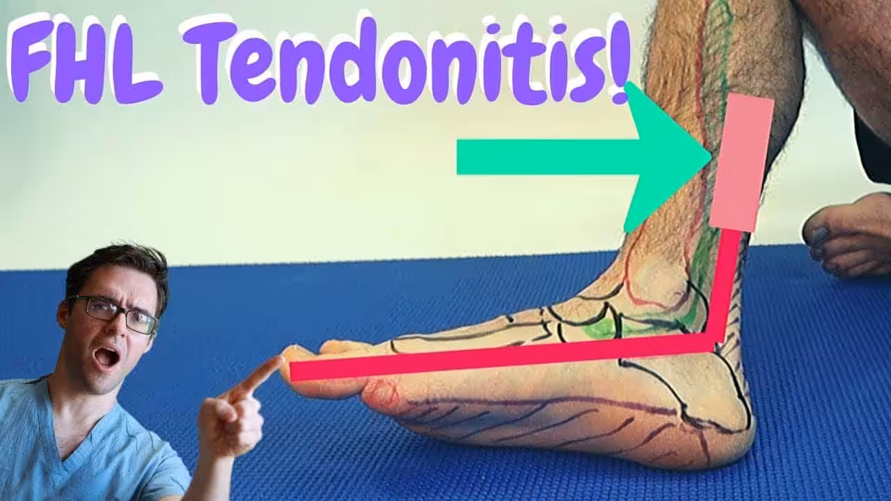
The flexor hallucis longus originates in the posterior leg, travels behind the fibula and tibia, passes through a fibro-osseous tunnel at the posterior ankle (between the medial and lateral talar tubercles), continues beneath the sustentaculum tali of the calcaneus, and inserts into the distal phalanx of the great toe. Its primary function is plantarflexion of the great toe — critical for push-off during walking and running, and for the relevé and pointe positions in ballet. The narrow fibro-osseous tunnel at the posterior ankle is the most common site of tendinopathy and entrapment: even mild tendon thickening from overuse can create a triggering phenomenon where the tendon catches in the tunnel, producing the characteristic “hallux saltans” or trigger toe.
Causes and Risk Factors
FHL tendonitis is an overuse injury driven by repetitive plantarflexion loading of the great toe. It is the quintessential dance injury — up to 25% of professional ballet dancers develop FHL tendonitis during their careers, with the forced plantarflexion of pointe and demi-pointe positions creating the highest FHL loads. Beyond dancers, we see FHL tendonitis in runners (particularly those with a strong push-off pattern), triathletes, soccer players, and gymnasts. Risk factors include a prominent posterior talar process (os trigonum syndrome frequently coexists), hyperpronation that shifts load to the medial forefoot, rapid increases in training volume, and hard flooring surfaces. Accessory ossicles (os trigonum) adjacent to the posterior ankle tunnel are present in up to 10% of the population and can dramatically narrow the FHL’s passage, predisposing to tendon impingement with minimal additional stress.
Symptoms and Diagnosis
FHL tendonitis produces a very specific pain pattern that distinguishes it from other posterior ankle conditions. The primary symptoms are posteromedial ankle pain (behind and slightly medial to the medial malleolus), pain with passive great toe extension or resisted plantarflexion, and occasionally a triggering sensation in the big toe — the tendon catching in its tunnel producing a snapping or locking phenomenon (hallux saltans). Pain is worst during push-off, stair descent, and the relevé position. Swelling in the retromalleolar groove may be palpable.
Diagnosis is primarily clinical: tenderness with direct palpation of the FHL tendon in the retromalleolar groove, reproduction of symptoms with the Stretch Test (passive great toe dorsiflexion with the ankle in plantarflexion), and the Triggering Test (passive great toe flexion-extension through range). MRI identifies tendon thickening, partial tears, and fluid in the tendon sheath (tenosynovitis). Diagnostic ultrasound in skilled hands demonstrates tendon caliber changes, tears, and dynamic triggering in real time. Weight-bearing foot and ankle X-rays rule out posterior talar process fracture, os trigonum, and adjacent bone pathology.
Treatment Options
Activity Modification and Relative Rest (Week 1–4)
The foundation of FHL tendonitis management is reducing the repetitive plantarflexion loading that caused the injury. Dancers must temporarily avoid pointe and demi-pointe work. Runners should reduce mileage by 50% and eliminate sprint, hill, and speed work. The ankle can be supported in a posterior heel lift (10–15 mm) in athletic footwear to reduce tension on the FHL at rest. NSAIDs (ibuprofen 400–600 mg three times daily with food for 7–10 days) reduce acute tenosynovitis. Ice application to the retromalleolar groove for 15 minutes after activity is helpful in the acute phase.
Stretching and Physical Therapy (Week 2–10)
The FHL is tightly coupled to the gastrocnemius-soleus complex — tight calves dramatically increase FHL strain. Aggressive calf stretching (both straight-knee gastrocnemius and bent-knee soleus components) twice daily is the most impactful flexibility intervention. For the FHL itself, gentle great toe extension stretching — performed seated, pulling the toe toward the shin while keeping the ankle neutral — stretches the tendon within its tunnel and maintains mobility. Physical therapy adds eccentric FHL strengthening (toe curls against resistance, towel scrunching), manual therapy to the ankle and subtalar joints, and proprioceptive retraining that is essential for dancers returning to work.
Ultrasound-Guided Corticosteroid Injection (Week 6–12)
When conservative measures haven’t adequately controlled tenosynovitis pain by 6–8 weeks, ultrasound-guided corticosteroid injection into the FHL tendon sheath provides targeted anti-inflammatory relief. We specifically inject the tendon sheath (peritendinous), never the tendon substance itself — intratendinous steroid injection risks partial or complete tendon rupture. In our hands, a single well-placed injection provides 3–6 months of significant relief in the majority of patients, during which physical therapy and activity modification can rehabilitate the underlying overuse pathology. We do not perform more than 2 sheath injections per year.