
Medically reviewed by Dr. Tom Biernacki, DPM
Board-certified podiatric surgeon | Balance Foot & Ankle, Howell & Bloomfield Hills, MI
Last reviewed: May 2026
Quick answer: Metatarsalgia Causes can significantly impact your daily life and mobility. Our Michigan podiatrists provide expert evaluation and evidence-based treatment — from conservative care to minimally invasive procedures — to relieve your symptoms and restore function. Same-day appointments available in Howell and Bloomfield Hills, MI.

| Cause | Mechanism | Key Finding | Treatment |
|---|---|---|---|
| Prominent metatarsal heads | Long or plantarflexed 2nd metatarsal concentrates load | Pain under 2nd–3rd MTP; callus under head | Metatarsal pad, custom orthotic, metatarsal shortening surgery |
| Hammertoe / claw toe | MTP subluxation depresses metatarsal head plantarward | Concurrent toe deformity; dorsal corn | Toe correction (surgical or conservative) |
| High heels / fashion footwear | Forefoot overload from elevated heel | Bilateral; directly correlates with heel height | Heel height reduction; forefoot cushion insole |
| Obesity / excess weight | Proportionally higher forefoot ground reaction force | Often bilateral; diffuse ball-of-foot pain | Weight management; cushioned orthotics |
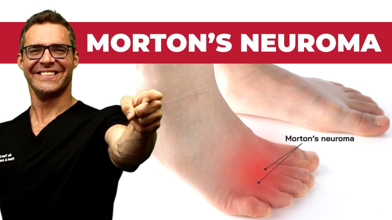
| Morton’s neuroma | Interdigital nerve compression causes forefoot pain | Mulder’s click; 3rd–4th web space burning | Metatarsal pad, injection, cryotherapy, surgery |
| Stress fracture (metatarsal) | Cyclic overload of metatarsal shaft or neck | Focal point tenderness over bone; edema | CAM boot 6–8 weeks; no weight-bearing |
| Sesamoiditis | Sesamoid bone inflammation under first MTP | Tenderness specifically at sesamoids | Dancer’s pad; offloading orthotic; boot if fracture |
| Fat pad atrophy | Age-related loss of plantar cushioning | Thin plantar skin; palpable bones; common >55 yrs | Extra-cushioned insole; cushioned footwear |
| Rheumatoid arthritis | MTP joint synovitis → subluxation → metatarsal head prominence | Multiple joints; ESR/CRP elevated; symmetric | Rheumatology co-management; accommodative orthotics |
| Treatment | Best For | Evidence | Notes |
|---|---|---|---|
| Metatarsal pad (proximal to heads) | All forefoot pain; first-line | Strong | Must be placed PROXIMAL to metatarsal heads, not under them |
| Extra-cushioned insole (PEBA foam) | Fat pad atrophy, obesity, diffuse pain | Moderate–Strong | HOKA, New Balance Fresh Foam platforms also help |
| Custom orthotics | Structural causes (long 2nd met, pronation) | Strong | With metatarsal bar; DPM-prescribed |
| Rocker-sole shoe | All metatarsalgia; reduces peak forefoot pressure | Very strong | Hoka One One, MBT style; 20–50% pressure reduction |
| Corticosteroid injection | Inflammatory metatarsalgia, neuroma | Moderate | Into MTP joint or web space; temporary relief |
| Metatarsal osteotomy (Weil) | Long or plantarflexed 2nd–4th metatarsal | Strong | Shortens/elevates metatarsal; predictable relief |
Quick answer:Metatarsalgia (ball-of-foot pain) is overloading of the metatarsal heads, causing burning or aching pain that worsens with standing and improves with rest. Treatment: metatarsal pad placed just behind the 2nd-3rd metatarsal heads, wider shoes, orthotics. Underlying causes (hammertoe, cavus foot) must be addressed for lasting relief. Call (810) 206-1402.
![Metatarsalgia Treatment [BEST Ball of Foot Pain RELIEF 2024]](https://www.michiganfootdoctors.com/wp-content/cache/flying-press/d7857672675f2bf191a8e896b2f7208b.jpg)
Watch: Metatarsalgia Treatment [BEST Ball of Foot Pain RELIEF 2024] — MichiganFootDoctors YouTube
Foot pain isn't resolving?
Same-week appointments at podiatrist in Howell & podiatrist in Bloomfield Hills
Quick Answer: What Causes Metatarsalgia?
Metatarsalgia is aching or burning pain under the ball of the foot caused by overloading the metatarsal heads — the five rounded bones just behind your toes. The most common causes are high-impact activity, biomechanical imbalances (high arch, flat foot, or a longer second metatarsal), footwear with narrow toe boxes or high heels, and age-related fat pad thinning. Addressing the underlying cause — not just the symptom — is the key to lasting relief.
You’ve probably noticed it most on hard floors in the morning — that nagging ache under the front of your foot, like you’re walking on a bruise. Or maybe it hits during a run, forcing you to compensate with a limp. In our clinic at Balance Foot & Ankle, metatarsalgia is one of the top five complaints we see in active adults and older patients alike. It sounds like a single condition, but it’s really a symptom — pain localized to the metatarsal heads — with several distinct causes that require different treatments. Getting the cause right is everything.
What Is Metatarsalgia

Metatarsalgia — from the Greek metatarsal (foot bones) and algos (pain) — is a clinical term for pain and inflammation at the metatarsal heads, the rounded ends of the five metatarsal bones that bear your weight with every step. It is not a diagnosis of why your foot hurts; it is a description of where it hurts. The underlying cause could be mechanical, structural, or a secondary consequence of another foot problem — and each responds to different treatment. In simple terms, metatarsalgia means the front of your foot is taking more load than it can handle.
The condition is extremely common, affecting an estimated 8–12% of the general population and considerably higher rates among runners, women who wear heels regularly, and adults over 60. It accounts for up to 6% of all sports medicine foot injuries. Because it’s so common and often misattributed to plantar fasciitis treatment or Morton’s neuroma, it frequently goes untreated or undertreated for months before patients reach a specialist.
The Anatomy Behind the Pain
Understanding why metatarsalgia develops requires a quick look at forefoot biomechanics. The five metatarsal bones connect the midfoot to the toes and form the front arch of the foot (the transverse arch). Each metatarsal head sits on a small fat pad — a natural shock absorber — and is surrounded by joint capsule, collateral ligaments, and flexor tendon pulleys. During normal walking, load distributes roughly as follows: metatarsal heads 2–4 carry about 57% of forefoot load collectively, while the 1st metatarsal handles about 40% thanks to the sesamoid bones beneath it.
This balance is fragile. Two anatomical variants set people up for metatarsalgia before they even put on their shoes:
| Anatomical Variant | Description | Effect on Load Distribution |
|---|---|---|
| Index Plus (Greek Foot) | 2nd metatarsal longer than 1st | 2nd metatarsal head bears disproportionate load — highest-risk pattern for 2nd MT stress fracture and plantar plate tear |
| Index Minus (Morton’s Foot) | 1st metatarsal shorter than 2nd | 1st metatarsal can’t offload effectively; load shifts to central metatarsals |
| High Arch (Pes Cavus) | Rigid, underpronating foot | Poor shock absorption; forefoot and heel take full impact instead of distributing across midfoot |
| Flat Foot (Pes Planus) | Overpronating, flexible arch | Forefoot splays on impact, spreading metatarsal heads and stretching plantar plate; increased 2nd–4th MT loading |
Primary Causes of Metatarsalgia
Primary metatarsalgia develops independently — it’s not caused by another foot condition. It accounts for the majority of cases we diagnose at Balance Foot & Ankle and responds well to conservative treatment when identified correctly.
1. Overuse and High-Impact Activity
Runners, high-impact aerobic exercisers, and people who suddenly increase training volume are prime candidates for metatarsalgia. With each mile run, the metatarsal heads absorb forces 2–3x body weight. A 180-pound runner generates approximately 450–540 pounds of force through the forefoot per step. Multiply that by 1,500 steps per mile and the cumulative loading becomes enormous. The fat pads compress, the joint capsules inflame, and the surrounding soft tissue develops microtears — all producing the characteristic aching bruised sensation. In our clinic, we typically see this in runners who jumped from 20 to 35+ miles per week within a single training cycle, or athletes returning from injury who ramped too fast.
2. Footwear with High Heels or Narrow Toe Boxes
This is the most common cause in women. A 2-inch heel shifts 57% of body weight onto the forefoot — the same forefoot that should be carrying only about 40% during level walking. At 3 inches, that rises to 76%. The narrow toe box common in fashionable heels simultaneously compresses the metatarsal heads together laterally, increasing intermetatarsal pressure and creating the ideal conditions for both metatarsalgia and Morton’s neuroma. We see the consequences every day: professional women who’ve worn heels for decades present with severely flattened fat pads and chronic forefoot inflammation that takes months to reverse even with perfect footwear changes.
3. Age-Related Fat Pad Atrophy
The fat pads under the metatarsal heads — nature’s shock absorbers — thin significantly with age. By age 60, many patients have lost 30–50% of their original pad thickness. The collagen and elastin fibers in the pad also lose resilience, so the remaining tissue doesn’t bounce back as effectively between steps. This leaves the metatarsal heads with little cushioning, making hard floors, tile, and even thin-soled shoes feel punishing. Fat pad atrophy is one reason older adults often feel immediate relief from thick-soled athletic shoes even without any other intervention — they’re replacing the lost biological cushioning with synthetic foam.
4. Biomechanical Imbalance (Foot Type)
Both high-arched (cavus) and flat (planus) foot types increase metatarsal loading but through different mechanisms. Cavus feet are rigid and underpronating — they absorb very little shock through midfoot motion, concentrating impact at the heel and forefoot. Flat feet overpronate, causing the forefoot to splay outward on impact and loading the 2nd through 4th metatarsals. In both cases, a functional orthotic — not just arch support — that addresses the specific kinematic fault is critical to long-term relief. Gel insoles from the drugstore address cushioning but not the mechanical imbalance, which is why so many patients get only partial relief from over-the-counter products.
Secondary (Structural) Causes of Metatarsalgia
Secondary metatarsalgia develops as a consequence of another foot problem that alters load distribution. These causes require treating the underlying condition — addressing the forefoot pain alone provides only temporary relief.
Hallux Valgus (Bunion) — Load Transfer to 2nd Metatarsal
A bunion is a progressive deformity in which the big toe drifts toward the second toe and the 1st metatarsal deviates medially. As this happens, the 1st metatarsal progressively loses its ability to bear forefoot load — it migrates away from the ground contact point. The load that should be carried by the 1st metatarsal-sesamoid complex transfers to the 2nd and 3rd metatarsals. Patients with bunions frequently develop secondary metatarsalgia of the 2nd MT long before the bunion itself becomes debilitating. In our practice, we always assess the 1st ray function in any patient with central metatarsalgia — treating the forefoot pain without addressing the bunion only delays the inevitable.
Hallux Rigidus — First Ray Stiffness
Hallux rigidus is arthritis of the 1st metatarsophalangeal joint that limits big toe dorsiflexion. During normal walking, the big toe dorsiflexes 65° during the toe-off phase to allow the foot to push off efficiently (the Windlass mechanism). When this motion is restricted, the body compensates by supinating the foot and loading the outer metatarsals during push-off. The lateral forefoot takes repeated abnormal stress with every step, producing metatarsalgia of the 4th and 5th metatarsals — a pattern that clinically distinguishes hallux rigidus-associated metatarsalgia from the more common central (2nd–3rd) pattern.
Rheumatoid Arthritis — Synovial Inflammation
Rheumatoid arthritis preferentially targets the metatarsophalangeal joints, often before it affects the hands. Synovial inflammation and joint erosion flatten the forefoot, displace the fat pads anteriorly, and leave the metatarsal heads exposed. Patients develop a characteristic broad, splayed forefoot with prominent MT heads and severe fat pad displacement. RA-associated metatarsalgia differs from mechanical causes in that it typically affects multiple metatarsals symmetrically in both feet and is accompanied by morning stiffness lasting >1 hour. Any bilateral symmetric forefoot pain warrants rheumatologic workup, especially with morning stiffness.
Previous Foot Surgery Altering Load Distribution
Hallux valgus correction (bunionectomy), hammertoe repair, or previous metatarsal surgery that shortens or elevates a metatarsal can significantly alter forefoot load distribution. A metatarsal that is surgically elevated or shortened no longer participates effectively in forefoot weight-bearing, transferring its share to adjacent metatarsals. Transfer lesions and calluses predictably form under adjacent MT heads post-operatively when metatarsal length or elevation relationships are disrupted. This is why experienced foot surgeons carefully assess metatarsal parabola (the relative length cascade of MT 1–5) before any forefoot surgery.
Metatarsalgia Risk Factors at a Glance
| Risk Factor | Mechanism | Modifiable? |
|---|---|---|
| High heels (>2 inches) | Shifts 57–76% of body weight to forefoot | Yes |
| Narrow toe box | Compresses metatarsal heads laterally | Yes |
| Sudden increase in activity | Exceeds tissue adaptation capacity | Yes |
| Age >50 | Fat pad atrophy, reduced collagen | No (mitigable) |
| High arch (cavus) | Rigid foot, poor shock absorption | No (manageable) |
| Flat foot (planus) | Forefoot splay, central MT overload | No (manageable) |
| Long 2nd metatarsal (index plus) | 2nd MT head bears disproportionate load | No (manageable) |
| Hallux valgus (bunion) | Transfers 1st ray load to central MT | Yes (surgical) |
| Rheumatoid arthritis | MTP synovitis, fat pad displacement | Medical management |
| Obesity (BMI >30) | Increased absolute ground reaction force | Yes |
Symptoms and How We Diagnose Metatarsalgia
Metatarsalgia produces a recognizable symptom cluster, but the precise pattern helps identify which metatarsal is involved and what’s causing it. The classic presentation is a burning, aching, or bruised sensation under the ball of the foot — specifically under the 2nd, 3rd, or 4th metatarsal heads (or occasionally all of them). The pain is worst when walking barefoot on hard surfaces like tile or hardwood, immediately upon standing after rest, and during push-off in athletic activities. It often improves with cushioned footwear and rest. In contrast to plantar fasciitis, which hurts most at heel strike first thing in the morning, metatarsalgia is worst at forefoot loading — when the toes are pushing off and all the weight is at the front of the foot.
Location clues by metatarsal: Pain under the 2nd MT head alone often indicates a plantar plate issue or stress fracture; under all central metatarsals suggests mechanical overloading or RA; pain specifically between the 3rd and 4th toes with a “click” points toward Morton’s neuroma rather than true metatarsalgia (the distinction matters for treatment). We use a combination of clinical examination — palpating each metatarsal head individually, performing the Mulder’s click test for neuroma, checking hallux range of motion for rigidus, and assessing foot type — along with X-rays to rule out stress fracture or cortical changes from Freiberg’s infraction, and ultrasound when soft tissue pathology (plantar plate, neuroma) is suspected.
Treatment Options That Address the Root Cause
Effective metatarsalgia treatment must address the underlying cause, not just cushion the pain. The treatment ladder progresses from conservative to interventional, with the majority of patients achieving lasting relief without surgery when the correct modality is selected.
Sale

PowerStep Pinnacle High Arch Orthotic Insoles, Plantar Fasciitis Relief, Supination Heel Pain, Arch Support, PowerStep Insoles for Women and Men, Made in USA (Men’s 10-10.5, Women’s 12)
- High Arch Support: PowerStep supination insoles deliver firm, flexible high arch support plus a deep heel cradle for comfort, stability & motion control, helping align feet, reduce pain, and protect against ball & heel pressure.
- All Day Comfort & Support: PowerStep Pinnacle High shoe inserts for women and men use premium dual layer cushioning to deliver heel to toe comfort and responsive bounce back with every step, without going flat.
- Relieves & Helps Prevent Pain: PowerStep Pinnacle High insoles for supination can help alleviate common foot conditions often linked to supination, including plantar fasciitis, Achilles tendonitis, fat pad atrophy, and Morton’s neuroma.
- No Trimming: PowerStep insoles move easily from shoe to shoe. Inserts are sized by shoe size for footwear with removable factory insoles. Designed for walking, running, work & casual dress shoes; pairs well with best walking shoes for women and men.
- Made in the USA: We stand behind our PowerStep Insoles for women and men. Proudly made in the USA & backed by a 30-day money-back guarantee. HSA & FSA Eligible
Footwear Modification (First Priority)
Wide toe box, low heel (<1 inch), and firm midsole with adequate forefoot cushioning is the foundation. Athletic shoes in half a size larger than usual allow the foot to function without compression. For specific models — including women’s, work, and wide-toe-box picks — see our podiatrist-ranked best shoes for metatarsalgia. Barefoot walking and thin-soled flats should be avoided during the acute phase — even 2–4 weeks of appropriate footwear can dramatically reduce inflammation. For professional women who cannot eliminate heels entirely, limiting heel wear to <3 hours per day and keeping spare athletic shoes at the office represents a pragmatic compromise that often prevents further fat pad damage.
Metatarsal Pad Placement
A metatarsal pad offloads the MT heads by placing a slight dome proximal to (behind) them, redistributing load to the metatarsal shafts. This simple intervention, when placed correctly, can reduce metatarsal head pressure by 40–50% according to force platform studies. The critical detail — which we cover in the “Most Common Mistake” section below — is that the pad must go BEHIND the metatarsal heads, not under them. Most commercially available insoles with built-in metatarsal support actually position the pad correctly; the problem arises when patients place adhesive felt pads themselves without guidance.
Functional Orthotics
For patients with significant biomechanical contributors (cavus, planus, hypermobile 1st ray, hallux valgus), a functional orthotic addresses the root kinematic fault. Unlike simple arch supports, functional orthotics control rearfoot motion, control forefoot splay, and can be built with incorporated metatarsal domes. In our practice, we prescribe functional orthotics for patients who have failed conservative treatment at 6–8 weeks, have clearly identifiable foot type contributors, or are high-volume athletes who need long-term prevention. Custom orthotics are more expensive but provide significantly more precision than OTC versions for structural contributors.
Corticosteroid Injection
For severe or recalcitrant metatarsalgia with significant inflammation, a small-volume corticosteroid injection into the most symptomatic MTP joint capsule can provide rapid relief. We use this sparingly — repeated injections risk further fat pad atrophy, worsening the underlying problem. One to two injections maximum in a treatment episode, always combined with footwear and orthotic intervention, is our standard approach. Ultrasound guidance allows precise placement with minimal collateral tissue effect.
Surgical Options (Last Resort)
Surgery is rarely necessary for isolated primary metatarsalgia. Weil osteotomy (surgically shortening and elevating the offending metatarsal) is effective for severe index plus deformity or long-standing metatarsalgia that has failed all conservative measures. For secondary metatarsalgia from hallux valgus, bunion correction can dramatically improve central forefoot loading. Surgical intervention should only be considered after 6–12 months of well-executed conservative treatment. The evidence for Weil osteotomy shows good-to-excellent results in 85–90% of carefully selected patients (Migues et al., 2004).
The Best Insoles for Metatarsalgia: PowerStep Pinnacle
PowerStep Pinnacle Insoles — Foundation Wellness Partner
Of all the OTC insoles we evaluate in clinic, PowerStep Pinnacle is the one we recommend most for metatarsalgia. It combines a semi-rigid arch shell (critical for controlling overpronation and cavus-type loading), adequate metatarsal arch support positioned correctly just behind the metatarsal heads, and deep heel cup for rearfoot control. Unlike gel-only insoles that address cushioning but not mechanics, Pinnacle addresses both simultaneously.
Why it helps metatarsalgia specifically: The built-in metatarsal arch dome is positioned at the correct anatomical location — not so far forward that it sits under the MT heads. The firm EVA top cover distributes load rather than letting the foot sink through a soft pad. For patients with mild-to-moderate biomechanical contributors to metatarsalgia, Pinnacle often provides the mechanical correction that expensive custom orthotics would otherwise require.
Not Ideal For:
- Patients with RA-associated metatarsalgia (need extra-depth footwear, not just insoles)
- Post-surgical forefoot reconstruction (requires custom device)
- Severe flat feet requiring maximum motion control orthotic
- Acute stress fracture (requires offloading boot, not insole)
Most Common Mistake: Placing the Pad in the Wrong Spot
The single most common mistake we see in patients who’ve attempted self-treatment is placing the metatarsal pad directly under the metatarsal heads instead of immediately behind (proximal to) them. When the pad is under the heads, it actually increases pressure at the most symptomatic location — the opposite of what’s intended. The pad’s purpose is to create a gentle dorsal force that transfers load from the MT heads to the shafts, 2–3 centimeters proximal. This can only happen if the dome of the pad sits behind the heads. A correctly placed pad makes walking on hard floors feel dramatically better within minutes. An incorrectly placed pad makes it worse. If someone has tried a metatarsal pad and says it didn’t help, the first question we ask is: “Where did you put it?”
Red Flags: When Metatarsalgia Requires Urgent Evaluation
⚠️ See a Podiatrist Promptly If You Have:
- Swelling and bruising over one metatarsal — possible stress fracture requiring X-ray within 24–48 hours
- Pain worse at rest than with activity — atypical pattern suggesting tumor, infection, or inflammatory arthritis rather than mechanical cause
- Bilateral symmetric forefoot pain with morning stiffness >1 hour — rheumatoid arthritis workup needed
- Numbness or burning radiating into toes — may indicate Morton’s neuroma or peripheral neuropathy, not simple metatarsalgia
- Visible metatarsal head prominence or toe deformity — joint subluxation or dislocation (plantar plate rupture) requiring imaging
- Pain not improving after 4–6 weeks of appropriate footwear changes — underlying structural or inflammatory cause likely; imaging and specialist evaluation indicated
In-Office Treatment at Balance Foot & Ankle
At Balance Foot & Ankle, Dr. Tom Biernacki, Dr. Carl Jay, and Dr. Daria Gutkin evaluate and treat all causes of metatarsalgia at our Howell and Bloomfield Hills locations. Our assessment combines physical examination, foot pressure analysis, digital X-ray, and ultrasound as needed to identify the precise cause before recommending treatment. We offer same-day appointments for acute forefoot pain. For patients with secondary metatarsalgia from bunions, hammertoes, or hallux rigidus, we provide comprehensive surgical planning with a focus on restoring normal load distribution across the entire forefoot — not just addressing the most symptomatic point. Request an appointment or call (810) 206-1402.
Frequently Asked Questions
Sources
- Espinosa N, Brodsky JW, Maceira E. “Metatarsalgia.” Journal of the American Academy of Orthopaedic Surgeons. 2010;18(8):474-485.
- DiPreta JA. “Metatarsalgia, lesser toe deformities, and associated disorders of the forefoot.” Medical Clinics of North America. 2014;98(2):233-251.
- Migues A, Slullitel G, Bilbao F, et al. “Floating-toe deformity as a complication of the Weil osteotomy.” Foot & Ankle International. 2004;25(9):609-613.
- Besse JL. “Metatarsalgia.” Orthopaedics & Traumatology: Surgery & Research. 2017;103(1S):S29-S39.
- Waldecker U. “Metatarsalgia in hallux valgus deformity: a pedographic analysis.” Journal of Foot and Ankle Surgery. 2002;41(5):300-308.
Ball of Foot Pain Affecting Your Daily Life?
Dr. Tom Biernacki identifies the exact cause of your metatarsalgia — mechanical, structural, or inflammatory — and delivers targeted treatment, not guesswork. Same-day appointments available.
📞 (810) 206-1402 | Howell & Bloomfield Hills, MI
Related Conditions & Resources
For more on related conditions and treatments:
- Metatarsalgia exercises & massage
- Sesamoiditis treatment 2026
- Capsulitis of the second toe
- Foot pain when walking: causes by location
- Hammer toe causes: why toes curl and buckle
- Howell podiatrist office
- Bloomfield Hills podiatrist office
Need to see a podiatrist? Call (810) 206-1402 or book online. Same-week availability.
Watch: Dr. Tom Biernacki Explains Metatarsalgia
Ready to feel better?
Same-week appointments available in Howell and Bloomfield Hills, Michigan.
If home treatment isn’t providing relief for your metatarsalgia, our podiatry team at Balance Foot & Ankle can help with same-day evaluations and advanced in-office care.
Ready to Get Relief?
Same-day appointments available in Howell & Bloomfield Hills, MI
4.9★ | 1,123 Reviews | 3,000+ Surgeries
Or call: (810) 206-1402
Dr. Tom Biernacki, DPM is a board-certified foot & ankle surgeon (ABFAS & ABPM) at Balance Foot & Ankle Specialists in Southeast Michigan. With over a decade of clinical experience, he specializes in heel pain, bunions, diabetic foot care, sports injuries, and minimally invasive surgery. Dr. Biernacki is a member of the APMA and ACFAS, and his patient education content on MichiganFootDoctors.com and YouTube has made him one of the most-followed foot & ankle educators on YouTube.