


Foot pronation affects the entire kinetic chain — and overpronation can trigger knee, hip, and back pain that gets misdiagnosed for months. Addressing the foot mechanics often resolves the upstream problems.
You’re in the right place. Dr. Tom Biernacki, DPM, FACFAS — board-certified foot & ankle surgeon with 3,000+ surgeries — explains exactly what foot pronation and the kinetic chain means and what works. Call (810) 206-1402 for same-day appointment at Howell or Bloomfield Hills.
Quick answer: Foot Pronation Biomechanics Kinetic Chain Knee Hip Pain has multiple potential causes including mechanical, neurological, vascular, and inflammatory. The patterns we see most often are overuse, poorly-fitted shoes, and biomechanical imbalance. Red flags requiring urgent evaluation: warmth/redness (infection), inability to bear weight (fracture), and unilateral swelling without injury (DVT). Call (810) 206-1402.
Medically reviewed by Dr. Tom Biernacki, DPM · Board-Certified Podiatric Surgeon · Last reviewed: April 2026 · Editorial Policy
The most important clinical decision with Foot Pronation Biomechanics Kinetic Chain Knee Hip Pain isn’t which treatment to start with — it’s identifying the correct subtype. That changes everything. Call (810) 206-1402.
Quick Answer
Foot Pronation and the Kinetic Chain: How Overpronation Affe relates to foot pain — typically caused by overuse, footwear, or biomechanics. Most patients improve in 6-12 weeks with conservative care. Same-week appointments in Howell + Bloomfield Hills: (810) 206-1402.

Medically reviewed by Dr. Tom Biernacki, DPM — Board-Certified Podiatric Surgeon — Balance Foot & Ankle, Howell & Bloomfield Hills, MI. Last updated April 2026.
▶ Watch
Medically Reviewed by Dr. Tom Biernacki, DPM — Board-Certified Podiatrist, Balance Foot & Ankle Specialists, Michigan. Last updated April 2026.
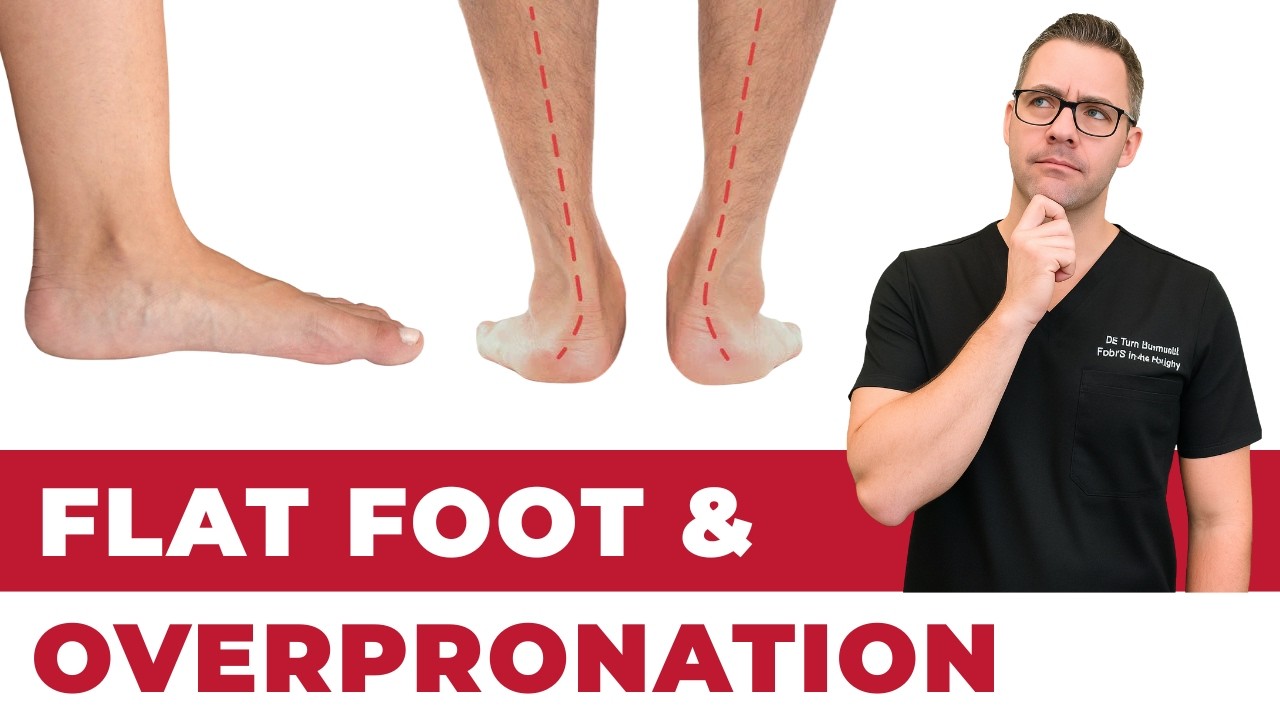
The foot is the foundation of the musculoskeletal kinetic chain — every ground reaction force that the body must absorb, redirect, and propel from begins at foot contact. Excessive or prolonged pronation at the subtalar joint does far more than cause foot pain; it initiates a cascade of rotational and angular forces that propagate proximally through the ankle, tibia, knee, hip, and lumbar spine. Understanding this kinetic chain relationship is central to podiatric biomechanics and explains why custom orthotics can meaningfully reduce pain not only in the foot but also at the knee, hip, and lower back in properly selected patients.
Normal Pronation: Essential Shock Absorption
Pronation — the combination of eversion, abduction, and dorsiflexion at the subtalar joint — is a normal and essential motion during the loading phase of gait. Following initial heel contact, controlled pronation unlocks the midtarsal joints, allowing the foot to conform to irregular ground surfaces, elongate and flatten slightly to dissipate impact forces, and position the forefoot for stable push-off. Approximately 4–6° of subtalar eversion during early stance is biomechanically normal and necessary. The pathology lies not in pronation itself but in excessive magnitude (>6–8° of eversion), excessive duration (pronation that continues past mid-stance into terminal stance when the foot should be supinating for push-off), or inadequate muscular control of the rate of pronation (pronation velocity).
Tibial Internal Rotation and Knee Mechanics
Subtalar joint eversion is mechanically coupled to tibial internal rotation through the talocalcaneal joint geometry — as the calcaneus everts, the talus adducts and plantarflexes, driving the tibia into internal rotation. During normal gait, subtalar eversion in loading response produces approximately 8–12° of tibial internal rotation. In overpronators, excess subtalar eversion produces proportionally greater tibial internal rotation, which continues later into stance than normal. This excessive and prolonged tibial internal rotation places the femur in relative external rotation (or abduction) relative to the tibia — increasing dynamic valgus at the knee, elevating patellofemoral joint contact stress, and increasing iliotibial band tension. Research demonstrates that overpronation is a significant contributing factor to patellofemoral pain syndrome (PFPS), iliotibial band syndrome, medial tibial stress syndrome (shin splints), and tibial stress fractures in runners and active patients.
Hip and Lumbar Effects
Tibial internal rotation propagates to the femur as internal femoral rotation — increasing hip adduction and medial rotation during stance, which alters hip joint loading, increases the moment arm for hip abductor muscles (requiring greater gluteus medius activation to maintain pelvic stability), and increases peak hip contact stress medially. Persistent femoral internal rotation with ipsilateral pelvic drop creates a functional leg length discrepancy and asymmetric lumbar loading that contributes to sacroiliac joint dysfunction and lower back pain. Notably, these proximal effects are amplified by foot asymmetry — unilateral overpronation creates consistently asymmetric tibial rotation and pelvic tilt throughout thousands of steps daily.
Custom Orthotics and Kinetic Chain Correction
Custom foot orthotics designed from a weight-bearing or non-weight-bearing 3D digital scan address overpronation by posting the rearfoot and forefoot to limit excessive subtalar eversion and correct its timing. Well-constructed custom orthotics demonstrably reduce tibial internal rotation during running, decrease patellofemoral contact stress, reduce gluteus medius activation demand, and alleviate plantar fascia strain simultaneously. The clinical evidence for orthotic benefit in patellofemoral pain, iliotibial band syndrome, plantar fasciitis, and medial tibial stress syndrome is supported by multiple systematic reviews and randomized controlled trials. Dr. Biernacki at Balance Foot & Ankle fabricates custom 3D-scanned orthotics following comprehensive biomechanical gait analysis. Medicare and most major insurance plans cover custom orthotics when medically indicated. Call (810) 206-1402 to schedule a biomechanical evaluation.
📧 Get Dr. Tom’s Free Lab Test Guide
Discover the 5 lab tests every person over 35 should ask their doctor about — explained in plain English by a board-certified physician.
📍 Located in Michigan?
Our board-certified podiatrists treat this condition at two convenient locations. Same-day appointments often available.
Insurance Accepted
BCBS · Medicare · Aetna · Cigna · United Healthcare · HAP · Priority Health · Humana · View All →
Howell Office
4330 E Grand River Ave
Howell, MI 48843
Get Directions →
Bloomfield Hills Office
43494 Woodward Ave, #208
Bloomfield Hills, MI 48302
Get Directions →
Your Board-Certified Podiatrists


Ready to Get Back on Your Feet?
Same-week appointments available at both locations.
Book Your AppointmentFrequently Asked Questions
When should I see a doctor?
See a podiatrist if pain persists past 2 weeks, prevents normal activity, or is accompanied by red-flag symptoms (warmth, swelling, numbness, inability to bear weight).
Can I treat this at home?
Mild cases respond to RICE protocol (rest, ice, compression, elevation), supportive shoes, and OTC anti-inflammatories. Persistent symptoms need professional evaluation.
How long does it take to heal?
Most soft tissue injuries resolve in 2-6 weeks with appropriate care. Bone injuries take 6-12 weeks. Chronic conditions need longer-term management.
What is Foot pain?
Foot pain is a common foot/ankle condition that affects mobility and quality of life. Understanding the underlying cause is the first step in successful treatment. Our podiatrists at Balance Foot & Ankle perform a hands-on biomechanical exam, review your activity history, and use diagnostic imaging when appropriate to identify the root cause—not just treat the symptom. Many patients have been told to “rest and ice” without a deeper diagnostic workup; our approach is different.
Symptoms and warning signs
Common signs of foot pain include pain that worsens with activity, morning stiffness, swelling, tenderness when palpated, and difficulty bearing weight. If you experience sudden severe pain, inability to walk, visible deformity, numbness or color change, contact our office the same day or visit urgent care—these can signal a more serious injury such as a fracture, tendon rupture, or vascular compromise. Diabetics with any foot wound should seek same-day care.
Conservative treatment options
Most cases of foot pain respond to non-surgical care: structured rest, supportive footwear changes, custom orthotics, targeted stretching and strengthening protocols, anti-inflammatory medications when medically appropriate, and in-office procedures such as ultrasound-guided injections. We also offer advanced therapies including MLS laser therapy, EPAT/shockwave, regenerative injections, and image-guided procedures. Treatment is sequenced from least invasive to most invasive, and we explain the rationale at every step.
When is surgery considered?
Surgery is reserved for cases that fail 3-6 months of well-structured conservative care, when there is structural pathology (severe deformity, complete tear, advanced arthritis), or when imaging shows damage that will not heal without intervention. Our surgeons have performed 3,000+ foot and ankle procedures and prioritize minimally-invasive techniques whenever appropriate. We discuss recovery timelines, return-to-activity milestones, and realistic outcome expectations before any procedure is scheduled.
Recovery timeline and prevention
Recovery from foot pain varies based on severity and chosen treatment path. Conservative cases often improve within 4-8 weeks with consistent adherence to the protocol. Post-procedural recovery may range from a few days (in-office procedures) to several months (reconstructive surgery). Long-term prevention involves footwear assessment, activity modification, structured strengthening, and regular check-ins with your podiatrist if you have a history of recurrence. We provide written home-exercise plans and digital follow-up support.
Ready to feel better?
Same-week appointments available in Howell and Bloomfield Hills, Michigan.
Book Your VisitIn-Office Treatment at Balance Foot & Ankle
If home treatment isn’t providing relief for your foot pain, our podiatry team at Balance Foot & Ankle can help with same-day evaluations and advanced in-office care.
Same-day appointments available. (810) 206-1402
Doctor Hoy’s Natural Pain Relief Gel
Natural topical pain relief I use in our clinic. Arnica + camphor formula — apply directly to the area 3–4x daily. ($20–25)
Shop Doctor Hoy’s →Ready for Expert Care?
Same-day appointments in Howell & Bloomfield Hills, MI.
4.9★ | 1,123 Reviews | 3,000+ Surgeries
Or call: (810) 206-1402
Dr. Tom Biernacki, DPM is a board-certified foot & ankle surgeon (ABFAS & ABPM) at Balance Foot & Ankle Specialists in Southeast Michigan. With over a decade of clinical experience, he specializes in heel pain, bunions, diabetic foot care, sports injuries, and minimally invasive surgery. Dr. Biernacki is a member of the APMA and ACFAS, and his patient education content on MichiganFootDoctors.com and YouTube has made him one of the most-followed foot & ankle educators on YouTube.