
Medically reviewed by Dr. Tom Biernacki, DPM
Ready to be seen? Book a real appointment time online, 24/7 — Howell or Bloomfield Township. Takes about a minute, 24/7.
Board-certified podiatric surgeon | Balance Foot & Ankle, Howell & Bloomfield Township, MI
Last reviewed: May 2026

Quick answer: A syndesmotic (high) ankle sprain injures the ligaments joining the two shin bones just above the ankle. It is slower to recover than a common lateral sprain, and stability — not pain level — is what decides between a boot and surgical fixation. Call (810) 206-1402.
Medically reviewed by Dr. Tom Biernacki, DPM
Board-certified podiatric surgeon | Balance Foot & Ankle
Last reviewed: April 2026
‘High ankle sprain’ is the common name for a syndesmotic injury — and despite being less well-known than the typical lateral ankle sprain, it’s significantly more serious. In our clinic, we frequently see athletes who were told they ‘just sprained their ankle’ but are still not back on the field 10 weeks later. In many of these cases, the syndesmosis was injured and was never properly identified or treated.
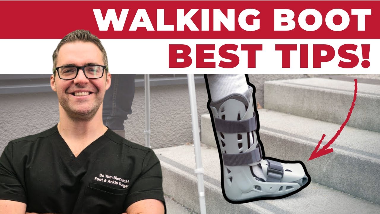
A high ankle sprain is a different injury from the common lateral sprain — it takes longer to settle, and the question that decides between a boot and surgery is whether the joint is actually unstable. Book online in Howell or Bloomfield Township — the booking form takes about a minute, 24/7.
A high ankle sprain is a different injury from the common lateral sprain — it takes longer to settle, and the question that decides between a boot and surgery is whether the joint is actually unstable. Book online in Howell or Bloomfield Township — the booking form takes about a minute, 24/7.
What Is the Syndesmosis?
The distal tibiofibular syndesmosis is the fibrous articulation between the distal tibia and fibula just above the ankle joint. Unlike a synovial joint (which has a joint space and fluid), the syndesmosis is held together by four ligaments and an interosseous membrane:
- Anterior inferior tibiofibular ligament (AITFL) — the most commonly injured syndesmotic ligament; runs diagonally from the fibula to the tibia anteriorly
- Posterior inferior tibiofibular ligament (PITFL) — the strongest syndesmotic ligament; posterior counterpart to the AITFL
- Interosseous ligament (IOL) — continuation of the interosseous membrane at the distal tibiofibular level
- Inferior transverse ligament — deepest posterior component; provides significant stability
The syndesmosis maintains the ‘mortise’ — the precise relationship between the tibia and fibula that creates a stable socket for the talus. Even minor widening (diastasis) of the mortise dramatically reduces contact area between the tibia/fibula and talus, causing rapid cartilage degeneration.
High Ankle Sprain vs. Lateral Ankle Sprain
- Mechanism: lateral sprain — inversion with plantarflexion; syndesmotic injury — external rotation of the foot or dorsiflexion with axial load
- Pain location: lateral sprain — below/at lateral malleolus; syndesmosis — above the ankle along the fibula and between the tibia and fibula
- Recovery time: lateral Grade 2 — 4–6 weeks; syndesmotic injury — 8–16+ weeks
- Sports most affected: lateral sprains — basketball, soccer; syndesmotic — football, hockey, alpine skiing
- Stability implications: lateral sprains rarely require surgery; unstable syndesmotic injuries often do
Diagnosing a Syndesmosis Injury
Clinical tests that suggest syndesmotic injury:
- Squeeze test — compressing the tibia and fibula together at mid-calf reproduces pain at the ankle syndesmosis
- External rotation stress test — foot in dorsiflexion, externally rotating the foot while stabilizing the lower leg reproduces syndesmotic pain
- Cotton test — manual lateral translation of the talus in the mortise; abnormal translation indicates instability
- Fibula translation test — anterior-posterior translation of the distal fibula relative to the tibia
Imaging: weight-bearing ankle X-rays assess the tibiofibular space and medial clear space. CT scan provides 3D assessment of the mortise. MRI is the gold standard for visualizing the syndesmotic ligaments.
Key takeaway: The squeeze test is the most reliable bedside test for syndesmotic injury — pain at the ankle when you squeeze the tibia and fibula together at mid-calf is highly specific for syndesmotic damage. If a patient fails this test after an ankle injury, we order weight-bearing X-rays and MRI before allowing return to sport.
Treatment of Syndesmosis Injuries
Stable Syndesmotic Injuries (No Diastasis)
Walking boot immobilization for 4–6 weeks followed by progressive rehabilitation. More structured and prolonged than lateral ankle sprain rehab. Return to sport: 8–12 weeks with proper rehabilitation focusing on proprioception and sport-specific loading.
Unstable Syndesmotic Injuries (Diastasis Present)
Surgical fixation is required to restore the mortise anatomy. Options include:
- Syndesmotic screw fixation — one or two screws placed across the fibula into the tibia, holding the joint reduced while ligaments heal; screws may be removed at 3 months
- TightRope/Endobutton fixation — flexible implant that allows normal ankle motion while maintaining reduction; lower re-dislocation risk than screws; no removal typically needed
- Suture tape augmentation — newer technique reinforcing the AITFL with a non-absorbable suture tape for dynamic stabilization
⚠️ Seek Immediate Evaluation If:
- Ankle injury involved a rotational mechanism (not just rolling inward)
- Pain is located above the ankle along the fibula or between the shin bones
- Positive squeeze test — significant pain when mid-calf is compressed
- You are an athlete with an ankle injury and need clearance to return to play
Frequently Asked Questions
How long does a syndesmosis injury take to heal?
Stable syndesmotic injuries typically require 8–12 weeks before return to sport. Surgically treated unstable injuries average 12–16 weeks. This is significantly longer than lateral ankle sprains — the syndesmosis must heal completely before athletic loading, or the mortise widens and cartilage damage follows.
Can syndesmosis injuries heal without surgery?
Stable syndesmotic sprains (ligament injury without diastasis) heal without surgery with appropriate immobilization and rehabilitation. Unstable injuries with mortise widening (diastasis) almost always require surgical fixation — non-surgical treatment of instability leads to chronic pain and early ankle arthritis.
Why does a high ankle sprain take longer to heal than a regular sprain?
Syndesmotic ligaments heal more slowly because they’re fibrous structures under constant load — every step compresses and slightly widens the mortise, stressing the healing tissue. Lateral ankle ligaments don’t bear the same constant compressive load. Additionally, even small degrees of residual instability in the mortise cause cartilage damage that perpetuates symptoms.
Dr. Tom’s Recovery Kit
Menthol + arnica + magnesium for post-injury and post-surgical soreness. Apply 3-4x daily.
View on Amazon →
FTC Disclosure: As an Amazon Associate and Foundation Wellness affiliate, we earn from qualifying purchases. Dr. Biernacki only recommends products used in our clinic or personally vetted.
Ready to Get Relief?
Same-day appointments available in Howell & Bloomfield Township, MI
4.9★ | 1,123 Reviews | 3,000+ Surgeries
Or call: (810) 206-1402
Sources
- Norkus SA, Floyd RT. The anatomy and mechanisms of syndesmotic ankle sprains. J Athl Train. 2001.
- Sman AD, et al. Diagnostic accuracy of clinical tests for ankle syndesmosis injury. Br J Sports Med. 2015.
- Kortekangas T, et al. Suture-button versus screw fixation of the syndesmosis. J Bone Joint Surg Am. 2019.
- American Orthopaedic Foot & Ankle Society. High Ankle Sprain. aofas.org. 2025.
In-Office Treatment at Balance Foot & Ankle
If home treatment isn’t providing relief for your foot joint pain, our podiatry team at Balance Foot & Ankle can help with same-day evaluations and advanced in-office care.
American Academy of Orthopaedic Surgeons: High Ankle Sprain (Syndesmosis)
Ready to Get Relief?
Same-day appointments available in Howell & Bloomfield Township, MI
4.9★ | 1,123 Reviews | 3,000+ Surgeries
Or call: (810) 206-1402
High ankle sprain or syndesmosis injury?
These need the right diagnosis to heal properly — missed ones cause lasting instability. In Michigan? our specialists can help. Same-week visits in Howell & Bloomfield Township, most insurance accepted.
📅 Book an Appointment 📞 (810) 206-1402★ 4.9 from 1,123+ Google reviews · Same-week visits · We verify your insurance free — most visits are just a copay.

Dr. Tom Biernacki, DPM is a board-certified foot & ankle surgeon (ABFAS & ABPM) at Balance Foot & Ankle Specialists in Southeast Michigan. With over a decade of clinical experience, he specializes in heel pain, bunions, diabetic foot care, sports injuries, and minimally invasive surgery. Dr. Biernacki is a member of the APMA and ACFAS, and his patient education content on MichiganFootDoctors.com and YouTube has made him one of the most-followed foot & ankle educators on YouTube.