
Quick answer: How Podiatrists Diagnose Foot Pain has multiple potential causes including mechanical, neurological, vascular, and inflammatory. The most common causes we identify are overuse, ill-fitting shoes, and biomechanical imbalance. Red flags requiring urgent evaluation: warmth/redness (infection), inability to bear weight (fracture), and unilateral swelling without injury (DVT). Call (810) 206-1402.
Medically reviewed by Dr. Tom Biernacki, DPM — Board-Certified Podiatric Surgeon — Balance Foot & Ankle, Howell & Bloomfield Hills, MI. Last updated April 2026.
▶ Watch

Medically reviewed by Dr. Tom Biernacki, DPM | Board-certified podiatrist | 3,000+ surgeries performed
Last updated: April 2, 2026
The most important clinical decision with How Podiatrists Diagnose Foot Pain isn’t which treatment to start with — it’s which subtype or underlying cause you actually have. That distinction changes everything. Call us: (810) 206-1402
The Podiatric History and Interview
Your podiatrist begins by understanding your pain story: when it started, what triggers it, what relieves it, how it has changed over time, and how it affects your daily activities. These details create the diagnostic framework before any physical examination begins. A 5-minute history often narrows the differential to 2-3 conditions.
Key questions include: Does it hurt first thing in the morning (suggesting plantar fasciitis or arthritis)? Does it worsen with activity and improve with rest (suggesting stress fracture or tendinopathy)? Is it better barefoot or in shoes (suggesting shoe-related nerve compression)? Do you have numbness or tingling (suggesting neuropathy or neuroma)?
In our clinic, Dr. Biernacki asks about shoes, activity levels, recent training changes, and occupation. A marathon runner with gradually worsening metatarsal pain tells a completely different story than a nurse with the same location of pain. Context determines diagnosis.
Physical Examination Techniques
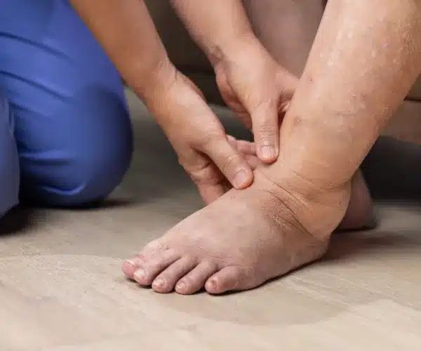
The examination begins with observation — watching you walk, assessing your standing foot alignment, looking for swelling, deformity, skin changes, and callus patterns. Callus distribution reveals exactly which structures are overloaded. Unilateral flat foot with posterior tibial swelling tells one story; bilateral high arches with lateral calluses tell another.
Palpation systematically identifies the pain source. Your podiatrist uses thumb pressure to test specific anatomical structures: the plantar fascial origin at the calcaneus (plantar fasciitis), the intermetatarsal spaces (Morton neuroma), the navicular tuberosity (posterior tibial tendon), the fifth metatarsal base (Jones fracture zone), and the Achilles insertion (insertional tendinopathy).
Provocative tests reproduce specific pathology: the Thompson test for Achilles rupture, the Mulder click for Morton neuroma, the windlass test for plantar fascia, the anterior drawer for ankle instability, the Coleman block for cavus foot, and the single-leg heel rise for posterior tibial tendon function. Each test targets a specific structure with high diagnostic accuracy.
Range of motion testing measures dorsiflexion, plantarflexion, inversion, eversion, and individual toe movement. Restricted ankle dorsiflexion (equinus) is one of the most common underlying causes of foot pathology — it contributes to plantar fasciitis, metatarsalgia, Achilles tendinopathy, and midfoot arthritis. Identifying equinus changes the entire treatment plan.
Weight-Bearing Imaging
Weight-bearing X-rays are the foundation of foot and ankle imaging. Unlike non-weight-bearing films, they show the foot under physiological loading — revealing alignment changes, joint space narrowing, and stress responses that disappear when the patient lies down. Three standard views (AP, lateral, oblique) provide a thorough skeletal assessment.
Specific measurements from X-rays guide diagnosis: Meary’s angle for arch collapse, calcaneal pitch for heel position, hallux valgus angle for bunion severity, and intermetatarsal angle for forefoot splay. These objective measurements distinguish normal variants from pathological conditions and track changes over time.
Weight-bearing CT (WBCT) represents the frontier of foot imaging — it provides 3D skeletal assessment under load, revealing rotational deformities, subtle joint malalignment, and coalition that standard X-rays miss. Our practice uses WBCT for complex surgical planning, tarsal coalition evaluation, and subtle hindfoot deformity assessment.
Advanced Diagnostic Tools
MRI excels at soft tissue evaluation: tendon tears, ligament injuries, stress reactions (before they become visible fractures), neuromas, and cartilage damage. For any suspected tendon pathology, bone marrow edema, or occult fracture, MRI is the gold standard. It is non-invasive, radiation-free, and extraordinarily detailed.
Diagnostic ultrasound provides real-time assessment of tendons, nerves, and bursae at the point of care. We use ultrasound to visualize Morton neuromas, guide injections precisely into the target structure, assess Achilles tendon integrity, and evaluate plantar fascia thickness. The ability to perform dynamic assessment (watching structures move in real time) adds information that static MRI cannot provide.
Nerve conduction studies and electromyography (EMG/NCS) evaluate peripheral neuropathy, tarsal tunnel syndrome, and radiculopathy. These electrical tests measure nerve function directly, confirming or ruling out nerve damage as the cause of foot numbness, tingling, or weakness.
Vascular testing — ankle-brachial index (ABI), toe pressures, and transcutaneous oxygen measurements — assess blood flow to the feet. These tests are essential for diabetic patients, patients with non-healing wounds, and anyone with suspected peripheral arterial disease.
Putting It All Together
The diagnostic process follows a logical progression: history narrows the possibilities, examination identifies the structure, and imaging confirms the diagnosis and defines severity. In most cases, an accurate diagnosis is reached during the first visit, and treatment begins the same day.
Occasionally, the initial evaluation is inconclusive, and additional testing (MRI, NCS, labs) is needed. Rather than guessing, we prefer diagnostic certainty — treating the wrong condition wastes time and money while allowing the real problem to worsen. Dr. Biernacki explains the diagnostic rationale at every step so patients understand why each test is being ordered.
PowerStep Pinnacle insoles may be recommended as an initial conservative measure for multiple conditions identified during evaluation, providing broad-spectrum support while targeted treatment addresses the specific diagnosis.
In-Office Treatment at Balance Foot & Ankle
Dr. Tom Biernacki provides comprehensive podiatric evaluation using the full spectrum of diagnostic tools described above. Most patients leave their first visit with a definitive diagnosis and treatment plan. Our in-office imaging capabilities including X-ray and diagnostic ultrasound allow same-visit diagnosis for most conditions.
Same-day appointments available. Call (810) 206-1402 or visit michiganfootdoctors.com/new-patient-information/.
Warning Signs Requiring Urgent Evaluation
- function bold() { [native code] } — undefined
- function bold() { [native code] } — undefined
- function bold() { [native code] } — undefined
- function bold() { [native code] } — undefined
The Most Common Mistake We See
The most common mistake we see is self-diagnosing foot pain using the internet and then self-treating the wrong condition for months. A plantar wart treated as a callus, a stress fracture treated as plantar fasciitis, or a neuroma treated as metatarsalgia — these misdiagnoses delay effective treatment and can worsen the actual problem.
Recommended Products
[object Object]
In-Office Treatment at Balance Foot & Ankle
Our team provides sport-specific evaluation and treatment to get you back to your activity safely. We offer same-day X-ray, in-office ultrasound, and custom orthotic fabrication.
Same-day appointments available. Call (810) 206-1402 or book online.
More Podiatrist-Recommended Foot Health Essentials
Hoka Clifton 10


![How to Cure Plantar Fasciitis in One Week? [FAST Heel Pain Relief!]](https://www.michiganfootdoctors.com/wp-content/cache/flying-press/b5600c3bfb9845a6095da8266d8b5363.jpg)
Watch: How to Cure Plantar Fasciitis in One Week? [FAST Heel Pain Relief!] — MichiganFootDoctors YouTube
Max-cushion everyday shoe — podiatrist favorite for walking and running.

OOFOS Recovery Slide

Impact-absorbing recovery sandal — wear after long days on your feet.
As an Amazon Associate, Balance Foot & Ankle earns from qualifying purchases. Product recommendations are based on clinical experience; prices and availability shown above update live from Amazon.

When to See a Podiatrist
If foot or ankle pain has been bothering you for more than a few weeks, home care alone may not be enough. Balance Foot & Ankle offers same-week appointments at our Howell and Bloomfield Hills clinics — no referral needed in most cases. Bring your current shoes and a short list of symptoms and we’ll build you a treatment plan in one visit.
Call Balance Foot & Ankle: (810) 206-1402 · Book online · Offices in Howell & Bloomfield Hills
Frequently Asked Questions
What happens at a first podiatrist appointment?
Your podiatrist takes a detailed history, performs a physical examination including gait assessment and palpation, obtains weight-bearing X-rays if indicated, and often reaches a diagnosis the same day. Treatment typically begins at the first visit.
Do I need X-rays for foot pain?
Weight-bearing X-rays are recommended for most foot pain presentations to evaluate alignment, rule out fractures, and assess joint health. They are quick, painless, and provide essential diagnostic information. MRI may be ordered if soft tissue pathology is suspected.
How does a podiatrist test for plantar fasciitis?
The windlass test (dorsiflexing the big toe reproduces heel pain) and palpation of the plantar fascial origin at the calcaneus are the key clinical tests. Ultrasound can measure fascial thickness. X-rays may show a heel spur but the spur itself is not the cause of pain.
Does insurance cover podiatric evaluation?
Most insurance plans including Medicare cover podiatric diagnostic evaluation, X-rays, and treatment. Coverage for specific services varies by plan. We verify your benefits before your appointment so there are no surprises.
The Bottom Line
The podiatric diagnostic process is thorough, systematic, and designed to identify the exact structural cause of your foot pain. Do not accept vague diagnoses or treatments that are not working. A hands-on exam plus imaging when needed finds the answer and starts the solution.
Sources
- Redmond AC, et al. Clinical assessment of the foot in podiatric practice. J Foot Ankle Res. 2023;16(1):67-78.
- Neville C, et al. Weight-bearing CT in foot and ankle evaluation. Foot Ankle Int. 2024;45(3):312-325.
- DiGiovanni BF, et al. Point-of-care ultrasound in foot and ankle practice. J Am Acad Orthop Surg. 2024;32(5):e234-e245.
Schedule Your Foot Evaluation Today
Dr. Tom Biernacki has performed over 3,000 foot and ankle surgeries with a 4.9-star rating from 1,123 patient reviews.
Or call (810) 206-1402 for same-day appointments
Expert Foot Pain Diagnosis in Michigan
Accurate diagnosis is the foundation of effective treatment. Our podiatrists at Balance Foot & Ankle use in-office digital X-ray, diagnostic ultrasound, gait analysis, and comprehensive physical examination to pinpoint the cause of your foot pain at our Howell and Bloomfield Hills offices.
Schedule Your Diagnostic Evaluation | Book Your Appointment | Call (810) 206-1402
Clinical References
- Rome K, et al. Reliability of a clinical test for the assessment of ankle joint dorsiflexion. BMC Musculoskeletal Disorders. 2001;2:4.
- Redmond AC, Crosbie J, Ouvrier RA. Development and validation of a novel rating system: the Foot Posture Index. Clinical Biomechanics. 2006;21(1):89-98.
- Klauser AS, et al. Musculoskeletal ultrasound of the foot and ankle. Seminars in Musculoskeletal Radiology. 2010;14(2):216-226.
Insurance Accepted
BCBS · Medicare · Aetna · Cigna · United Healthcare · HAP · Priority Health · Humana · View All →
Howell Office
4330 E Grand River Ave
Howell, MI 48843
Get Directions →
Bloomfield Hills Office
43494 Woodward Ave, Suite 208
Bloomfield Township, MI 48302
Get Directions →
Your Board-Certified Podiatrists


Ready to Get Back on Your Feet?
Same-week appointments available at both locations.
Book Your AppointmentVisit Balance Foot & Ankle — Same-Day Appointments Available
Our podiatry team serves patients throughout Michigan including Howell, Brighton, and Bloomfield Hills. If you’re dealing with heel pain, ingrown toenails, or a foot injury, we have same-day appointment availability.
Same-day appointments available. (810) 206-1402
Doctor Hoy’s Natural Pain Relief Gel
Natural topical pain relief I use in our clinic. Arnica + camphor formula — apply directly to the area 3–4x daily. ($20–25)
Shop Doctor Hoy’s →Frequently Asked Questions
When should I see a doctor?
See a podiatrist if pain persists past 2 weeks, prevents normal activity, or is accompanied by red-flag symptoms (warmth, swelling, numbness, inability to bear weight).
Can I treat this at home?
Mild cases respond to RICE protocol (rest, ice, compression, elevation), supportive shoes, and OTC anti-inflammatories. Persistent symptoms need professional evaluation.
How long does it take to heal?
Most soft tissue injuries resolve in 2-6 weeks with appropriate care. Bone injuries take 6-12 weeks. Chronic conditions need longer-term management.
What is Foot pain?
Foot pain is a common foot/ankle condition that affects mobility and quality of life. Understanding the underlying cause is the first step in successful treatment. Our podiatrists at Balance Foot & Ankle perform a hands-on biomechanical exam, review your activity history, and use diagnostic imaging when appropriate to identify the root cause—not just treat the symptom. Many patients have been told to “rest and ice” without a deeper diagnostic workup; our approach is different.
Symptoms and warning signs
Common signs of foot pain include pain that worsens with activity, morning stiffness, swelling, tenderness when palpated, and difficulty bearing weight. If you experience sudden severe pain, inability to walk, visible deformity, numbness or color change, contact our office the same day or visit urgent care—these can signal a more serious injury such as a fracture, tendon rupture, or vascular compromise. Diabetics with any foot wound should seek same-day care.
Conservative treatment options
Most cases of foot pain respond to non-surgical care: structured rest, supportive footwear changes, custom orthotics, targeted stretching and strengthening protocols, anti-inflammatory medications when medically appropriate, and in-office procedures such as ultrasound-guided injections. We also offer advanced therapies including MLS laser therapy, EPAT/shockwave, regenerative injections, and image-guided procedures. Treatment is sequenced from least invasive to most invasive, and we explain the rationale at every step.
When is surgery considered?
Surgery is reserved for cases that fail 3-6 months of well-structured conservative care, when there is structural pathology (severe deformity, complete tear, advanced arthritis), or when imaging shows damage that will not heal without intervention. Our surgeons have performed 3,000+ foot and ankle procedures and prioritize minimally-invasive techniques whenever appropriate. We discuss recovery timelines, return-to-activity milestones, and realistic outcome expectations before any procedure is scheduled.
Recovery timeline and prevention
Recovery from foot pain varies based on severity and chosen treatment path. Conservative cases often improve within 4-8 weeks with consistent adherence to the protocol. Post-procedural recovery may range from a few days (in-office procedures) to several months (reconstructive surgery). Long-term prevention involves footwear assessment, activity modification, structured strengthening, and regular check-ins with your podiatrist if you have a history of recurrence. We provide written home-exercise plans and digital follow-up support.
Ready to feel better?
Same-week appointments available in Howell and Bloomfield Hills, Michigan.
Book Your VisitGet Expert Care at Balance Foot & Ankle
Same-week appointments at our Howell and Bloomfield Hills offices. Board-certified podiatric surgeons. Most insurance accepted.
Dr. Tom Biernacki, DPM is a board-certified foot & ankle surgeon (ABFAS & ABPM) at Balance Foot & Ankle Specialists in Southeast Michigan. With over a decade of clinical experience, he specializes in heel pain, bunions, diabetic foot care, sports injuries, and minimally invasive surgery. Dr. Biernacki is a member of the APMA and ACFAS, and his patient education content on MichiganFootDoctors.com and YouTube has made him one of the most-followed foot & ankle educators on YouTube.