
Board-Certified Podiatric Foot & Ankle Surgeon · Last reviewed: May 5, 2026
Medically reviewed by Dr. Tom Biernacki, DPM
Board-certified podiatric surgeon | Balance Foot & Ankle
Last reviewed: April 2026
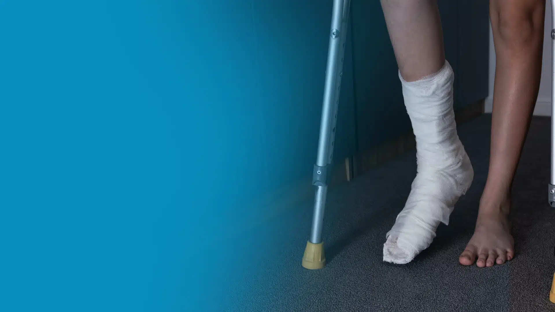
You landed awkwardly on the outside of your foot during a basketball game, or felt a sharp snap mid-stride on a long run. Maybe it was a simple stumble off a curb. However it happened, you now have pain and swelling along the outer edge of your foot — and you’re wondering whether this will just walk off, or whether you need to get it checked out immediately.
If the pain is centered at or just below the bony bump on the outside of your foot — the base of your fifth metatarsal — there is a meaningful chance you have a Jones fracture. Of all the foot fractures we see at Balance Foot & Ankle, Jones fractures are among the most commonly mismanaged. They’re frequently mistaken for a simple ankle sprain or a minor avulsion fracture, which leads to delayed treatment, chronic pain, and in some cases, a fracture that never fully heals.

In this guide, Dr. Tom Biernacki walks you through the exact anatomy of a Jones fracture, how it differs from an avulsion fracture — a critical distinction that completely changes treatment — your full range of treatment options, and a realistic recovery timeline based on what we see in practice every day.
The most important clinical decision with Jones Fracture isn’t which treatment to start with — it’s identifying the correct subtype. That changes everything. Call (810) 206-1402.
Table of Contents
- What Is a Jones Fracture?
- Jones Fracture Symptoms
- What Causes a Jones Fracture?
- How Is a Jones Fracture Diagnosed?
- Jones Fracture vs. Avulsion Fracture: A Critical Distinction
- Jones Fracture Treatment Options
- Recovery Timeline
- Warning Signs: When to See a Podiatrist
- The Most Common Mistake We See
- Frequently Asked Questions
- The Bottom Line
- Sources

What Is a Jones Fracture?
A Jones fracture is a specific type of break that occurs at a precise location on the fifth metatarsal — the long bone running along the outer edge of your foot that connects to your little toe. Understanding exactly where this fracture occurs is not merely academic; it determines everything about how difficult healing will be and what treatment is required.
The fifth metatarsal is divided into three anatomical zones. Zone 1 is the very tip of the base — the prominent bony bump you can feel on the outside of your foot. Zone 2 is the proximal diaphysis, a narrow transitional region just beyond that tuberosity where the bone transitions from its wider base to the narrower shaft. Zone 3 is the proximal shaft itself, farther down the bone toward the toes. A true Jones fracture occurs in Zone 2 — the proximal diaphysis — and this precise location is what makes it so clinically significant.
The reason Zone 2 matters so much comes down to blood supply. The base of the fifth metatarsal receives blood from branches of the peroneal artery. The shaft has its own blood supply from the nutrient artery entering further down. But Zone 2 sits in a vascular watershed area — a gap between these two blood supplies where neither feeds the bone reliably. When you fracture this zone, the bone fragments struggle to mount an adequate healing response. Without reliable blood flow delivering oxygen, nutrients, and bone-forming cells, healing can stall or fail entirely — a complication called nonunion.
The fracture was first described in 1902 by British surgeon Sir Robert Jones, who fractured his own fifth metatarsal while dancing at a military party — making this one of the few orthopedic conditions named after a physician who personally experienced it. Over a century later, the challenge he identified remains just as relevant in modern sports medicine.
In our clinic at Balance Foot & Ankle, we see Jones fractures regularly — not just in elite athletes, but in recreational runners, weekend basketball players, and even people who simply stepped off a curb the wrong way. The injury does not discriminate by fitness level, but the consequences of mismanaging it can affect anyone equally.
Key takeaway: A Jones fracture is not just any fifth metatarsal fracture — it specifically involves Zone 2, a vascular watershed area where bone healing is inherently slow and unreliable. Getting the diagnosis right is the first step toward getting the treatment right.
Jones Fracture Symptoms
Jones fracture symptoms typically begin immediately after injury — though in the case of stress fractures, they develop insidiously over days to weeks of increased activity. Recognizing the specific pattern of symptoms helps distinguish a Jones fracture from a lateral ankle sprain, which presents similarly and is far more common.
The hallmark symptom is sharp, localized pain along the outer edge of the foot, specifically at or just below the prominent bony bump at the base of the fifth metatarsal. Unlike lateral ankle sprains — which produce tenderness over the lateral ankle ligaments (anterior talofibular and calcaneofibular ligaments above and slightly posterior to this area) — Jones fracture pain is lower on the foot, further toward the arch side and slightly forward of the ankle. This anatomical distinction is clinically critical and separable on examination.
The most common symptoms we see in our clinic include:
- Immediate, localized pain at the outer midfoot — not centered at the ankle itself
- Rapid swelling along the lateral foot within minutes to hours of injury
- Bruising (ecchymosis) spreading along the outer foot, sometimes toward the ankle within 24–48 hours
- Difficulty bearing weight — most patients can limp but cannot walk normally without pain
- Point tenderness directly over Zone 2 when pressed with a finger
- Worsening pain with push-off motions — stepping up stairs, walking quickly, or rising on tiptoe
- An audible or felt “snap” at the time of injury in acute fractures (not always present in stress fractures)
Stress fractures of Zone 2 — sometimes called “march fractures” of the fifth metatarsal — present differently. They develop gradually over a training cycle where mileage or intensity has increased. Pain typically begins as a dull ache that worsens with activity and improves with rest, then progressively becomes more constant as the fracture propagates. Many patients with stress fractures of Zone 2 walk into our office having trained through the pain for weeks before seeking care.
| Symptom | Acute Jones Fracture | Zone 2 Stress Fracture |
|---|---|---|
| Onset | Immediate after specific trauma | Gradual over days to weeks |
| Pain quality | Sharp, intense immediately | Dull ache → progressively sharper |
| Swelling | Rapid and significant | Mild to moderate, builds over time |
| Bruising | Common within 24–48 hours | Rare or absent |
| Weight-bearing | Painful but usually possible | Manageable early, worsens with activity |
| Audible snap | Often present | Absent |
| Activity history | Single traumatic event | Increased training load over weeks |
What Causes a Jones Fracture?
Jones fractures occur through two distinct mechanisms: a single acute traumatic event, or the cumulative stress of repetitive loading. Understanding which mechanism caused your fracture matters because it affects how we approach treatment and how long recovery will realistically take.
The most common acute mechanism is a forced inversion (rolling inward) of the ankle combined with a plantar-flexed foot. When you land with your foot rolled inward on the outer edge — as happens when you come down awkwardly from a jump in basketball, or step into a hole while running — the peroneal muscles attached to the fifth metatarsal experience a sudden violent tensile force. Simultaneously, the bone absorbs compressive forces from impact. Zone 2 is the mechanically weakest point in this equation, and it fails first.
A second acute mechanism is direct blow or lateral compression of the outer foot, seen in contact sports, vehicle accidents, or objects falling on the foot. The fracture pattern may vary slightly, but Zone 2 remains the most vulnerable point anatomically.
Stress fractures of Zone 2 develop through cumulative bone fatigue from repetitive loading. When bone is stressed cyclically — through running, jumping, dancing, or marching — microscopic cracks develop faster than the bone can repair them. Over time these accumulate into a partial or complete stress fracture. This is why Zone 2 stress fractures are so prevalent in:
- Basketball and soccer players — high-frequency push-off and lateral cutting forces
- Long-distance runners, especially those who recently increased mileage or transitioned to minimalist footwear
- Ballet and modern dancers who spend extended time in demi-pointe
- Military recruits during basic training with rapid activity escalation
- Gymnasts with repetitive high-impact landings
Several risk factors significantly increase susceptibility to both types. Cavus foot (high-arched foot structure) is among the most important — this foot type transfers disproportionate force to the lateral column with every step, chronically overloading Zone 2. Vitamin D and calcium deficiency reduce bone density and the ability to repair microdamage between training sessions. Poor or worn footwear with inadequate lateral support compounds the mechanical load at the fifth metatarsal base.
The presentation we see most commonly in our clinic is an “acute-on-chronic” pattern: a patient who has had low-grade lateral foot pain for weeks or months of heavy training, then experiences a final traumatic event that completes the fracture. This is actually a stress fracture that has finally fully snapped. These fractures have the most challenging healing because the bone was already compromised before the acute event, and sclerosis (hardening of the fracture zone) is often visible on the initial X-ray.
Key takeaway: High-arched (cavus) foot structure is one of the strongest risk factors for Jones fractures and refractures. Addressing lateral column overloading with custom orthotics is essential to long-term prevention after any fifth metatarsal Zone 2 injury.
How Is a Jones Fracture Diagnosed?
Diagnosing a Jones fracture accurately — and distinguishing it precisely from an avulsion fracture of Zone 1 — requires a systematic clinical examination followed by appropriate imaging. Getting the zone right changes treatment completely, so we approach every lateral foot injury with a structured diagnostic sequence.
Clinical Examination
We begin with a thorough history: mechanism of injury, exact onset, any prior episodes of lateral foot pain (suggesting pre-existing stress changes), training volume, and relevant medical history including nutritional status and prior foot conditions. The difference between an acute twisting mechanism and a gradual onset during increased training immediately directs our differential diagnosis.
Physical examination focuses on precise palpation. The key maneuver is applying direct fingertip pressure over Zone 2 — the narrow region just distal to the tuberosity, at the metadiaphyseal junction. Exquisite point tenderness at this specific location, clearly distinct from tenderness over the lateral ankle ligaments above, strongly suggests a Jones fracture. We also assess for concurrent ligamentous injury, as lateral ankle sprains and fifth metatarsal fractures frequently coexist.
X-Ray (Standard First-Line Imaging)
Weight-bearing foot X-rays in three views — AP (anteroposterior), lateral, and oblique — are the first imaging study ordered for any suspected Jones fracture. In acute fractures, a visible fracture line through Zone 2 is typically identifiable on the oblique view. However, early stress fractures frequently appear normal on initial X-ray, and a negative film does not rule out significant bone pathology.
The oblique view is most important: it provides the clearest visualization of the fifth metatarsal base and allows precise zone classification. We also look for medullary sclerosis — whitish, hardened bone within the fracture zone — which indicates chronicity and significantly worsens the prognosis for non-surgical healing.
CT Scan (When X-Ray Is Equivocal)
When X-ray findings are equivocal — a thin fracture line that may or may not cross Zone 2, or questions about displacement or comminution — CT provides superior bony detail. CT can precisely characterize fracture location, degree of fragmentation, displacement, and the extent of any sclerosis. We order CT preoperatively for virtually every Jones fracture heading toward surgical repair, as screw sizing depends on accurate measurement of the medullary canal diameter.
MRI (Gold Standard for Stress Fractures)
When clinical suspicion for a stress fracture is high but X-rays are negative, MRI is the definitive diagnostic tool. MRI detects bone marrow edema — the earliest sign of bone stress — before a frank fracture line appears on plain film. This is critical for athletes: catching a stress reaction early and modifying training can prevent progression to a complete fracture, potentially avoiding surgery altogether. A negative MRI reliably rules out significant osseous pathology and redirects workup toward soft-tissue causes.
Differential Diagnosis
Several conditions mimic Jones fractures on history and examination and must be considered in every evaluation:
- Zone 1 avulsion fracture (pseudo-Jones fracture) — the most critical distinction; treated far more conservatively with weight-bearing as tolerated
- Lateral ankle sprain — most common cause of acute lateral foot and ankle pain; no bony injury on X-ray
- Peroneal tendon injury — tendinopathy or partial tear at the peroneus brevis insertion on the fifth metatarsal tuberosity
- Cuboid fracture — less common; pain more centralized on the dorsal midfoot and cuboid facet
- Os vesalianum or os peroneum — accessory ossicles near the fifth metatarsal base that can be mistaken for fracture fragments on X-ray
- Lisfranc ligament injury — pain more medial, at the tarsometatarsal joint complex; separate injury pattern
Jones Fracture vs. Avulsion Fracture: A Critical Distinction
No distinction in foot fracture management carries more clinical weight than the difference between a Jones fracture and an avulsion fracture of the fifth metatarsal base. These two injuries look similar on presentation, occur from overlapping mechanisms, and affect adjacent zones of the same bone — yet their treatment, prognosis, and risk of complications are completely different. Treating an avulsion fracture like a Jones fracture is unnecessary overtreatment. Treating a Jones fracture like an avulsion fracture risks nonunion, refracture, and years of chronic lateral foot pain.
| Feature | Jones Fracture (Zone 2) | Avulsion Fracture (Zone 1) |
|---|---|---|
| Location on 5th metatarsal | Proximal diaphysis (shaft-base junction) | Tip of the tuberosity at the very base |
| Common nickname | “True Jones fracture” | “Pseudo-Jones fracture” |
| Primary mechanism | Ankle inversion + push-off; stress loading | Acute ankle inversion; peroneus brevis pull |
| Blood supply to fracture zone | Watershed — poor and unreliable | Good — proximal to watershed |
| Nonunion risk | High — up to 25–30% without surgery | Very low — heals reliably in most cases |
| Typical treatment | NWB cast or surgical screw fixation | Walking boot or hard-soled shoe; WBAT |
| Expected healing time | 6–20 weeks conservative; 10–14 weeks surgical | 4–8 weeks |
| Surgery required? | Often yes — especially in athletes | Rarely if ever |
On an oblique foot X-ray, the fracture line of an avulsion fracture runs perpendicular to the long axis of the fifth metatarsal — horizontally across the very tip of the tuberosity, often appearing as a small chip. A Jones fracture line runs across the metadiaphyseal junction, clearly distal to the tuberosity and at or within the shaft. An experienced podiatrist can usually make this distinction reliably on plain X-ray, but borderline cases — particularly when the fracture line is at the very boundary of Zones 1 and 2 — warrant CT for definitive classification.
Jones Fracture Treatment Options
Jones fracture treatment depends on three factors: the acuity of the fracture (acute traumatic vs. chronic stress), the patient’s activity demands, and imaging characteristics including the presence or absence of medullary sclerosis. At Balance Foot & Ankle, we evaluate all three before recommending a treatment pathway — because there is no one-size-fits-all protocol for this fracture.
Non-Surgical Treatment
Non-surgical management is appropriate for low- to moderate-demand patients with an acute Jones fracture and no medullary sclerosis on X-ray — meaning this is a first-time, truly acute injury without pre-existing stress fracture changes. The protocol is strict:
- Non-weight-bearing (NWB) short leg cast for 6–8 weeks — the cornerstone of non-operative Jones fracture management. The cast must be non-weight-bearing; even partial weight-bearing in a boot without cast immobilization can disrupt healing at Zone 2.
- Crutches or knee scooter to maintain strict NWB compliance throughout the cast period
- X-ray at 4–6 weeks to assess early bridging callus formation and confirm healing is progressing
- Transition to a removable protective boot after confirmed healing on X-ray, with gradual progressive weight-bearing
- Physical therapy for peroneal strengthening, proprioceptive retraining, and sport-specific activity preparation before return to full activity
The important caveat with non-surgical treatment is that healing rates are variable and lower than most patients expect. Published data show healing rates of 72–93% with strict non-operative management — meaning up to 28% of patients will not heal adequately without eventually requiring surgery. Medullary sclerosis on the initial X-ray is a warning sign that non-surgical management is likely to fail; we counsel these patients honestly about the probability that surgery will ultimately be required.
A well-fitted fracture walker boot is essential during the rehabilitation phase after cast removal, providing the protection needed as bone consolidation completes and the patient transitions back to full weight-bearing.
Surgical Treatment: Intramedullary Screw Fixation
Surgical repair is the preferred treatment for athletes, highly active patients, anyone with sclerosis on X-ray indicating a chronic stress fracture, and patients who have already failed non-operative management. The gold standard procedure is intramedullary (IM) screw fixation — a minimally invasive technique where a cannulated stainless steel or titanium screw is inserted through a small incision at the base of the fifth metatarsal and threaded down the medullary canal, compressing the fracture site from within.
The procedure is typically performed as an outpatient surgery under general or regional anesthesia and takes 20–45 minutes. The biological rationale is that rigid interfragmentary compression at Zone 2 counteracts the shear forces that prevent healing in the watershed vascular zone. By stabilizing fracture fragments and promoting direct bone contact, the screw allows the bone’s limited blood supply to mount an effective healing response that it cannot achieve in an unstabilized environment.
Outcome data strongly support surgery for higher-demand patients:
- Return-to-sport averages 7–10 weeks with surgery vs. 14–26 weeks without
- Nonunion rate drops to under 5% with surgery vs. up to 28% without
- Refracture rate after surgical repair: approximately 10–15% (vs. up to 30% with non-surgical management)
- Patient satisfaction consistently high in published series
Larger-diameter screws (4.5–5.5 mm depending on canal size) provide better rotational stability and are associated with lower refracture rates than the smaller screws used in older studies. We determine screw size from preoperative CT measurement of the medullary canal, individualized to each patient’s anatomy.
Nutritional Optimization for Bone Healing
Regardless of whether treatment is surgical or non-surgical, optimizing bone-healing biology is essential and too often overlooked. We routinely assess and address three key nutritional factors:
- Vitamin D — deficiency is extremely common in patients who sustain stress fractures. We check serum 25-hydroxyvitamin D levels and supplement to achieve 40–60 ng/mL. Doses of 2,000–5,000 IU daily are often required.
- Calcium — 1,000–1,200 mg daily from dietary and supplemental sources combined supports the mineralization phase of fracture healing
- Protein — at least 1.2–1.6 g/kg body weight per day supports the collagen matrix that forms the scaffold for new bone
Biomechanical Correction: Addressing the Root Cause
In patients with cavus foot or other biomechanical contributors to lateral column overloading, addressing the underlying foot structure is essential to preventing refracture. Custom orthotic insoles designed to redistribute plantar pressure away from the lateral foot are a cornerstone of long-term Jones fracture management. Without biomechanical correction, refracture rates remain elevated even after technically successful surgical fixation.
Jones Fracture Recovery Timeline
Recovery from a Jones fracture is often longer and less predictable than patients expect. We set clear expectations from the outset because the most common reason for poor outcomes is non-compliance with non-weight-bearing restrictions — driven by frustration at the slow pace of healing. The following timeline applies to both surgical and non-surgical pathways.
| Phase | Non-Surgical Timeline | Surgical Timeline |
|---|---|---|
| Weeks 0–2 | NWB short leg cast applied; pain and swelling management; elevation above heart level; strict crutch use | Outpatient surgery; NWB splint; elevation and ice; oral analgesia; wound check at 10–14 days |
| Weeks 2–6 | Continued NWB cast; X-ray at 4 weeks to assess early callus; no weight-bearing at all | Transition to NWB boot at 2 weeks; X-ray confirms screw position and early bone response |
| Weeks 6–10 | X-ray shows bridging callus → transition to walking boot; progressive weight-bearing begins; PT starts | X-ray at 6–8 weeks shows callus; protected weight-bearing begins in boot; formal PT initiated |
| Weeks 10–14 | Full weight-bearing in boot; transition to supportive shoe with orthotic; PT progresses to sport-specific work | Full weight-bearing in shoe with orthotic; sport-specific training begins under PT supervision |
| Weeks 14–26 | Gradual return to low-impact activity; serial X-ray monitoring; return to sport contingent on imaging and functional testing | Return to full sport activity for most athletes; final X-ray to confirm complete healing |
| 6–12 months | Full healing confirmed; long-term orthotic use for high-risk patients; bone density optimization ongoing | Screw removal only if symptomatic hardware irritation; long-term orthotic; bone density optimization |
The single most important variable affecting non-surgical recovery time is the presence or absence of medullary sclerosis on initial X-ray. Patients with sclerosis face prolonged timelines and substantially elevated nonunion risk. When we see sclerosis on a patient’s first X-ray — combined with a history of weeks of lateral foot pain before the acute fracture event — we have an honest conversation: non-surgical management will likely fail, and proceeding to surgery from the outset is more efficient and results in a shorter overall recovery.
Key takeaway: For competitive athletes, surgery provides faster return-to-sport (7–10 weeks vs. 14–26 weeks), dramatically lower nonunion rates, and superior long-term outcomes. The evidence supporting early surgical fixation in athletes is consistent across decades of sports medicine literature.
Warning Signs: When to See a Podiatrist
⚠️ See a podiatrist urgently if you experience:
- Sudden sharp pain on the outer edge of your foot following a traumatic event, fall, or awkward landing
- An audible “crack” or “pop” at the outer foot during injury, followed by swelling and inability to walk normally
- Lateral foot pain that has been building over more than 2–3 weeks of increased training and is progressively worsening
- Outer foot pain present at rest or that wakes you from sleep — this level of bone stress requires urgent evaluation
- Swelling, bruising, or visible deformity over the outer midfoot after any trauma
- Renewed or increased pain in a previously diagnosed Jones fracture site — refracture is common and requires prompt reassessment
- Numbness, tingling, or color changes in toes following outer foot injury — though rare, vascular or nerve injury warrants same-day evaluation
If you are unsure whether you have a Jones fracture or a simple sprain, the correct decision is always to get evaluated with an X-ray. The cost of imaging is trivial compared to the cost of missing a Jones fracture and allowing a nonunion to develop over weeks or months of inadequate treatment.
The Most Common Mistake We See
The single most common mistake we see with Jones fractures is treating them like avulsion fractures — or managing them the way most lateral ankle sprains are handled. This happens at urgent care centers, emergency departments, and primary care offices where the subtle zone distinction on X-ray is missed or not recognized as clinically significant.
The scenario is familiar: a patient arrives with lateral foot pain after rolling their ankle. An X-ray is taken. A fifth metatarsal base fracture is identified and correctly diagnosed. But instead of zone classification and specific counseling about the poor vascularity of Zone 2, the patient is placed in a walking boot and told they can put weight on it. This is the correct management for a Zone 1 avulsion fracture. For a Jones fracture, it is undertreatment that can result in delayed union or complete nonunion.
By the time these patients reach our office — often 8 to 12 weeks later with persistent, unresolved lateral foot pain — they have a Zone 2 fracture with varying degrees of sclerosis and failed bone healing. At that point, treatment is substantially more complex than it would have been at initial presentation. Surgery is almost always required, recovery is longer, and bone quality at the fracture site is compromised from weeks of inadequate immobilization. The original “wait and see in a boot” approach has added months to the recovery trajectory.
The second most common mistake is non-weight-bearing non-compliance. Patients consistently underestimate how important strict NWB is to Jones fracture healing. We regularly see patients who walked without crutches “just a few times” because crutches are difficult, the boot felt supportive enough, or they were told by someone else that full non-weight-bearing wasn’t necessary. Even occasional loading of Zone 2 during the healing period can disrupt the nascent fracture callus and restart the healing clock. We are explicit with every non-operative Jones fracture patient: this is a strict non-weight-bearing protocol, not a “comfortable weight-bearing as tolerated” situation.
Frequently Asked Questions
Can a Jones fracture heal without surgery?
Yes — for low-demand patients without medullary sclerosis on initial X-ray, strict non-weight-bearing in a short leg cast for 6–8 weeks produces adequate healing in approximately 72–93% of cases. However, athletes, highly active individuals, and anyone with evidence of pre-existing bone stress (sclerosis) on X-ray have substantially lower healing rates with non-surgical management and are generally better served by surgical fixation at the outset. The decision should be individualized based on imaging findings and activity demands.
How long does Jones fracture recovery take?
With non-surgical treatment, expect 6–20 weeks from injury to return to full activity, depending on healing response. Surgical fixation significantly compresses this timeline — most athletes with surgical repair return to sport in 7–14 weeks. The wide range in non-surgical recovery reflects the inherent variability of healing in Zone 2’s vascular watershed territory. Patients with sclerosis on X-ray should plan for the longer end of both ranges.
What happens if a Jones fracture doesn’t heal?
A Jones fracture that fails to heal is called a nonunion. Clinically, nonunion presents as persistent lateral foot pain, functional limitation with activity, and on X-ray, a persistent fracture gap with surrounding sclerosis. Treatment for established nonunion almost universally requires surgery — typically intramedullary screw fixation combined with bone grafting to stimulate the healing response at the previously failed fracture site. This is a more complex, higher-risk procedure than primary fixation and carries a longer recovery. Early and appropriate treatment at initial presentation is the most reliable way to avoid this outcome.
Can I drive with a Jones fracture?
If the fracture is in your right foot, driving is not safe during the non-weight-bearing phase — you cannot operate brake and gas pedals safely with an NWB restriction. If the fracture is in your left foot, driving an automatic transmission vehicle may be permissible at your treating podiatrist’s discretion. Clarify driving restrictions explicitly at your first appointment, as the answer depends on your specific fracture management plan and local traffic regulations.
Will I need the screw removed after Jones fracture surgery?
Most patients do not require screw removal. The intramedullary screw is a permanent implant in the majority of cases and causes no problems once healing is complete. A minority of patients develop hardware irritation — often related to the screw head prominence near the fracture site — and may request removal once the bone has fully healed, typically no sooner than 12 months post-surgery. Routine screw removal is not standard practice; we remove hardware only when the patient has symptoms attributable specifically to the implant.
Is a Jones fracture the same as a broken fifth metatarsal?
Not exactly. “Broken fifth metatarsal” describes any fracture along the entire fifth metatarsal bone, including all three zones and the shaft. A Jones fracture is a specific subtype — a Zone 2 fracture at the proximal diaphysis — with a distinctly challenging prognosis. Other fifth metatarsal fractures include Zone 1 avulsion fractures, mid-shaft spiral fractures (dancer’s fracture), and distal shaft fractures. Each has a different treatment plan and prognosis. Zone classification on X-ray is the essential step that determines which type you have and how it should be treated.
The Bottom Line
A Jones fracture is one of the most consequential “small” injuries in foot and ankle medicine. It occurs in a specific zone of the fifth metatarsal with notoriously poor blood supply, heals slowly and unpredictably, and carries a genuine risk of nonunion and refracture if not properly managed from the first visit. The most critical first step is accurate diagnosis — specifically, zone classification that distinguishes a Jones fracture from the far more common and forgiving avulsion fracture of Zone 1.
Treatment selection must account for activity demands, imaging characteristics (especially the presence of sclerosis), and realistic expectations about healing timelines. For most athletes and active patients, surgical intramedullary screw fixation offers significantly faster return to sport, dramatically lower nonunion rates, and better long-term outcomes than non-surgical management. For lower-demand patients with clean imaging, strict non-weight-bearing gives the fracture the best possible chance to heal without surgery.
If you have lateral foot pain after trauma — or persistent outer foot pain building through increased training — do not wait and hope it resolves. Get evaluated with an X-ray, get a precise zone classification, and make a treatment decision based on your specific situation. Catching a Jones fracture early and treating it correctly is the difference between a 10-week recovery and a year of chronic pain.
Sources
- Japjec M, et al. “Jones fracture — surgical treatment: a systematic review.” Injury. 2021;52(Suppl 5):S87–S92.
- Roche AJ, Calder JD. “Treatment and return to sport following a Jones fracture of the fifth metatarsal: a systematic review.” Knee Surgery, Sports Traumatology, Arthroscopy. 2013;21(6):1307–1315.
- Murawski CD, Kennedy JG. “Percutaneous internal fixation of proximal fifth metatarsal Jones fractures with Charlotte Carolina screw and bone marrow aspirate concentrate.” The American Journal of Sports Medicine. 2011;39(6):1295–1301.
- Porter DA, et al. “Fifth metatarsal Jones fractures in the athlete.” Foot & Ankle International. 2005;26(3):204–210.
- Lawrence SJ, Botte MJ. “Jones’ fractures and related fractures of the proximal fifth metatarsal.” Foot & Ankle. 1993;14(7):358–365.
- Bigsby E, et al. “Epidemiology of fifth metatarsal fractures.” The Annals of The Royal College of Surgeons of England. 2014;96(7):519–521.
Lateral Foot Pain After an Injury? Get an Accurate Diagnosis Today.
Same-day appointments for foot fracture evaluation in Howell & Bloomfield Hills, MI. On-site X-ray. Expert surgical and non-surgical Jones fracture management.
4.9★ | 1,123 Reviews | 3,000+ Surgeries | Dr. Tom Biernacki, DPM
Or call: (810) 206-1402
Dr. Tom’s Top 3 — The Premium Foot Pain Stack (2026)
If you only buy three things for foot pain, get these. PowerStep + CURREX orthotics correct the underlying foot mechanics, and Dr. Hoy’s pain gel delivers fast topical relief. This is the exact stack Dr. Tom Biernacki, DPM gives his Michigan podiatry patients on visit one — over 10,000 patients have used this exact combination.
Dr. Tom Biernacki, DPM is a board-certified podiatrist + Amazon Associate. Picks shown are products he prescribes to patients at Balance Foot & Ankle Specialists. We earn a commission on qualifying purchases at no extra cost to you. All products independently tested + reviewed for 30+ days minimum. Last verified: April 28, 2026.

PowerStep Pinnacle MaxxDr. Tom’s #1 Brand
Dr. Tom’s most-prescribed OTC orthotic. Lateral wedge corrects overpronation that causes 90% of foot pain. Deep heel cradle stabilizes the ankle. Built by podiatrists, used by patients worldwide.
- Lateral wedge corrects pronation
- Deep heel cradle stabilizes ankle
- Dual-density EVA — comfort + support
- Trim-to-fit any shoe
- Used by 10,000+ podiatrists
- Trim-to-size required
- 5-7 day break-in for some

CURREX RunProDr. Tom’s #1 Brand
3 arch heights for custom fit (Low/Med/High). Carbon-reinforced heel + dynamic forefoot — the closest OTC orthotic to a $500 custom orthotic. Engineered in Germany.
- 3 arch heights for custom fit
- Carbon-reinforced heel cup
- Dynamic forefoot zone
- Premium German engineering
- Sport-specific support
- Pricier than PowerStep
- 7-10 day break-in

Dr. Hoy’s Natural Pain Relief GelDr. Tom’s #1 Brand
Menthol-based natural pain relief — Dr. Tom’s #1 brand for fast relief without greasy residue. Safe for diabetics + daily use. Cleaner formula than Voltaren or Biofreeze.
- Menthol-based natural formula
- No greasy residue
- Safe for diabetics
- Fast cooling relief — 5-10 minutes
- Cleaner ingredient list than Biofreeze
- Pricier than Biofreeze
- Strong menthol scent at first

What is Stress fracture?
Stress fracture is a common foot/ankle condition that affects mobility and quality of life. Understanding the underlying cause is the first step in successful treatment. Our podiatrists at Balance Foot & Ankle perform a hands-on biomechanical exam, review your activity history, and use diagnostic imaging when appropriate to identify the root cause—not just treat the symptom. Many patients have been told to “rest and ice” without a deeper diagnostic workup; our approach is different.
Symptoms and warning signs
Common signs of stress fracture include pain that worsens with activity, morning stiffness, swelling, tenderness when palpated, and difficulty bearing weight. If you experience sudden severe pain, inability to walk, visible deformity, numbness or color change, contact our office the same day or visit urgent care—these can signal a more serious injury such as a fracture, tendon rupture, or vascular compromise. Diabetics with any foot wound should seek same-day care.
Conservative treatment options
Most cases of stress fracture respond to non-surgical care: structured rest, supportive footwear changes, custom orthotics, targeted stretching and strengthening protocols, anti-inflammatory medications when medically appropriate, and in-office procedures such as ultrasound-guided injections. We also offer advanced therapies including MLS laser therapy, EPAT/shockwave, regenerative injections, and image-guided procedures. Treatment is sequenced from least invasive to most invasive, and we explain the rationale at every step.
When is surgery considered?
Surgery is reserved for cases that fail 3-6 months of well-structured conservative care, when there is structural pathology (severe deformity, complete tear, advanced arthritis), or when imaging shows damage that will not heal without intervention. Our surgeons have performed 3,000+ foot and ankle procedures and prioritize minimally-invasive techniques whenever appropriate. We discuss recovery timelines, return-to-activity milestones, and realistic outcome expectations before any procedure is scheduled.
Recovery timeline and prevention
Recovery from stress fracture varies based on severity and chosen treatment path. Conservative cases often improve within 4-8 weeks with consistent adherence to the protocol. Post-procedural recovery may range from a few days (in-office procedures) to several months (reconstructive surgery). Long-term prevention involves footwear assessment, activity modification, structured strengthening, and regular check-ins with your podiatrist if you have a history of recurrence. We provide written home-exercise plans and digital follow-up support.
Ready to feel better?
Same-week appointments available in Howell and Bloomfield Hills, Michigan.
Book Your VisitIn-Office Treatment at Balance Foot & Ankle
If home treatment isn’t providing relief for your foot and ankle injuries, our podiatry team at Balance Foot & Ankle can help with same-day evaluations and advanced in-office care.
Same-day appointments available. (810) 206-1402
Doctor Hoy’s Natural Pain Relief Gel
Natural topical pain relief I use in our clinic. Arnica + camphor formula — apply directly to the area 3–4x daily. ($20–25)
Shop Doctor Hoy’s →Related Conditions
Frequently Asked Questions
What injuries require a walking boot?
Walking boots are used for: stress fractures of the metatarsals or calcaneus, acute ankle sprains (grade 2–3), Jones fractures, Lisfranc sprains, posterior tibial tendon insufficiency, plantar fasciitis refractory to other treatments, Achilles tendinopathy, post-surgical protection, and Charcot foot. The common thread is controlled immobilization that allows walking while protecting healing tissue. Each condition has a different expected duration in the boot and different weight-bearing instructions.
How long do I have to wear a walking boot?
Duration varies by diagnosis: metatarsal stress fracture 4–6 weeks, Jones fracture 6–8 weeks, severe ankle sprain 3–6 weeks, Achilles tendinopathy exacerbation 2–4 weeks. The boot duration is a starting point — we reassess at each visit and extend or progress based on clinical and imaging findings. Coming out of the boot too early is the single most common cause of re-injury. We establish clear criteria (pain level, imaging, strength testing) for when boot progression is appropriate.
Should I wear the walking boot all day, including when sleeping?
For most fractures: yes, including sleeping, for the first 2–4 weeks. The rationale — nighttime movement without the boot can undo the day’s protected healing. Some patients sleep more comfortably without it after the initial acute phase, which is fine for stable stress fractures but not for unstable fractures or acute injuries. We’ll give you specific sleeping instructions based on your injury. If not told otherwise, wearing it to bed is always the safer default.
Can I drive with a walking boot on my right foot?
We advise against it — and many insurance companies consider it comparable to impaired driving. A boot on the right foot significantly slows braking reaction time. If your boot is on the right foot, arrange alternative transportation for the boot period. Left-foot boots don’t affect driving mechanics in most vehicles. Automatic transmission cars with a left-foot boot are generally manageable; standard transmission is more complex. When in doubt, don’t drive — your safety and legal liability are at stake.
What is an Aircast boot vs. a standard walking boot?
Aircast and similar air-bladder boots (CAM walkers) allow inflation around the ankle for customizable compression and stability — particularly useful for ankle sprains and soft tissue injuries where swelling fluctuates. Standard rigid boots offer fixed immobilization more appropriate for fractures requiring strict positional control. We select the boot type based on injury mechanism and healing requirements. For most fractures, a rigid CAM boot is standard; for ankle ligament injuries, an air stirrup design is often preferred.
Will I lose muscle while wearing a walking boot?
Yes — disuse atrophy begins within 48–72 hours of immobilization. Calf muscle volume can decrease 3–5% per week in a boot. This is normal and expected. Upper-body workouts, swimming, and seated exercises maintain cardiovascular fitness during boot wear. After boot removal, a structured rehabilitation protocol (typically 4–8 weeks of progressive calf loading and balance training) rebuilds strength. Patients who do formal physical therapy post-boot return to full function 4–6 weeks faster than those who just stop wearing the boot.
How do I keep my other leg and back from hurting while in a boot?
The boot’s heel height (typically 3–4cm) creates a limb length discrepancy that stresses the opposite knee, hip, and lower back. Two solutions: (1) Use a boot with a rocker bottom sole to reduce gait compensation; (2) Add a heel lift to the opposite shoe to equalize leg lengths. Most patients who develop contralateral knee or back pain during boot wear benefit immediately from a 1–2cm heel lift in the non-booted shoe. We provide these at your boot fitting appointment.
What is a stress fracture and why does it need a boot?
A stress fracture is a micro-crack in bone caused by repetitive loading rather than acute trauma — common in the 2nd and 3rd metatarsals, calcaneus, and navicular in runners and active individuals. Unlike a full fracture, stress fractures don’t always show on X-ray initially; MRI is the gold standard diagnosis. The boot protects the healing fracture from the repetitive stress that caused it, allowing the micro-crack to fill in. Continuing to load an unprotected stress fracture risks complete fracture, which may require surgery.
Can I shower with a walking boot?
Most walking boots are not waterproof — the foam lining holds moisture, which softens skin and creates maceration risk. Remove the boot for showering, using a shower chair or crutches for balance if non-weight-bearing. Wrap the leg in a plastic bag secured above the knee for protection if needed. Completely dry the foot and liner before replacing. Some patients use a waterproof boot cover (DryPro) to shower with the boot on — acceptable for stable injuries but not for acute fractures where positioning matters.
When can I return to sports after using a walking boot?
Return-to-sport timing depends entirely on the diagnosis. For stress fractures: typically 4–8 weeks after X-ray or MRI confirms healing, then a graduated 4–6 week return-to-run program. For ankle sprains: functional testing (single-leg hop, agility) guides return rather than time alone. We use a structured protocol: walking → jogging → running → sports-specific drills → full return. There’s no universal timeline — we establish return criteria at your initial visit so you have a roadmap.
📋 Dr. Tom Biernacki, DPM, FACFAS answers:
This is one of the most important decisions in foot fracture management. For non-athletes and low-demand patients, a Jones fracture can be treated non-surgically in a non-weight-bearing cast for 6–8 weeks — but non-union rates of 15–25% mean the bone may fail to heal, requiring surgery anyway. For athletes, competitive individuals, or anyone wanting the fastest reliable return to activity, I strongly recommend surgical fixation with an intramedullary screw: it reduces non-union risk to under 5%, allows earlier weight-bearing, and cuts recovery time nearly in half. The decision hinges on activity level and the patient’s tolerance for a prolonged non-weight-bearing period. A re-fractured Jones fracture after failed conservative treatment is far harder to fix than the original injury — this is one of the few foot fractures where I proactively recommend surgery as first-line for most active patients.
What is a Jones fracture?
A Jones fracture is a specific break at the metaphyseal-diaphyseal junction of the fifth metatarsal — the area where the base meets the shaft, approximately 1.5–2 cm from the tuberosity. This zone has a tenuous blood supply that makes healing unpredictable and places Jones fractures at high risk for delayed union, nonunion, and refracture.
Who is at risk for a Jones fracture?
Jones fractures are most common in athletes who participate in cutting sports (basketball, football, soccer) and in individuals with a cavovarus foot type, which places increased lateral column loading. They can occur acutely from a sudden inversion force or as a stress fracture from repetitive mechanical overload. Vitamin D deficiency and low bone density are additional risk factors.
When does a Jones fracture need surgery?
Surgical fixation with an intramedullary screw is strongly recommended for competitive athletes and active individuals because it accelerates healing, significantly reduces nonunion risk, and allows return to sport in 6–8 weeks versus 3–6 months with casting. Nonsurgical management with strict non-weight-bearing in a cast for 6–8 weeks is an option for low-activity patients.
Dr. Tom Biernacki, DPM is a board-certified foot & ankle surgeon (ABFAS & ABPM) at Balance Foot & Ankle Specialists in Southeast Michigan. With over a decade of clinical experience, he specializes in heel pain, bunions, diabetic foot care, sports injuries, and minimally invasive surgery. Dr. Biernacki is a member of the APMA and ACFAS, and his patient education content on MichiganFootDoctors.com and YouTube has made him one of the most-followed foot & ankle educators on YouTube.