


Lateral ankle ligament anatomy explains why grades of sprains heal at different rates — the ATFL is most often torn, the CFL second, and the PTFL only in severe injuries.
You’re in the right place. Dr. Tom Biernacki, DPM, FACFAS — board-certified foot & ankle surgeon with 3,000+ surgeries — explains exactly what lateral ankle ligament anatomy means and what works. Call (810) 206-1402 for same-day appointment at Howell or Bloomfield Hills.
Medically reviewed by Dr. Tom Biernacki, DPM · Board-Certified Podiatric Surgeon · Last reviewed: April 2026 · Editorial Policy
The most important clinical decision with Lateral Ankle Ligament Anatomy Three Grades Ankle Sprain isn’t which treatment to start with — it’s identifying the correct subtype. That changes everything. Call (810) 206-1402.
Quick Answer
Lateral Ankle Ligament Anatomy and the Three Grades of Ankle relates to foot/ankle injury — typically caused by trauma or twist. Most patients improve in 4-8 weeks with conservative care. Same-week appointments in Howell + Bloomfield Twp: (810) 206-1402.
Medically reviewed by Dr. Tom Biernacki, DPM — Board-certified foot & ankle surgeon, 3,000+ surgeries performed. Updated April 2026 with current clinical evidence. This article reflects real practice experience from Balance Foot & Ankle Specialists in Howell and Bloomfield Hills, Michigan.
Quick Answer
An ankle sprain is a stretch or tear of the lateral ligaments caused by an inward roll of the foot. Grades 1-2 respond to RICE, bracing, and progressive loading within 2-4 weeks. See a podiatrist same-day if you cannot bear weight, have bone tenderness, or severe swelling within 1 hour (Ottawa Rules).
Watch: Dr. Tom Biernacki, DPM
Medically reviewed by Dr. Tom Biernacki, DPM — Board-Certified Podiatric Surgeon — Balance Foot & Ankle, Howell & Bloomfield Hills, MI. Last updated April 2026.
▶ Watch
Medically Reviewed by Dr. Tom Biernacki, DPM — Board-Certified Podiatrist, Balance Foot & Ankle Specialists, Michigan. Last updated April 2026.
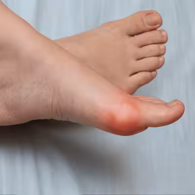
Lateral ankle sprain is the most common musculoskeletal injury in sports and recreational activity, accounting for 15–25% of all sports injuries and representing 1 million emergency department visits annually in the United States. Despite its common occurrence, lateral ankle sprain is frequently undertreated — a complete understanding of the ligament anatomy involved, accurate clinical grading, and appropriate rehabilitation is essential to prevent the 40% of patients who develop chronic ankle instability after inadequately managed initial sprains.
Lateral Ankle Ligament Anatomy
Three lateral ankle ligaments: the anterior talofibular ligament (ATFL) — originates from the anterior fibula, inserts on the talar neck; the primary restraint against anterior talar translation; the weakest of the three; injured first in inversion sprains. The calcaneofibular ligament (CFL) — originates from the fibular tip, inserts on the lateral calcaneus; restrains calcaneal inversion; injured second in progressive inversion mechanism. The posterior talofibular ligament (PTFL) — the strongest; injured only in severe sprains and fracture-dislocations. Injury sequence: ATFL tears first with progressive plantarflexion-inversion; CFL tears with more vertical force vector; combined ATFL + CFL tears produce clinically significant instability requiring more aggressive rehabilitation and carrying higher risk of chronic instability.
Grading, Diagnosis, and Management
Grade I: ATFL sprain/stretch, no macroscopic tear; minimal swelling and tenderness; full weight-bearing maintained; anterior drawer negative. Grade II: partial ATFL tear ± partial CFL involvement; moderate swelling and ecchymosis; painful weight-bearing; anterior drawer weakly positive. Grade III: complete ATFL tear + complete CFL tear; significant swelling and ecchymosis; unable to bear weight; anterior drawer strongly positive (>10mm) and talar tilt positive (>5–10°). Imaging: Ottawa rules (absence of bone tenderness over the fibula, malleoli, navicular, and 5th metatarsal base — allows clinical exclusion of fracture without X-ray in 95% of cases). MRI: for Grade III injuries, suspected OCD (40% prevalence with high-grade sprains), or chronic instability evaluation. RICE protocol (Grade I–III initial): rest, ice (15–20 minutes every 2 hours for 48–72 hours), compression, elevation. Early mobilization: superior to immobilization for Grades I–II — functional rehabilitation with balance training and peroneal strengthening reduces re-sprain rate by 50%. Dr. Biernacki at Balance Foot & Ankle evaluates ankle sprains clinically and with imaging and provides structured rehabilitation and Brostrom repair for chronic instability. Call (810) 206-1402 at our Bloomfield Hills or Howell office.
📧 Get Dr. Tom’s Free Lab Test Guide
Discover the 5 lab tests every person over 35 should ask their doctor about — explained in plain English by a board-certified physician.
📍 Located in Michigan?
Our board-certified podiatrists treat this condition at two convenient locations. Same-day appointments often available.
In-Office Treatment at Balance Foot & Ankle
If home care isn’t resolving your ankle pain, a visit with a board-certified podiatrist is the fastest path to accurate diagnosis and a personalized plan. At Balance Foot & Ankle Specialists, Dr. Tom Biernacki, Dr. Carl Jay, and Dr. Daria Gutkin offer same-day and next-day appointments at both our Howell and Bloomfield Hills offices. We perform on-site diagnostic ultrasound, digital X-ray, conservative care, advanced regenerative treatments, and minimally invasive surgery when indicated.
Call (810) 206-1402 or request an appointment online. Most insurance plans accepted, including Medicare, Blue Cross Blue Shield, Aetna, Cigna, and United Healthcare.
More Podiatrist-Recommended Ankle Sprain Essentials
Stability Walking/Running Shoe
Brooks Adrenaline GTS 25 — lateral support during recovery walking.
KT Tape for Ankle Support
KT Tape — proprioceptive support for athletic return-to-play.
Supportive Insole

Watch: Fix TWISTED Ankle, ROLLED Ankle or SPRAINED Ankle Ligaments FASTER! — MichiganFootDoctors YouTube
PowerStep Pinnacle — arch support reduces re-injury risk during recovery.
As an Amazon Associate, Balance Foot & Ankle earns from qualifying purchases. Product recommendations are based on clinical experience; prices and availability shown above update live from Amazon.
When to See a Podiatrist
A sprain that hasn’t fully recovered after 6 weeks often has residual ligament laxity or occult fracture that keeps the ankle unstable. Balance Foot & Ankle X-rays and stress-tests every lingering sprain — if the ligament is torn, we offer bracing, PRP, and (for chronic instability) minimally-invasive repair. Don’t keep re-rolling the same ankle; let us stabilize it properly.
Call Balance Foot & Ankle: (810) 206-1402 · Book online · Offices in Howell & Bloomfield Hills
Frequently Asked Questions
How do I know if I sprained or broke my ankle?
Both cause pain, swelling, and difficulty walking. Key differences: fractures often cause more immediate severe pain, tenderness directly over bone (not just ligament), and inability to bear any weight. X-rays and the Ottawa Ankle Rules help determine if imaging is needed.
How long does an ankle sprain take to heal?
Grade I (mild): 1–2 weeks. Grade II (moderate): 3–6 weeks. Grade III (complete tear): 2–3 months. Chronic instability from improperly treated sprains can persist and may require surgery.
What is the best treatment for a sprained ankle?
RICE protocol (Rest, Ice, Compression, Elevation) for the first 48–72 hours, followed by protected weight-bearing as tolerated. Physical therapy rehabilitation is critical for high-grade sprains to restore strength and proprioception and prevent chronic instability.
Need Treatment at Balance Foot & Ankle?
Dr. Tom Biernacki, Dr. Carl Jay, and Dr. Daria Gutkin see patients at our Howell and Bloomfield Township offices.
Book Online or call (810) 206-1402
Insurance Accepted
BCBS · Medicare · Aetna · Cigna · United Healthcare · HAP · Priority Health · Humana · View All →
Howell Office
3980 E Grand River Ave, Suite 140
Howell, MI 48843
Get Directions →
Bloomfield Hills Office
43700 Woodward Ave, Suite 207
Bloomfield Hills, MI 48302
Get Directions →
Your Board-Certified Podiatrists


Ready to Get Back on Your Feet?
Same-week appointments available at both locations.
Book Your AppointmentDifferential Diagnosis: What Else Could It Be?
Several conditions share symptoms with Ankle Sprain and are commonly misdiagnosed in the first office visit. Considering these alternatives is part of every Balance Foot & Ankle exam:
- Peroneal tendon tear. Snapping behind the lateral malleolus or weakness everting the foot.
- High-ankle (syndesmosis) sprain. Pain over the syndesmosis with squeeze + external rotation — needs longer recovery.
- Lateral malleolus fracture. Bone-point tenderness positive on Ottawa rules — get an X-ray.
If your symptoms don’t fit the textbook pattern, ask your podiatrist which differentials they ruled out — that conversation often shortcuts months of trial-and-error treatment.
In Our Clinic
Most of our ankle sprains are acute — a patient comes in the same day or within 48 hours after rolling the ankle. We apply the Ottawa Ankle Rules first: bone tenderness at the posterior malleolus, navicular, or base of the 5th metatarsal, or inability to bear weight for 4 steps, means we image immediately to rule out fracture. For a clean grade 1–2 lateral ligament sprain, we use a short period of boot immobilization if needed, then transition into an ankle brace + proprioception training. The mistake we often see: patients skip the rehab phase and re-sprain within a year.
Most Common Mistake We See
The most common mistake we see is: Returning to sport as soon as the pain resolves. Fix: first pass a 30-second single-leg balance test with eyes closed and complete a graded return-to-sport progression.
Warning Signs That Need Same-Day Care
Seek immediate evaluation at Balance Foot & Ankle if you experience any of the following:
- Unable to bear weight for four steps
- Bone tenderness at the ankle bones (Ottawa)
- Severe swelling within one hour of injury
- Numbness or tingling in the foot
Call (810) 206-1402 — same-day and next-day appointments at our Howell and Bloomfield Hills offices.
Watch: Dr. Tom explains
Podiatrist-recommended products
As an Amazon Associate, Dr. Tom earns from qualifying purchases.
Protects lateral ligament complex.
View on Amazon →Neutralizes subtalar posture.
View on Amazon →Acute lateral ligament injury.
View on Amazon →Topical for tender ligaments.
View on Amazon →Related resources
Ready to solve this? Book today.
Same-week appointments · Howell & Bloomfield Hills · 4.9★ (1,123+ reviews)
☎ (810) 206-1402Book Online →Watch: Dr. Tom explains
Podiatrist-recommended products
As an Amazon Associate, Dr. Tom earns from qualifying purchases.
Acute icing
View on Amazon →Grade 3 immobilization
View on Amazon →Recovery support
View on Amazon →Topical relief
View on Amazon →Related resources
Ready to solve this? Book today.
Same-week appointments · Howell & Bloomfield Hills · 4.9★ (1,123+ reviews)
☎ (810) 206-1402Book Online →Pros & Cons of Conservative Care for foot care
Advantages
- ✓ Conservative care first
- ✓ Same-week appointments
- ✓ Multiple insurance accepted
Considerations
- ✗ Self-treatment can mask issues
- ✗ See a podiatrist if pain >2 weeks
Dr. Tom’s Recommended Products for foot care
Affiliate disclosure: As an Amazon Associate, Balance Foot & Ankle earns from qualifying purchases. We only recommend products we use with patients.
Footnanny Heel Cream Dr. Tom’s Pick
Best for: Daily moisturizer for cracked heels
Ready to Get Back on Your Feet?
Same-day appointments in Howell + Bloomfield Twp. Most insurance accepted. Dr. Tom Biernacki, DPM & team.
Book Today — Same-Day Appointments Available
Call Now: (810) 206-1402
About Your Care Team at Balance Foot & Ankle
Dr. Tom Biernacki, DPM · Board-Certified Foot & Ankle Surgeon. Specializes in conservative-first care, minimally invasive bunion surgery, and complex reconstruction.
Dr. Carl Jay, DPM · Accepting new patients. Specializes in sports medicine, athletic injuries, and routine podiatric care.
Dr. Daria Gutkin, DPM, AACFAS · Accepting new patients. Specializes in surgical reconstruction and pediatric podiatry.
Locations: 4330 E Grand River Ave, Howell, MI 48843 · 43494 Woodward Ave Suite 208, Bloomfield Twp, MI 48302
Hours: Mon–Fri 8:00 AM – 5:00 PM · (810) 206-1402
Frequently Asked Questions
When should I see a podiatrist?
If symptoms persist past 2 weeks, affect your normal activity, or are accompanied by red-flag symptoms (warmth, redness, swelling, inability to bear weight).
What does treatment cost?
Most diagnostic visits and conservative treatments are covered by Medicare and major insurers. Out-of-pocket costs vary by your specific plan.
How quickly can I get an appointment?
Most non-urgent cases see us within 5 business days. Urgent cases (sudden pain, possible fracture) typically same or next business day.
Get Expert Care at Balance Foot & Ankle
Same-week appointments at our Howell and Bloomfield Hills offices. Board-certified podiatric surgeons. Most insurance accepted.
Ready for Expert Care?
Same-day appointments in Howell & Bloomfield Hills, MI.
4.9★ | 1,123 Reviews | 3,000+ Surgeries
Or call: (810) 206-1402
Dr. Tom Biernacki, DPM is a double board-certified podiatrist and foot & ankle surgeon at Balance Foot & Ankle Specialists in Southeast Michigan. With over a decade of clinical experience, he specializes in heel pain, bunions, diabetic foot care, sports injuries, and minimally invasive surgery. Dr. Biernacki is a member of the APMA and ACFAS, and his patient education content on MichiganFootDoctors.com and YouTube has reached over one million views.