


Medically Reviewed by Dr. Tom Biernacki, DPM, FACFAS — Board-certified podiatrist & foot surgeon | Balance Foot & Ankle | Last updated: May 2026
Quick Answer: Lumbar Spinal Stenosis and Foot Pain
Lumbar spinal stenosis compresses nerve roots in the lower spine, producing a characteristic pattern of leg and foot pain called neurogenic claudication — cramping, aching, or weakness in the legs and feet that worsens with walking and standing, and relieves with sitting or bending forward. Foot symptoms include numbness, tingling, weakness, and in severe cases foot drop from L4–L5 nerve root compression. Podiatric management addresses the mechanical foot consequences; spinal management (injections, decompression surgery) addresses the source. Accurate diagnosis distinguishes spinal from peripheral nerve or vascular causes.
Foot pain that originates in the spine is one of the most commonly missed diagnoses in podiatry — and for good reason. The foot symptoms are real, the foot examination may show sensory changes and weakness, and without a careful history exploring the relationship between posture, walking distance, and symptom onset, lumbar spinal stenosis can easily be attributed to peripheral neuropathy or other foot pathology.
As a podiatrist in Howell and Bloomfield Hills, I maintain a high index of suspicion for spinal causes in patients presenting with bilateral leg and foot symptoms, symptoms that reliably worsen with walking a consistent distance (neurogenic claudication), and symptoms that are relieved by sitting or the typical grocery-cart lean (forward trunk flexion that opens the spinal canal). These patients need spine evaluation alongside podiatric care.
Neurogenic vs. Vascular Claudication: Key Distinctions
| Feature | Neurogenic Claudication (Spinal Stenosis) | Vascular Claudication (PAD) |
|---|---|---|
| Pain character | Cramping, aching, weakness, numbness; bilateral | Calf cramping/tightening; often unilateral |
| Onset trigger | Walking AND prolonged standing; worse downhill | Walking only; severity proportional to distance |
| Relief position | Sitting, forward trunk flexion, lying down | Standing still (rest, not posture-dependent) |
| Cycling | Tolerated well (flexed posture opens canal) | Limited by same ischemic mechanism |
| Pulses | Normal foot pulses | Reduced or absent pedal pulses |
| Diagnosis | MRI lumbar spine, EMG/NCS | ABI (ankle-brachial index), vascular duplex |
Foot Manifestations of Lumbar Spinal Stenosis
The specific foot symptoms in spinal stenosis depend on which nerve root level is compressed. L4 compression produces weakness of ankle dorsiflexion (difficulty lifting the foot) and sensory changes on the inner ankle. L5 compression — the most common level in lumbar stenosis — produces weakness of foot and toe extension plus sensory changes on the top of the foot and great toe. S1 compression affects the outer foot and heel, producing weakness of plantarflexion (push-off) and sensory changes on the outer foot and small toe. Multi-level stenosis produces combined patterns. Foot drop from L5 radiculopathy in spinal stenosis is clinically indistinguishable from peroneal nerve palsy without electrodiagnostic and imaging evaluation.
Custom orthotics and appropriate footwear are appropriate management for the mechanical foot consequences of spinal stenosis — gait compensation patterns, altered weight distribution, and reduced proprioception. They do not treat the underlying nerve compression. Patients with neurogenic claudication who receive only foot-level treatment continue to experience progressive functional limitation and fall risk from ongoing nerve compression. When I identify a spinal pattern in a patient’s presentation, I refer for spine consultation (physiatry, neurosurgery, or orthopedic spine) and manage the foot component in parallel — not instead of the primary cause.
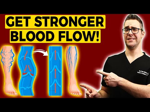
Watch: Circulation, Nerve Pain & Leg Pain — Dr. Tom Biernacki
Book an evaluation → | (810) 206-1402
Frequently Asked Questions
Can spinal stenosis cause numbness in the feet?
Yes — numbness and tingling in the feet are common presentations of lumbar spinal stenosis. The distribution follows the compressed nerve root level: L5 produces dorsal foot and great toe numbness; S1 produces outer foot and heel numbness; L4 produces inner ankle numbness. Bilateral foot numbness that worsens with walking or standing and improves with sitting is a strong indicator of neurogenic claudication from lumbar stenosis, distinguishing it from peripheral neuropathy (which does not change with posture) or vascular disease (which does not change with posture either).
Does spinal stenosis cause foot drop?
Severe L4–L5 nerve root compression from spinal stenosis can cause foot drop — weakness or inability to lift the front of the foot. This occurs when the L5 nerve root (which innervates the tibialis anterior and toe extensor muscles) is severely compressed. Foot drop from spinal stenosis may be sudden (from acute disc herniation) or gradual (from progressive stenosis). It requires urgent spine evaluation when acute. An AFO brace restores foot clearance during gait while the underlying spinal cause is addressed.
What exercises help spinal stenosis foot symptoms?
Flexion-based exercises that open the posterior spinal canal are the mainstay of conservative physical therapy for lumbar spinal stenosis. These include knee-to-chest stretches, stationary cycling (maintains flexed posture), aquatic therapy, and lumbar flexion exercises in supine. Extension exercises (backward bending) worsen stenosis symptoms and should be avoided. Walking with a slightly forward trunk lean (as if pushing a cart) naturally flexes the lumbar spine and extends comfortable walking distance for most stenosis patients.
How is spinal stenosis distinguished from peripheral neuropathy?
Both can cause bilateral foot numbness, tingling, and weakness. Key distinguishing features: neuropathy symptoms are constant or worse at night (not posture-dependent); spinal stenosis symptoms worsen with walking/standing and improve with sitting. Nerve conduction studies and EMG test both the peripheral nerves and can show the pattern of radiculopathy (spine origin) versus peripheral neuropathy. MRI of the lumbar spine and blood work (for metabolic neuropathy causes like diabetes) together complete the diagnostic picture. I refer patients with suspected spinal patterns for appropriate workup rather than treating presumed neuropathy.
When should I see a podiatrist vs. spine specialist for foot pain?
See a podiatrist first for foot pain that is localized, provoked by foot-specific activities, and involves local tenderness. See a spine specialist (or ask your podiatrist for referral) when foot symptoms are bilateral, worsen with walking a consistent distance, are relieved by sitting or forward bending, are accompanied by back or buttock pain, or include significant weakness. Balance Foot & Ankle evaluates for spinal patterns at initial consultation and provides appropriate referrals — call (810) 206-1402.
Foot & Leg Pain Evaluation — Spinal, Vascular, Neurological
Comprehensive foot pain workup including spinal pattern screening — Howell & Bloomfield Hills, MI
Related: Nerve Pain in Foot | Custom Orthotics Michigan | Foot Drop Treatment
Dr. Tom Biernacki, DPM is a double board-certified podiatrist and foot & ankle surgeon at Balance Foot & Ankle Specialists in Southeast Michigan. With over a decade of clinical experience, he specializes in heel pain, bunions, diabetic foot care, sports injuries, and minimally invasive surgery. Dr. Biernacki is a member of the APMA and ACFAS, and his patient education content on MichiganFootDoctors.com and YouTube has reached over one million views.
- Diagnosis and Treatment of Plantar Fasciitis (PubMed / AAFP)
- Heel Pain (APMA)
- Hallux Valgus (Bunions): Evaluation and Management (PubMed)
- Bunions (Mayo Clinic)
Recommended Products from Dr. Tom