
Medically reviewed by Dr. Tom Biernacki, DPM
Board-certified podiatric surgeon | Balance Foot & Ankle, Howell & Bloomfield Township, MI
Last reviewed: June 2026

Common peroneal nerve compression at the outside of the knee can cause foot drop, numbness, and tingling on the top of the foot — and it often responds to bracing, position changes, or simple decompression surgery.
You’re in the right place. Dr. Tom Biernacki, DPM, FACFAS — board-certified foot & ankle surgeon with 3,000+ surgeries — explains exactly what common peroneal nerve compression means and what works. Call (810) 206-1402 for same-week appointment at Howell or Bloomfield Township.
Quick answer: Common peroneal nerve compression happens when the nerve is pinched at the outside of the knee (the fibular head), causing foot drop, numbness, and tingling across the top of the foot and outer lower leg. Most cases improve with conservative care — activity and position changes, an ankle-foot orthosis (AFO) brace, and physical therapy to take pressure off the nerve — with surgical decompression reserved for severe or non-improving cases. Seek prompt care if you develop a sudden foot drop or rapidly worsening weakness.
What Is Common Peroneal Nerve Compression?
The common peroneal nerve (also called the common fibular nerve) is a branch of the sciatic nerve that winds around the fibular neck — the bony prominence on the outer side of the knee — before dividing into the superficial and deep peroneal nerves that supply sensation and motor function to the lateral leg and entire dorsal foot. This anatomical position makes it the most commonly injured peripheral nerve in the lower extremity.
Common peroneal nerve compression (also called peroneal nerve entrapment) at the fibular head produces a recognizable syndrome: weakness of dorsiflexion (foot drop), weakness of eversion (turning the foot outward), and numbness or tingling over the dorsal (top) surface of the foot and lateral leg. Foot drop — the inability to lift the foot during the swing phase of walking — is the most functionally disabling manifestation.
The reason this one spot is so vulnerable is simple anatomy: at the fibular neck, the nerve sits directly against bone with only skin and a thin layer of fascia protecting it. There is no muscle padding. Anything that presses on the outside of the knee — a crossed leg, a cast edge, a hospital bed rail, even rapid weight loss removing the fat cushion — presses the nerve against bone.
Symptoms: How Peroneal Nerve Compression Feels
Symptoms depend on how severely and how long the nerve has been compressed. Most patients notice some combination of:
- Numbness or tingling on the top of the foot and the outer side of the lower leg — often the earliest sign. (If your numbness involves the sole or both feet, see our guide to foot numbness causes — the pattern points to a different diagnosis.)
- Foot drop — difficulty lifting the front of the foot, catching your toes when you walk, or slapping the foot down with each step.
- Weak eversion — trouble turning the foot outward, which can make the ankle feel unstable on uneven ground.
- A “steppage” gait — lifting the knee higher than normal so the dangling foot clears the floor.
- Shooting pain or electric tingling when the outside of the knee is tapped or pressed (Tinel’s sign).
Pain is often mild or absent — many patients have painless weakness and numbness, which is exactly why the condition gets ignored until the foot drop becomes obvious. Location matters: numbness on the top of the foot points to the peroneal nerve, while burning in both feet that starts at the toes suggests peripheral neuropathy, and numbness radiating from the back down the leg suggests a lumbar nerve root problem. Our location-based numbness guide walks through the patterns.
Causes and Risk Factors
The nerve’s superficial position at the fibular head makes it vulnerable to: habitual leg crossing (the single most common preventable cause), plaster cast or brace compression, prolonged squatting (construction workers, gardeners), weight loss exposing the nerve to pressure, ankle fracture or dislocation, proximal fibula fracture, knee surgery complications, and space-occupying lesions (ganglion cysts, lipomas, osteochondromas at the fibular head).
In the foot and ankle context, podiatrists most commonly encounter peroneal nerve issues secondary to ankle fractures, ankle bracing, or chronic ankle instability. Awareness of the nerve’s vulnerable anatomy is essential to avoid iatrogenic injury during lateral ankle surgery.
Higher-risk groups include people who have recently lost significant weight (including after bariatric surgery), patients hospitalized or bed-bound for long periods, diabetics (whose nerves tolerate compression poorly), distance drivers and desk workers who sit cross-legged for hours, and anyone in a long-leg cast or knee brace with a tight upper edge.
Foot Drop: The Symptom That Brings Patients In
Foot drop is not a diagnosis — it is a sign that the muscles that lift the foot (primarily the tibialis anterior) have lost their nerve supply. Common peroneal nerve compression at the fibular head is the most common cause, but the same picture can come from an L4–L5 disc herniation, sciatic nerve injury, or a tibialis anterior tendon rupture — which is a tendon problem, not a nerve problem, and is treated completely differently.
Foot drop has many possible causes beyond the peroneal nerve — including stroke, multiple sclerosis, L5 nerve-root compression, and muscle disease. For the full differential, see our complete guide to foot drop causes and treatment.
Sorting these out is the first job of the exam. With a peroneal nerve lesion at the fibular head, dorsiflexion and eversion are weak but inversion is spared (that muscle is supplied by a different nerve). With an L5 radiculopathy, inversion is weak too, and there is usually back pain. With a tendon rupture, the muscle fires but nothing moves the foot, and there is often a visible gap or lump at the front of the ankle. An accurate diagnosis here changes everything that follows — which is why guessing is a bad strategy and an in-person exam matters.
Diagnosis: How We Confirm It
Clinical diagnosis involves documenting the pattern of weakness (dorsiflexion, eversion) and sensory loss. Tinel’s sign at the fibular head (tapping the nerve reproduces shooting pain or paresthesias down the leg) confirms the level of compression. Electrodiagnostic studies (EMG/NCS) confirm the diagnosis, localize the injury level, and provide prognostic information about axonal loss vs. demyelination.
EMG/NCS timing matters: testing earlier than 3–4 weeks after the injury can miss axonal changes, so we often examine first, protect the foot, and schedule electrodiagnostics at the right window. If a mass is suspected at the fibular head — a ganglion cyst, lipoma, or bony prominence — ultrasound or MRI of the knee identifies it. Blood work (glucose, B12, thyroid) is added when the story suggests the compression is happening on top of an underlying neuropathy.
Treatment: From Position Changes to Surgery
Step 1 — Remove the cause. Stop crossing the legs, pad or trim the offending cast or brace edge, change squatting habits. For positional compression this alone is the cure: most cases recover fully within 3–6 months once the pressure is gone.
Step 2 — Protect the foot while the nerve recovers. An ankle-foot orthosis (AFO) holds the foot up during the swing phase so you can walk safely and don’t trip. This is not optional with significant foot drop — a fall with a fractured wrist or hip is a far worse outcome than the nerve injury itself.
Step 3 — Physical therapy. Daily ankle range-of-motion work prevents the Achilles tendon from contracting while the dorsiflexors are weak, and progressive strengthening rebuilds the muscles as the nerve recovers. Gait retraining reduces fall risk.
Step 4 — Surgical decompression is indicated for mass lesions (ganglion cysts, bony prominences), failed conservative care at 3 months for non-positional causes, or severe axonal loss on EMG. The nerve is decompressed at the fibular neck through a 4–6cm incision, with removal of any compressive pathology. Recovery of function follows nerve regeneration at approximately 1mm/day from the compression site. For long-standing foot drop that does not recover, tendon transfer surgery can restore the ability to lift the foot.
Recovery Timeline: What to Expect
| Injury type | What happened to the nerve | Expected recovery |
|---|---|---|
| Demyelinating (mild compression) | Insulation damaged, axons intact | Full recovery in 6–12 weeks |
| Positional compression (leg crossing, cast) | Usually demyelinating | Full recovery in 3–6 months once cause removed |
| Axonotmesis (axonal damage) | Axons damaged, nerve intact | Regrowth at ~1mm/day — 6–12 months for foot-level function |
| Neurotmesis (severed nerve) | Nerve transected | Surgical repair required; recovery variable |
The single best predictor is the EMG: a pure conduction block (demyelination) means full recovery is expected, while severe axonal loss means recovery will be slow and possibly partial. This is why we get electrodiagnostics on every foot drop of unknown cause before promising anyone a timeline.
Dr. Tom’s Product Recommendations

PowerStep Pinnacle Insoles
⭐ Highly Rated | Foundation Wellness Partner
For patients with common peroneal nerve compression and associated foot drop, ankle instability, or dorsiflexion weakness, a supportive insole helps improve gait mechanics during nerve recovery. PowerStep Pinnacle provides the arch and heel stability that compensates for reduced ankle and foot dorsiflexion control.
Nerve recovery phase, gait support, ankle stability during weakness
Severe foot drop requiring full AFO; does not substitute for nerve decompression
Disclosure: We earn a commission if you purchase through our links at no extra cost to you.
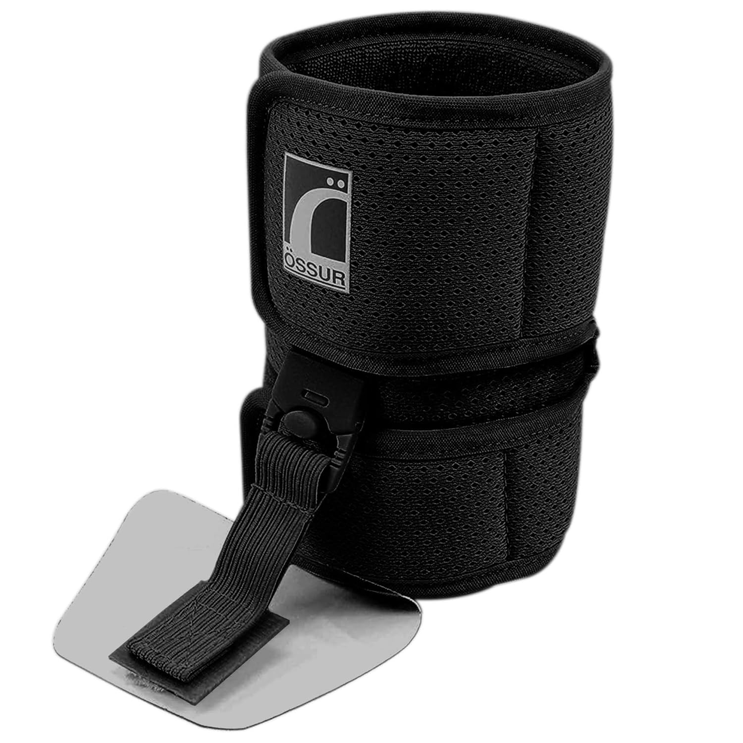
Ossur Foot-Up Drop Foot AFO Brace
⭐ Highly Rated | Lightweight Ankle-Foot Orthosis
The single most important device for foot drop while the nerve recovers. A lightweight ankle-foot orthosis (AFO) like the Ossur Foot-Up holds the toes up during the swing phase of walking so the foot doesn’t catch or slap — preventing the trips and falls that are the real danger of peroneal nerve palsy. It is discreet enough to wear inside a normal shoe.
Active foot drop, fall prevention, walking safely during nerve recovery
Severe/fixed foot drop needing a rigid carbon-fiber AFO; get fitted by your podiatrist
Disclosure: We earn a commission if you purchase through our links at no extra cost to you.
✅ Pros / Benefits
- Positional causes (leg crossing) recover fully after decompression
- Surgical decompression highly effective for mass lesions and structural compression
- AFO provides excellent functional compensation during nerve recovery
- Most cases recover within 3–6 months without surgery
❌ Cons / Risks
- Severe axonal loss requires months for nerve regeneration (1mm/day)
- Foot drop significantly impairs walking and creates fall risk
- Electrodiagnostic studies needed for prognosis but take 3–4 weeks post-injury to show changes
- Surgery does not guarantee full recovery if axonal loss is severe
Dr. Tom Biernacki’s Recommendation
Common peroneal nerve compression is one of the most preventable causes of foot drop I see. A surprising number of cases are from habitual leg crossing — something I remind every patient with unilateral foot numbness or weakness to change immediately. For patients who arrive with foot drop from an unknown cause, my first step is always EMG/NCS to assess axonal integrity. The prognosis varies enormously between a pure demyelinating conduction block (full recovery expected) and severe axonal loss (partial recovery at best).
— Dr. Tom Biernacki, DPM | Board-Certified Podiatric Surgeon | Balance Foot & Ankle, Michigan
When to Seek Care Urgently
Most peroneal nerve compression develops gradually and can be evaluated within a week or two. See a doctor promptly — within days, not weeks — if you have sudden complete foot drop, foot drop after a knee injury or fracture, a growing lump at the outside of the knee, or progressive weakness that is getting worse week over week. The longer a severely compressed nerve waits, the more axonal damage accumulates and the less complete the recovery. If the foot is also cold, pale, or pulseless, that is a circulation emergency, not a nerve problem — go to the ER.
Frequently Asked Questions
What causes numbness on the top of the foot?
Common peroneal nerve compression is the most common nerve cause of dorsal foot numbness. Other causes include extensor tendinitis from tight shoes, L4-L5 disc herniation affecting the peroneal nerve root, and superficial peroneal nerve entrapment in the lateral compartment.
Can common peroneal nerve compression cause foot drop?
Yes — weakness of the tibialis anterior (dorsiflexor) is the hallmark of common peroneal nerve palsy. Patients drag their foot during walking and are at high risk of tripping and falling.
Will peroneal nerve damage heal on its own?
Positional compression injuries (leg crossing, cast pressure) almost always recover fully within 3–6 months once the compression is removed. Traumatic injuries with severe axonal loss have more variable recovery.
How long does peroneal nerve recovery take?
Demyelinating injuries (mild compression): full recovery in 6–12 weeks. Axonotmesis (axonal damage without nerve transection): recovery at 1mm/day from the compression site — for foot-level function, this means 6–12 months. Neurotmesis (severed nerve): surgical repair required.
Do I need a brace for foot drop?
If your foot catches or slaps when you walk, yes — an ankle-foot orthosis (AFO) is the standard of care while the nerve recovers. It holds the foot up during the swing phase so you can walk safely and protects you from falls. Your podiatrist will fit the right style; soft carbon-fiber AFOs work for most compression-related foot drop.
How is peroneal nerve compression different from sciatica?
Both can cause weakness and numbness below the knee, but sciatica and L5 radiculopathy usually come with back or buttock pain and weaken foot inversion as well. Peroneal compression at the fibular head spares inversion and has no back pain — and tapping the outside of the knee reproduces the tingling. EMG/NCS distinguishes the two definitively.
Should I see a podiatrist or neurologist for peroneal nerve compression?
Both. A podiatrist or orthopedic surgeon manages structural causes and surgical decompression. A neurologist or physiatrist performs electrodiagnostic studies and guides nerve recovery rehabilitation.
Michigan Foot Pain? See Dr. Biernacki In Person
4.9★ rated | 1,123 Reviews | 3,000+ Surgeries | Howell & Bloomfield Township
Same-week appointments available. Board-certified podiatric surgeon.
📞 (810) 206-1402 Book Online →References

Dr. Tom Biernacki, DPM is a board-certified foot & ankle surgeon (ABFAS & ABPM) at Balance Foot & Ankle Specialists in Southeast Michigan. With over a decade of clinical experience, he specializes in heel pain, bunions, diabetic foot care, sports injuries, and minimally invasive surgery. Dr. Biernacki is a member of the APMA and ACFAS, and his patient education content on MichiganFootDoctors.com and YouTube has made him one of the most-followed foot & ankle educators on YouTube.