
You are in the right place. Dr. Tom Biernacki, DPM, FACFAS — board-certified foot & ankle surgeon with 3,000+ surgeries — explains exactly what this foot or ankle issue means and what works. Call (810) 206-1402 for same-day appointment at Howell or Bloomfield Hills.
Medically reviewed by Dr. Tom Biernacki, DPM · Board-Certified Podiatric Surgeon · Last reviewed: April 2026 · Editorial Policy
The most important clinical decision with Metatarsalgia Differential Diagnosis Mortons Plantar Plate Stress Fracture isn’t which treatment to start with — it’s identifying the correct subtype. That changes everything. Call (810) 206-1402.
Quick Answer
Metatarsalgia Differential Diagnosis: Morton’s Neuroma relates to plantar fasciitis — typically caused by tight calves and arch overload. Most patients improve in 6-12 weeks with conservative care. Same-week appointments in Howell + Bloomfield Hills: (810) 206-1402.

Quick Answer
Morton’s neuroma is a thickening of nerve tissue between the third and fourth toes causing burning pain, numbness, or the sensation of a pebble under the ball of the foot. Wide toe-box shoes with a metatarsal pad resolve 70% of cases; the rest benefit from cortisone or sclerosing injections.
Medically Reviewed by Dr. Tom Biernacki, DPM — Board-Certified Podiatrist, Balance Foot & Ankle Specialists, Michigan. Last updated April 2026.
Metatarsalgia — pain localized to the plantar forefoot at or near the metatarsal heads — is a symptom, not a diagnosis, and encompasses at least seven distinct clinical entities, each requiring a specific treatment approach. Accurately differentiating Morton’s neuroma, plantar plate tear, MTP joint synovitis, metatarsal stress fracture, Freiberg’s disease, intermetatarsal bursitis, and sesamoid pathology based on history, physical examination, and targeted imaging prevents the repeated ineffective treatments that characterize misdiagnosed metatarsalgia.
Watch: Dr. Tom Biernacki, DPM
Clinical Differentiation of Common Causes
Morton’s neuroma: burning, electric, or shooting pain radiating into the toes (classic third web space, between 3rd and 4th metatarsal heads); Mulder’s click — a palpable and audible click with lateral compression of the metatarsals combined with direct web space pressure; pain with narrow shoes that resolves with shoe removal; no joint swelling or positive drawer test; MRI confirms an ovoid lesion >5mm in the web space. Plantar plate tear (second MTP instability): plantar pain at the second metatarsal head; positive drawer test (dorsal subluxation of the proximal phalanx on the metatarsal head with 2mm dorsal translation); second toe ‘V sign’ (medial deviation); history of progressive toe deviation rather than shoe-related symptoms; MRI confirms the tear. MTP joint synovitis: diffuse dorsal and plantar joint swelling; pain with passive MTP joint dorsiflexion AND plantarflexion; no specific shoe-type correlation; synovitis secondary to inflammatory arthropathy (RA, psoriatic, gout) — ESR, CRP, urate level; MRI shows synovial thickening and joint effusion. Metatarsal stress fracture: dorsal metatarsal shaft tenderness (not plantar head pain); focal tenderness with axial compression along the metatarsal; history of increased walking/running; MRI confirms bone marrow edema along the shaft; first and fifth metatarsal stress fractures require more aggressive management than central shaft fractures. Freiberg’s infraction: second or third MTP joint pain; younger patients (adolescent females most common); X-ray shows flattening and sclerosis of the metatarsal head; MRI shows osteonecrosis and subchondral collapse.
Systematic Examination Protocol
The examination protocol for metatarsalgia should include: (1) plantar palpation of each metatarsal head individually to localize the pain; (2) web space palpation for neuroma; (3) Mulder’s test; (4) drawer test at each MTP joint; (5) axial compression of each metatarsal shaft; (6) passive MTP joint range of motion; (7) V sign assessment for lateral toe deviation. Imaging algorithm: weight-bearing AP foot X-ray first (stress fracture, Freiberg’s, joint space); MRI for soft tissue differentiation (neuroma vs. plantar plate vs. synovitis) when examination is unclear. Targeted injection: fluoroscopic or ultrasound-guided injection of 1% lidocaine into the suspected structure (web space vs. MTP joint) provides diagnostic confirmation. Dr. Biernacki at Balance Foot & Ankle performs comprehensive forefoot examination and provides targeted treatment for all causes of metatarsalgia at our Bloomfield Hills and Howell offices. Call (810) 206-1402.
📧 Get Dr. Tom’s Free Lab Test Guide
Discover the 5 lab tests every person over 35 should ask their doctor about — explained in plain English by a board-certified physician.
📍 Located in Michigan?
Our board-certified podiatrists treat this condition at two convenient locations. Same-day appointments often available.
More Podiatrist-Recommended Neuroma Essentials
Wide Neutral Cushion Shoe
New Balance 1080 V14 — max forefoot room decompresses the pinched nerve.
Wide-Toe-Box Walking Shoe
New Balance 990v6 — prevents the forefoot compression that triggers Morton’s neuroma.
Orthotic with Met Pad Built-In


![Calcaneus Stress Fracture Treatment [Heel Stress Fracture RECOVERY!]](https://www.michiganfootdoctors.com/wp-content/cache/flying-press/cdfca81fb25b3ea56c6af7d70a9e3fb0.jpg)
Watch: Calcaneus Stress Fracture Treatment [Heel Stress Fracture RECOVERY!] — MichiganFootDoctors YouTube
PowerStep Pinnacle — arch support reduces nerve irritation between metatarsals.
As an Amazon Associate, Balance Foot & Ankle earns from qualifying purchases. Product recommendations are based on clinical experience; prices and availability shown above update live from Amazon.
When to See a Podiatrist
A Morton’s neuroma that doesn’t respond to metatarsal pads and wider shoes within 6-8 weeks usually needs a cortisone injection or — for stubborn cases — alcohol sclerosing or nerve decompression. Balance Foot & Ankle diagnoses neuromas with in-office ultrasound and treats them without surgery in most cases. Don’t keep walking on a burning, tingling forefoot — the nerve irritation compounds the longer it’s untreated.
Call Balance Foot & Ankle: (810) 206-1402 · Book online · Offices in Howell & Bloomfield Hills
Frequently Asked Questions
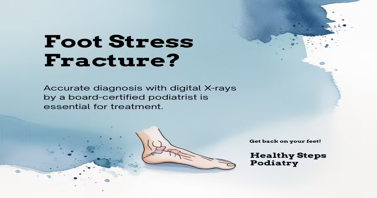
How long does a foot stress fracture take to heal?
Most foot stress fractures heal within 6–8 weeks with proper offloading. High-risk fractures (Jones fracture, navicular stress fracture) can take 3–6 months and sometimes require surgery. Premature return to activity is the most common cause of delayed healing.
How do I know if I have a stress fracture?
Stress fractures cause localized pain that worsens with activity and improves with rest, often with point tenderness over a specific bone. X-rays may be negative for 2–3 weeks after onset — MRI provides definitive diagnosis earlier.
Can you walk on a stress fracture?
This depends on the fracture location and severity. Many foot stress fractures allow limited walking in a protective boot. High-risk fractures (Jones, navicular) typically require non-weight-bearing. Walking on an unprotected stress fracture risks complete fracture.
Need Treatment at Balance Foot & Ankle?
Dr. Tom Biernacki, Dr. Carl Jay, and Dr. Daria Gutkin see patients at our Howell and Bloomfield Hills offices.
Book Online or call (810) 206-1402
Differential Diagnosis: What Else Could It Be?
Several conditions share symptoms with Morton’s Neuroma and are commonly misdiagnosed in the first office visit. Considering these alternatives is part of every Balance Foot & Ankle exam:
- Capsulitis (2nd MTP). Pain at 2nd-toe base rather than between toes; drawer test positive.
- Stress fracture. Single-point tenderness over a metatarsal shaft, not between toes.
- Freiberg’s infraction. AVN of metatarsal head, classic radiograph flattening.
If your symptoms don’t fit the textbook pattern, ask your podiatrist which differentials they ruled out — that conversation often shortcuts months of trial-and-error treatment.
In Our Clinic
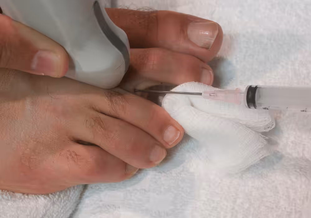
The classic Morton’s neuroma patient in our clinic is a 40- to 60-year-old woman who describes burning or “walking on a marble” in the 3rd intermetatarsal web space, often worsening in narrow or high-heeled shoes. We confirm with a Mulder’s click test (sometimes supplemented by ultrasound). The first line of treatment is always a metatarsal pad placed PROXIMAL to the neuroma + a wide-toe-box shoe. Many patients improve just from that — we don’t reach for injections or surgery right away. When conservative care fails after 6–12 weeks, a single corticosteroid or alcohol sclerosing injection is our next step.
Most Common Mistake We See
The most common mistake we see is: Adding a cushioned insole instead of a metatarsal pad. Fix: place the metatarsal pad PROXIMAL to (behind) the metatarsal heads — not directly under them.
Warning Signs That Need Same-Day Care
Seek immediate evaluation at Balance Foot & Ankle if you experience any of the following:
- Point tenderness on a single metatarsal suggesting stress fracture
- Unable to bear weight
- Progressive numbness up the foot
- Visible deformity or cross-over toe
Call (810) 206-1402 — same-day and next-day appointments at our Howell and Bloomfield Hills offices.
Watch: Dr. Tom explains
Podiatrist-recommended products
As an Amazon Associate, Dr. Tom earns from qualifying purchases.
Related resources
Ready to solve this? Book today.
Same-week appointments · Howell & Bloomfield Hills · 4.9★ (1,123+ reviews)
☎ (810) 206-1402Book Online →In-Office Treatment at Balance Foot & Ankle
When conservative care isn’t enough, Dr. Tom Biernacki and the team at Balance Foot & Ankle offer advanced, same-day options — including Foot & Ankle Fracture Repair Michigan at our Howell and Bloomfield Hills clinics.
Same-day appointments available. Call (810) 206-1402 or book online.
Pros & Cons of Conservative Care for plantar fasciitis
Advantages
- ✓ Conservative care resolves 90%+ of cases
- ✓ Multiple home treatment options
- ✓ Strong evidence base
- ✓ Imaging often not required
Considerations
- ✗ Recovery takes 6-12 weeks
- ✗ Mistakes prolong recovery
- ✗ Untreated can become chronic
- ✗ Can mimic other conditions
Dr. Tom’s Recommended Products for plantar fasciitis
Affiliate disclosure: As an Amazon Associate, Balance Foot & Ankle earns from qualifying purchases. We only recommend products we use with patients.
PowerStep Pinnacle Maxx Dr. Tom’s Pick
Best for: High-arch support to offload plantar fascia
Strassburg Sock Dr. Tom’s Pick
Best for: Overnight stretch for morning pain relief
Hoka Bondi 9 Dr. Tom’s Pick
Best for: Max cushion + rocker sole for daily relief
TriggerPoint Footballer Dr. Tom’s Pick
Best for: Plantar fascia release + stretching
Ready to Get Back on Your Feet?
Same-day appointments in Howell + Bloomfield Hills. Most insurance accepted. Dr. Tom Biernacki, DPM & team.
Book Today — Same-Day Appointments Available
Call Now: (810) 206-1402
About Your Care Team at Balance Foot & Ankle
Dr. Tom Biernacki, DPM · Board-Certified Foot & Ankle Surgeon. Specializes in conservative-first care, minimally invasive bunion surgery, and complex reconstruction.
Dr. Carl Jay, DPM · Accepting new patients. Specializes in sports medicine, athletic injuries, and routine podiatric care.
Dr. Daria Gutkin, DPM, AACFAS · Accepting new patients. Specializes in surgical reconstruction and pediatric podiatry.
Locations: 4330 E Grand River Ave, Howell, MI 48843 · 43494 Woodward Ave Suite 208, Bloomfield Hills, MI 48302
Hours: Mon–Fri 8:00 AM – 5:00 PM · (810) 206-1402
In-Office Treatment at Balance Foot & Ankle
If home treatment isn’t providing relief for your stress fractures, our podiatry team at Balance Foot & Ankle can help with same-day evaluations and advanced in-office care.
Same-day appointments available. (810) 206-1402
Frequently Asked Questions
When should I see a podiatrist?
If symptoms persist past 2 weeks, affect your normal activity, or are accompanied by red-flag symptoms (warmth, redness, swelling, inability to bear weight).
What does treatment cost?
Most diagnostic visits and conservative treatments are covered by Medicare and major insurers. Out-of-pocket costs vary by your specific plan.
How quickly can I get an appointment?
Most non-urgent cases see us within 5 business days. Urgent cases (sudden pain, possible fracture) typically same or next business day.
Ready to fix this for good?
Reading goes so far. The fastest path is a 30-minute office visit. Same-day Howell or Bloomfield Hills. Call (810) 206-1402.
Ready for Expert Care?
Same-day appointments in Howell & Bloomfield Hills, MI.
4.9★ | 1,123 Reviews | 3,000+ Surgeries
Or call: (810) 206-1402
Dr. Tom Biernacki, DPM is a board-certified foot & ankle surgeon (ABFAS & ABPM) at Balance Foot & Ankle Specialists in Southeast Michigan. With over a decade of clinical experience, he specializes in heel pain, bunions, diabetic foot care, sports injuries, and minimally invasive surgery. Dr. Biernacki is a member of the APMA and ACFAS, and his patient education content on MichiganFootDoctors.com and YouTube has made him one of the most-followed foot & ankle educators on YouTube.