
Medically reviewed by Dr. Tom Biernacki, DPM
Board-certified podiatric surgeon | Balance Foot & Ankle, Howell & Bloomfield Hills, MI
Last reviewed: May 2026
Balance Foot & Ankle offers same-day appointments for urgent foot and ankle conditions across Southeast Michigan — but the most important factor in outcomes isn’t getting seen quickly. Our podiatrists explain what to do in the first 24-48 hours before your appointment that most patients skip entirely. Call (810) 206-1402 — expert podiatric care across Michigan.

The most important clinical decision with Metatarsalgia Guide Michigan Podiatrist isn’t which treatment to start with — it’s identifying the correct subtype. That changes everything. Call (810) 206-1402.
Metatarsalgia: Differential Diagnosis by Location, Symptom, and Clinical Finding
Metatarsalgia is a symptom, not a diagnosis — forefoot pain at or near the metatarsal heads arising from any of seven distinct pathological mechanisms. The most critical clinical distinction is between PRIMARY metatarsalgia (mechanical overload of structurally normal metatarsals), SECONDARY metatarsalgia (forefoot pain caused by a specific pathology: Morton’s neuroma, Freiberg’s infarction, plantar plate tear, stress fracture), and REFERRED metatarsalgia (tarsal tunnel syndrome, L4-L5 radiculopathy mimicking forefoot pain). Treating mechanical metatarsalgia with offloading while missing an underlying Freiberg’s or stress fracture delays definitive care by months. The table below separates the seven most clinically significant forefoot pain generators across eight diagnostic features.
| Diagnosis | Classic Location | Pain Character | Aggravating Factor | Relieving Factor | Key Clinical Test | Imaging Finding | First-Line Treatment |
|---|---|---|---|---|---|---|---|
| Primary mechanical metatarsalgia (overload) | 2nd and 3rd metatarsal heads most common; plantar surface directly under head; bilateral pattern in high-arched (cavus) foot | Burning/aching; diffuse plantar forefoot; worse at end of day; feels like “walking on pebbles”; no neurological symptoms | Barefoot on hard floors; heeled shoes; prolonged standing; running/walking; tight toe box | Elevation; removing shoes; metatarsal pad proximal to the heads; barefoot on soft surface; rest | Metatarsal head tenderness on plantar palpation; callus formation directly under affected heads; assess foot type (cavus arch increases forefoot load) | Weight-bearing X-ray: relatively long 2nd metatarsal (index minus vs index plus pattern); no fracture; no joint changes | Metatarsal pad (Hapad or custom orthotic with met dome) placed just PROXIMAL to the symptomatic heads; offloads head by redirecting pressure to shaft; supportive wide-toe-box shoe; activity modification |
| Morton’s neuroma (interdigital neuroma) | 3rd web space (70%) or 2nd web space (15%); pain radiates INTO toes (3rd-4th most common; 2nd-3rd second most common); distinguishable from metatarsal head pain by radiation pattern | Sharp, burning, electric; sensation that “something is bunched in the shoe”; numbness/tingling in adjacent toes; pain radiates into toe tips; neurological symptoms distinguish from mechanical metatarsalgia | Tight narrow shoes; high heels (laterally compresses metatarsal heads, squeezing neuroma); prolonged walking; squeezing forefoot | Removing shoe and massaging forefoot; walking barefoot on soft surface; wide toe-box shoe | Mulder’s click: squeeze metatarsals medially while applying plantar pressure to web space — palpable click + reproduction of symptoms is 61-98% sensitive for Morton’s neuroma; web space tenderness vs metatarsal head tenderness | Ultrasound: hypoechoic mass in web space >5mm = Morton’s neuroma (92% sensitivity); MRI: T2-bright fusiform mass; X-ray typically normal (useful to exclude stress fracture) | Wide toe-box shoe + metatarsal pad to spread heads; corticosteroid injection into web space (60-70% short-term relief); sclerosing alcohol injections (4-7 series, 70-80% permanent resolution); surgical neurectomy if conservative fails × 6 months (90% cure) |
| Freiberg’s infarction (avascular necrosis of metatarsal head) | 2nd metatarsal head (68%); 3rd metatarsal head (27%); adolescent females most affected; insidious onset in distance runners; may present as acute or chronic forefoot pain | Deep, aching pain localized to single metatarsal head; dorsal swelling over MTP joint; stiffness and limited dorsiflexion at affected MTP; grinding (crepitus) with MTP range of motion in advanced cases | Push-off phase of gait; dorsiflexion of affected toe; running; prolonged standing; tight footwear compressing the head | Reduced weight-bearing; stiff-soled shoe; metatarsal offloading pad proximal to head | MTP joint stiffness (Freiberg’s limits dorsiflexion unlike mechanical metatarsalgia); dorsal joint-line tenderness at single MTP (not plantar as in mechanical); crepitus with passive MTP motion in Stages 3-5 | X-ray: Smillie staging (early flattening → sclerosis → fragmentation → collapse → DJD); MRI: bone marrow edema before X-ray changes — early diagnosis critical; early stage MRI may show changes when X-ray is normal | Stage 1-2: offloading + modified activity + stiff-soled shoe + casting if necessary; Stage 3-4: surgical debridement, synovectomy, osteotomy (dorsiflexion osteotomy to rotate viable cartilage into weight-bearing); Stage 5 (collapse with DJD): joint resection arthroplasty or MTP fusion |
| Plantar plate tear (2nd MTP instability) | 2nd MTP joint plantar surface (90% of plantar plate tears); presents as 2nd toe crossover deformity in chronic cases; the plantar plate is the primary stabilizer of the MTP joint | Plantar joint-line pain at 2nd MTP; instability sensation; progressive 2nd toe dorsal drift and medial deviation (crossover toe); swelling of 2nd MTP; difficulty finding comfortable shoe position for 2nd toe | Push-off; barefoot; gripping the floor with toes; dorsiflexion of 2nd toe under load (shoe toe box) | Buddy-taping 2nd toe to 3rd toe; metatarsal offloading; reduced activity; stiff-soled shoe | Lachman test of MTP joint (vertical drawer test): stabilize metatarsal, apply dorsal force to proximal phalanx — >2mm laxity = plantar plate insufficiency; compare to contralateral side. Valgus stress: medial deviation of 2nd toe on stress = lateral plate damage | MRI: plantar plate discontinuity or signal change at distal attachment to base of proximal phalanx; ultrasound: dynamic assessment shows plantar plate defect with dorsiflexion stress; X-ray: early stages normal; later stages show 2nd toe elevation and MTP joint space changes | Mild (Grade 1-2): dorsal buddy taping, metatarsal pad, stiff-soled shoe, activity modification × 6-12 weeks. Severe (Grade 3-4) or crossover toe: surgical plantar plate repair (direct suture repair through dorsal or plantar approach) + flexor tendon transfer to correct deformity |
| Metatarsal stress fracture | 2nd and 3rd shaft (most common in runners); 5th metatarsal base (Jones fracture zone in court athletes); 4th shaft; often bilateral in female athlete triad / low bone density | Insidious onset focal pain precisely over metatarsal shaft (not head, not web space); point tenderness on the bone, not plantar surface; worse during and after activity; no neurological symptoms; acute Jones fracture: sudden lateral foot pain with snap | Weight-bearing; running; jumping; prolonged walking; progressive — worsens with each workout rather than improving with warm-up | Strict non-weight-bearing; rest; elevation; ice. Unlike tendinopathy, does NOT warm up with activity — worsens consistently | Percussion test: tap metatarsal shaft longitudinally from toe to heel — reproduces fracture pain at fracture site (tuning fork vibration test similarly); point tenderness on bone shaft (not at head or web space) | X-ray: often NORMAL in first 2 weeks (sensitivity ~50%); MRI: periosteal edema, cortical signal change — diagnosis of choice in early presentation; bone scan: 100% sensitive but low specificity. Do NOT wait for X-ray changes in clinical stress fracture | 2nd-4th shaft: stiff-soled shoe or walking boot × 4-8 weeks; no running × 6 weeks; gradual return to activity. Jones fracture (Zone 2): non-weight-bearing boot × 6-8 weeks; surgical fixation (intramedullary screw) for athletes or displaced fractures — significantly faster return to sport vs conservative |
| Sesamoiditis / sesamoid stress fracture | Plantar 1st MTP joint; tibial sesamoid (medial) more commonly symptomatic than fibular (lateral); hallux push-off pain; localized to ball of foot under great toe, NOT the lesser metatarsal heads | Deep, focal aching directly under great toe at 1st MTP; worse with push-off and toe dorsiflexion; acute fracture: sudden severe pain with pop; chronic: gradual onset in dancer or runner | Barefoot; high heels (increases forefoot load under 1st MTP); running; dancing en pointe; ascending stairs | 1st MTP offloading; dancer’s pad (cutout under sesamoid); stiff-soled shoe | Sesamoid grind test: compress sesamoid against metatarsal head and rotate — crepitus + pain = sesamoiditis or arthrosis; tibial sesamoid tenderness on direct plantar palpation medial to 1st MTP joint | X-ray: bipartite sesamoid (normal variant) vs acute fracture (non-sclerotic jagged edges); MRI: bone marrow edema in sesamoid — distinguishes stress reaction from bipartite; bone scan: unilateral uptake in sesamoid confirms pathology in bipartite variant | Conservative (stress reaction): dancer’s pad with sesamoid cutout; activity modification; CAM boot if severe × 4-8 weeks. Fracture: non-weight-bearing × 8-12 weeks; sesamoidectomy if avascular necrosis or chronic nonunion (risk: transfer lesion to adjacent sesamoid) |
| Tarsal tunnel syndrome (referred metatarsalgia) | Diffuse plantar foot including forefoot; burning/tingling radiating from heel to forefoot and toes; does NOT localize to single web space or metatarsal head — distinguishing feature from Morton’s neuroma | Burning, tingling, numbness radiating from medial ankle through arch and into plantar forefoot; nocturnal symptoms (waking with foot tingling); worsens after prolonged standing; neurological distribution follows tibial nerve branches (medial and lateral plantar) | Prolonged standing; running; tight shoes; valgus foot deformity (stretches tarsal tunnel); activities at end of day | Elevation; removing shoes; rest; anti-inflammatory medication; ankle/foot stretching | Tinel’s sign over tarsal tunnel (posterior to medial malleolus): percussion → radiation into plantar foot = positive (66-70% sensitivity); Valleix sign: radiation proximally; dorsiflexion-eversion test (5-second position stress) reproduced symptoms | MRI: space-occupying lesion in tarsal tunnel (varicosities, ganglion, lipoma) or tunnel stenosis; EMG/NCS: reduced tibial sensory and motor conduction velocity across tarsal tunnel; X-ray: calcaneal spur or post-traumatic deformity narrowing tunnel | Conservative: orthotics for valgus correction (reduces tarsal tunnel tension); corticosteroid injection into tunnel; night splint; anti-inflammatory. Surgical: tarsal tunnel release (lacinate ligament division) + decompression of medial/lateral plantar branches if conservative fails × 6 months |
Metatarsalgia Treatment Protocol: Conservative to Surgical by Severity and Mechanism
| Treatment Tier | Intervention | Mechanism | Indication | Expected Outcome | When to Escalate |
|---|---|---|---|---|---|
| Tier 1 — Immediate offloading (Weeks 1-4) | Metatarsal pad (Hapad met pad or similar): adhesive foam dome placed 1cm PROXIMAL to the metatarsal heads, NOT under the heads; this redistributes pressure from the heads to the shafts. Must be PROXIMAL — placing under the head worsens pain | Reduces plantar pressure at metatarsal heads by 30-50% when correctly positioned; most cost-effective single intervention for mechanical metatarsalgia | Primary mechanical metatarsalgia; immediate pain relief while evaluation and orthotic fabrication proceed; adjunct to any other treatment | 60-70% report significant relief within 1-2 weeks; works best for 2nd and 3rd head overload; less effective for Morton’s neuroma | No relief at 4 weeks → evaluate for secondary cause (Morton’s, plantar plate, stress fracture); relief inconsistent → custom orthotic evaluation |
| Tier 1 — Footwear modification | Wide toe box (metatarsal width unrestricted); stiff rocker sole (reduces MTP dorsiflexion stress during push-off); low heel (reduces forefoot load); extra-depth shoe for Morton’s neuroma | Narrow toe boxes laterally compress metatarsal heads, worsening Morton’s neuroma and increasing interhead pressure; rocker sole reduces peak plantar pressure under metatarsal heads by 40-60% during late stance phase | All metatarsalgia causes as adjunct; especially critical for Morton’s neuroma (narrow shoe = direct cause); Freiberg’s (stiff sole protects compromised head) | Essential adjunct — no topical, injection, or orthotic treatment works long-term if footwear compresses the forefoot; footwear counseling required at every visit | Footwear corrected + 4 weeks no improvement → proceed to Tier 2 |
| Tier 2 — Custom orthotics with met dome | Custom functional orthotic with posted metatarsal dome; forefoot extension to MTP crease; accommodative padding for prominent heads; first-ray cutout for 1st MTP offloading (sesamoiditis); dancer’s pad (cutout under sesamoid) | Custom orthotic addresses underlying biomechanical driver (cavus arch, flexible flatfoot, hallux valgus transferring load to 2nd ray) that OTC pads cannot correct; also controls pronation that stresses 2nd MTP plantar plate | Failed OTC met pad × 4-6 weeks; recurrent/chronic metatarsalgia; bilateral metatarsalgia; identified biomechanical driver (cavus, hypermobile 1st ray, severe flatfoot); plantar plate instability | 70-80% long-term reduction in symptoms; addresses underlying mechanics rather than symptom only; reduces recurrence vs OTC padding | Custom orthotic + footwear + 3 months: insufficient relief → injection or procedure tier |
| Tier 3 — Corticosteroid injection | Corticosteroid (methylprednisolone 40mg or equivalent) injected into MTP joint capsule or web space; reduces acute inflammation, provides diagnostic confirmation (Morton’s: injection into correct web space should provide >50% relief within 48h) | Anti-inflammatory effect; Morton’s: reduces perineural edema around neuroma; mechanical metatarsalgia: reduces acute synovitis at overloaded MTP joint; also used for Freiberg’s (intra-articular) | Morton’s neuroma (first-line procedural; often 2-3 injections spaced 4-6 weeks = 60-70% sustained relief); acute inflammatory metatarsalgia; Freiberg’s Stage 1-2 with significant synovitis | Morton’s neuroma: 60-70% achieve satisfactory long-term relief with 1-3 injections; repeat injection if relief lasts >3 months and recurs; surgical threshold if relief lasts <3 months or requires >3 injections per year | Relief <6 weeks after 2nd injection → consider sclerosing alcohol (Morton’s) or surgical neurectomy |
| Tier 3 — Sclerosing alcohol injection (Morton’s neuroma) | 4% alcohol injected perineural under ultrasound guidance; 4-7 injections every 7-10 days; scleroses the neuroma tissue → permanent fibrous replacement; office-based, lower cost than surgery, avoids surgical risks | Selective neurolysis of the enlarged interdigital nerve without excision; eliminates neuroma pain by destroying conductive nerve tissue in the perineural fibrosis; does not cause stump neuroma (unlike surgical neurectomy) | Morton’s neuroma failed 1-2 corticosteroid injections; patient wishes to avoid surgery; neuroma <7mm (larger neuromas respond less well to sclerosing) | 70-80% report significant pain reduction at 6-month follow-up in prospective series; most effective for neuromas 5-7mm; comparable to surgical neurectomy in RCTs with lower complication rate | Failed 7 sclerosing injections → surgical neurectomy |
| Tier 4 — Surgical intervention | Morton’s neuroma: NEURECTOMY — dorsal or plantar approach; excise 1.5-2cm of digital nerve proximal to bifurcation; plantar approach allows more proximal resection but leaves plantar scar. Weil osteotomy (metatarsal shortening): shortens relatively long metatarsal by oblique osteotomy, reduces plantar pressure under that head. Plantar plate repair: direct suture repair of torn plantar plate via dorsal approach with metatarsal shortening to reduce tension at repair site. Freiberg’s: dorsiflexion osteotomy (Stages 3-4) or joint debridement/resection arthroplasty (Stage 5) | Neurectomy: definitive removal of pain-generating nerve tissue; Weil osteotomy: directly reduces pressure under the overloaded head by elevating and shortening it; plantar plate repair: restores primary MTP stabilizer preventing progressive crossover deformity | Morton’s neurectomy: failed 6 months of conservative including 2+ injections; Weil osteotomy: chronic mechanical overload under single head unresponsive to orthotics; plantar plate repair: Grade 3-4 tear with crossover toe deformity; Freiberg’s osteotomy: Stage 3-4 with residual viable cartilage | Morton’s neurectomy: 85-90% patient satisfaction; 5-10% develop stump neuroma (painful scar ball at resection site — most common complication); Weil osteotomy: 85-90% good/excellent results; complication: floating toe (toe lifts off floor post-op — 20-30% incidence, usually resolves); plantar plate repair: 80-90% restoration of alignment and pain relief | Neurectomy stump neuroma: re-excision, bury proximal nerve stump in intrinsic muscle belly to prevent recurrence; Weil floating toe: physical therapy MTP flexion exercises; persistent floating toe: flexor tendon transfer |

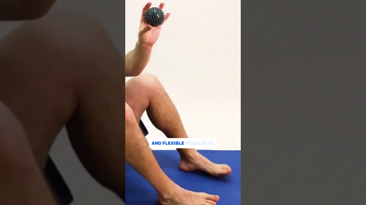
![Metatarsalgia Treatment [BEST Ball of Foot Pain RELIEF 2024]](https://www.michiganfootdoctors.com/wp-content/cache/flying-press/d7857672675f2bf191a8e896b2f7208b.jpg)
Watch: Metatarsalgia Treatment [BEST Ball of Foot Pain RELIEF 2024] — MichiganFootDoctors YouTube
Foot pain isn't resolving?
Same-week appointments at Howell & Bloomfield Hills
Medically Reviewed | Dr. Tom Biernacki, DPM | Board-Certified Podiatric Surgeon | Balance Foot & Ankle, Michigan

Metatarsalgia: Understanding and Treating Ball of Foot Pain
Metatarsalgia is a descriptive term — not a diagnosis — referring to pain in the forefoot at the metatarsal head region (ball of the foot). Like “headache” or “knee pain,” it describes a symptom location rather than a specific cause. This distinction matters clinically because multiple conditions cause forefoot pain at the metatarsal heads, and each requires a somewhat different management approach.
Dr. Tom Biernacki at Balance Foot & Ankle in Howell, Michigan approaches forefoot pain with systematic diagnosis before treatment. Determining whether metatarsalgia is mechanical overload, Morton’s neuroma, sesamoiditis, stress fracture, fat pad atrophy, or inflammatory arthritis changes the treatment pathway significantly.
Anatomy of Forefoot Pain
The forefoot region contains five metatarsal bones connecting the midfoot to the toes. The metatarsal heads — the rounded distal ends of these bones — bear significant weight during the propulsive phase of gait, particularly during the toe-off moment when body weight concentrates under the forefoot before the foot leaves the ground.
Each metatarsal head is cushioned by a plantar fat pad — fibrocartilaginous tissue that distributes pressure during loading. Between the metatarsal heads, digital nerves supply sensation to adjacent toes, running through the intermetatarsal spaces. Bursae (fluid-filled sacs) cushion the metatarsal heads from overlying soft tissue.
The metatarsal parabola — the arc of metatarsal head positions from first (shortest) through third (longest) and back to fifth — determines how forefoot pressure distributes. When this parabola is disrupted (by bunion, second metatarsal length discrepancy, or first ray hypermobility), pressure concentrates abnormally on specific metatarsal heads, creating overload and pain.
Causes of Metatarsalgia: The Differential Diagnosis
Mechanical/overload metatarsalgia is the most common pattern — excessive pressure concentrated on one or more metatarsal heads from biomechanical or footwear causes. High-heeled shoes that shift body weight to the forefoot, Morton’s foot anatomy (second toe longer than first), first ray hypermobility from bunion deformity, and activities with high forefoot loading (running, jumping) all contribute. This pattern responds best to pressure redistribution through metatarsal pads and footwear modification.
Morton’s neuroma is a perineural fibrosis (thickening) of the interdigital nerve, most commonly between the third and fourth metatarsal heads. Patients describe sharp, burning, or electric pain in the ball of the foot, often with a sensation of “walking on a pebble” or radiation into adjacent toes. Mulder’s sign — a painful click felt when squeezing the forefoot laterally while pressing on the affected interspace from plantar — is a classic clinical finding. Morton’s neuroma is frequently misdiagnosed as metatarsalgia and requires different treatment (targeted injections, neuroma excision) rather than simple pressure redistribution.
Sesamoiditis involves inflammation at the sesamoid bones beneath the first metatarsal head. Pain is specifically under the great toe joint, aggravated by activities requiring toe-off (running, dancing, walking uphill). Sesamoid stress fractures produce similar pain with more acute onset. This is addressed with sesamoid offloading orthotics and activity modification — very different from general metatarsal pad treatment.
Fat pad atrophy occurs in older adults and in patients with inflammatory arthritis or prior corticosteroid injections — the plantar fat pad thins, reducing natural cushioning under the metatarsal heads. This produces a characteristic “walking on bones” sensation. Treatment focuses on maximally cushioned footwear and accommodative orthotics with soft Poron or Plastazote padding rather than arch support or rigid correction.
MTP joint synovitis and predislocation — inflammatory changes in the second or third MTP joint — produce localized forefoot pain and swelling at the joint itself rather than diffusely in the ball of the foot. The drawer test (pulling the toe vertically while stabilizing the metatarsal head) detects plantar plate laxity or rupture. Predislocation syndrome, if not managed, progresses to hammer toe formation and painful toe dislocation.
Metatarsal stress fractures produce focal bone tenderness along the metatarsal shaft rather than at the head, aggravated by activity and relieved by rest. Plain X-rays are often negative for 2–3 weeks; bone scan or MRI confirms early stress fracture when clinical suspicion is high. This diagnosis requires activity modification and sometimes protected weight-bearing — not orthotics.
Clinical Evaluation: How Podiatrists Diagnose Forefoot Pain
Dr. Biernacki’s forefoot evaluation systematically identifies the responsible structure through physical examination and, when necessary, imaging and diagnostic injections.
Palpation mapping identifies the precise location of maximum tenderness — under specific metatarsal heads (mechanical metatarsalgia), between metatarsal heads (Morton’s neuroma), under the first metatarsal sesamoids (sesamoiditis), or along the metatarsal shaft (stress fracture). The specificity of forefoot palpation significantly narrows the differential diagnosis.
Provocative tests include Mulder’s sign for Morton’s neuroma, drawer test for MTP joint instability, and toe extension testing for sesamoiditis. Footwear analysis — examining wear patterns, toe box dimensions, and heel height — identifies contributing biomechanical factors.
Musculoskeletal ultrasound at Balance Foot & Ankle provides real-time visualization of Morton’s neuroma (hypoechoic intermetatarsal lesion), MTP joint effusion, plantar plate integrity, and metatarsal head fat pad thickness. Ultrasound-guided diagnostic injections with local anesthetic confirm the pain source when clinical examination is inconclusive — pain relief confirms the structure responsible.
Weight-bearing X-rays assess metatarsal parabola, joint space changes, sesamoid pathology, and stress fracture callus in longer-standing cases.
Conservative Treatment Protocols
Treatment is tailored to the specific metatarsalgia diagnosis. For mechanical overload metatarsalgia — the most common type — the evidence-based approach addresses footwear, pressure redistribution, and activity modification.
Metatarsal pads are the most effective conservative intervention for mechanical metatarsalgia — but only when placed correctly. The pad must be positioned 1–2cm proximal to the symptomatic metatarsal heads (behind the ball of the foot) to elevate and separate the metatarsal heads, redistributing pressure. Pads placed directly under the metatarsal heads increase rather than decrease pressure. Adhesive-backed pads can be repositioned during the first few uses until optimal placement is confirmed by symptom response.
Footwear modification includes increasing toe box depth and width to reduce dorsal forefoot pressure, reducing heel height to decrease propulsive forefoot loading, and ensuring adequate shoe length so toes don’t impinge against the shoe end. Rocker-bottom shoes accelerate the roll-through phase of gait, reducing time spent in the high-pressure toe-off moment — particularly beneficial for metatarsalgia in patients requiring more active footwear intervention.
Activity modification reducing high-impact forefoot loading (running, jumping, stair climbing) while maintaining lower-impact aerobic activity (cycling, swimming) allows pain reduction while maintaining fitness during recovery.
For Morton’s neuroma specifically, corticosteroid injection into the affected intermetatarsal space provides pain relief in 60–70% of patients. Injections may be repeated 2–3 times at 4–6 week intervals. Alcohol sclerosing injections offer an alternative for cases where corticosteroid benefit wanes — 4–6 injections of dilute alcohol under ultrasound guidance produces neuroma fibrosis and progressive symptom relief.
For sesamoiditis, dancer’s padding (donut-shaped pad around but not under the sesamoids) offloads the sesamoid region. Activity reduction in toe-off intensive activities is essential. Fracture cases may require brief cast immobilization or sesamoid-offloading orthosis.
Surgical Options for Metatarsalgia
When conservative management fails, surgical options address the specific diagnosed cause. For Morton’s neuroma refractory to injection therapy, surgical excision removes the affected nerve — providing complete pain resolution in 80–85% of cases, with the expected tradeoff of permanent numbness in the web space between affected toes. Decompression (releasing the intermetatarsal ligament without removing the nerve) is an alternative that preserves sensation with comparable pain outcomes in selected patients.
Metatarsal osteotomy for mechanical metatarsalgia shortens or elevates a prominent metatarsal head that is concentrating excessive forefoot pressure. This is indicated when the metatarsal parabola shows significant length discrepancy contributing to pathological pressure distribution and conservative measures have failed after appropriate duration.
Sesamoid surgery — sesamoidectomy (removal of one sesamoid) — is considered after failed conservative management of sesamoiditis or non-healing sesamoid fracture, accepting the tradeoff of reduced first toe push-off strength for relief from chronic pain.
MTP joint stabilization for predislocation syndrome and plantar plate tears prevents progressive toe dislocation and corrects existing deformity. Early treatment of predislocation syndrome with appropriate orthotics and activity modification may prevent progression to surgery; established plantar plate tears often require surgical repair combined with associated deformity correction.
Dr. Tom's Product Recommendations
Pedag Viva Metatarsal Pad — Self-Adhesive
⭐ Highly Rated
Adhesive-backed leather-covered foam metatarsal pad for ball of foot pain. Repositionable during initial placement to achieve optimal position proximal to metatarsal heads. Reduces peak forefoot pressure by redistributing load through the metatarsal shafts. Clinical first-line treatment for mechanical metatarsalgia.
Dr. Tom says: “Placed exactly where my podiatrist showed me — proximal to the painful area, not under it — and the relief was immediate. Proper placement makes all the difference.”
Mechanical metatarsalgia, general ball-of-foot pain, forefoot pressure redistribution
Morton’s neuroma (requires targeted interspace injection, not pad alone), sesamoiditis (needs donut padding around sesamoids specifically)
Disclosure: We earn a commission at no extra cost to you.
HOKA Bondi 8 Maximum Cushion Walking Shoe
⭐ Highly Rated
Maximum stack height cushioned shoe with 4mm heel drop and wide toe box. The extended midsole geometry reduces forefoot peak pressure during gait. Recommended by podiatrists for fat pad atrophy, chronic metatarsalgia, and patients requiring maximum shock absorption without orthopedic appearance.
Dr. Tom says: “My podiatrist told me maximum cushion was the key for my fat pad atrophy. These are the most comfortable shoes I’ve worn in years — the forefoot pain dropped substantially.”
Fat pad atrophy, chronic metatarsalgia, maximum cushioning needs
Patients requiring rigid arch correction (pair with appropriate insoles)
Disclosure: We earn a commission at no extra cost to you.
Silipos Gel Metatarsal Sleeve
⭐ Highly Rated
Soft gel sleeve worn around the forefoot providing circumferential metatarsal cushioning and support. Appropriate for runners and active patients with metatarsalgia who need consistent pad positioning during activity. More stable than adhesive pads for sports use.
Dr. Tom says: “Better than adhesive pads for my running — stays in position through a full 10K without shifting. My ball-of-foot pain is manageable again.”
Active patients, runners, metatarsalgia during athletic activity
Patients with circulation problems or diabetes requiring assessment before forefoot compression
Disclosure: We earn a commission at no extra cost to you.
Altra Escalante 3 Neutral Road Running Shoe
⭐ Highly Rated
Zero-drop neutral running shoe with foot-shaped toe box allowing natural toe splay and reducing metatarsal head compression during forefoot striking. A-Bound midsole provides responsive cushioning. Popular among runners transitioning from heel-strike to forefoot loading patterns.
Dr. Tom says: “After switching from pointed athletic shoes to these, my forefoot pain during runs decreased significantly. The width at the toe box makes the biggest difference.”
Runners with metatarsalgia, wide forefoot, natural toe splay needs
Patients requiring elevated heel drop for Achilles tightness
Disclosure: We earn a commission at no extra cost to you.
✅ Pros / Benefits
- M
- o
- s
- t
- m
- e
- t
- a
- t
- a
- r
- s
- a
- l
- g
- i
- a
- c
- a
- s
- e
- s
- r
- e
- s
- p
- o
- n
- d
- t
- o
- c
- o
- n
- s
- e
- r
- v
- a
- t
- i
- v
- e
- m
- e
- a
- s
- u
- r
- e
- s
- —
- p
- r
- o
- p
- e
- r
- l
- y
- p
- l
- a
- c
- e
- d
- m
- e
- t
- a
- t
- a
- r
- s
- a
- l
- p
- a
- d
- s
- a
- n
- d
- f
- o
- o
- t
- w
- e
- a
- r
- m
- o
- d
- i
- f
- i
- c
- a
- t
- i
- o
- n
- r
- e
- s
- o
- l
- v
- e
- t
- h
- e
- m
- a
- j
- o
- r
- i
- t
- y
- o
- f
- m
- e
- c
- h
- a
- n
- i
- c
- a
- l
- f
- o
- r
- e
- f
- o
- o
- t
- p
- a
- i
- n
- c
- a
- s
- e
- s
- w
- i
- t
- h
- o
- u
- t
- p
- r
- o
- c
- e
- d
- u
- r
- e
- s
- o
- r
- s
- u
- r
- g
- e
- r
- y
- .
- M
- o
- r
- t
- o
- n
- ‘
- s
- n
- e
- u
- r
- o
- m
- a
- r
- e
- s
- p
- o
- n
- d
- s
- t
- o
- c
- o
- r
- t
- i
- c
- o
- s
- t
- e
- r
- o
- i
- d
- i
- n
- j
- e
- c
- t
- i
- o
- n
- s
- i
- n
- 6
- 0
- –
- 7
- 0
- %
- o
- f
- c
- a
- s
- e
- s
- ,
- a
- v
- o
- i
- d
- i
- n
- g
- s
- u
- r
- g
- e
- r
- y
- f
- o
- r
- m
- o
- s
- t
- p
- a
- t
- i
- e
- n
- t
- s
- .
- F
- a
- t
- p
- a
- d
- a
- t
- r
- o
- p
- h
- y
- i
- s
- m
- a
- n
- a
- g
- e
- a
- b
- l
- e
- w
- i
- t
- h
- a
- p
- p
- r
- o
- p
- r
- i
- a
- t
- e
- f
- o
- o
- t
- w
- e
- a
- r
- s
- e
- l
- e
- c
- t
- i
- o
- n
- .
- T
- h
- e
- v
- a
- r
- i
- e
- t
- y
- o
- f
- e
- f
- f
- e
- c
- t
- i
- v
- e
- c
- o
- n
- s
- e
- r
- v
- a
- t
- i
- v
- e
- o
- p
- t
- i
- o
- n
- s
- m
- e
- a
- n
- s
- m
- o
- s
- t
- p
- a
- t
- i
- e
- n
- t
- s
- c
- a
- n
- a
- v
- o
- i
- d
- s
- u
- r
- g
- i
- c
- a
- l
- i
- n
- t
- e
- r
- v
- e
- n
- t
- i
- o
- n
- w
- i
- t
- h
- a
- p
- p
- r
- o
- p
- r
- i
- a
- t
- e
- d
- i
- a
- g
- n
- o
- s
- i
- s
- a
- n
- d
- t
- r
- e
- a
- t
- m
- e
- n
- t
- m
- a
- t
- c
- h
- i
- n
- g
- .
❌ Cons / Risks
- M
- e
- t
- a
- t
- a
- r
- s
- a
- l
- g
- i
- a
- d
- i
- a
- g
- n
- o
- s
- i
- s
- r
- e
- q
- u
- i
- r
- e
- s
- c
- l
- i
- n
- i
- c
- a
- l
- e
- x
- p
- e
- r
- t
- i
- s
- e
- t
- o
- d
- i
- s
- t
- i
- n
- g
- u
- i
- s
- h
- b
- e
- t
- w
- e
- e
- n
- m
- u
- l
- t
- i
- p
- l
- e
- c
- a
- u
- s
- e
- s
- —
- g
- e
- n
- e
- r
- i
- c
- t
- r
- e
- a
- t
- m
- e
- n
- t
- w
- i
- t
- h
- o
- u
- t
- s
- p
- e
- c
- i
- f
- i
- c
- d
- i
- a
- g
- n
- o
- s
- i
- s
- l
- e
- a
- d
- s
- t
- o
- f
- a
- i
- l
- e
- d
- c
- o
- n
- s
- e
- r
- v
- a
- t
- i
- v
- e
- m
- a
- n
- a
- g
- e
- m
- e
- n
- t
- a
- n
- d
- d
- e
- l
- a
- y
- s
- .
- M
- o
- r
- t
- o
- n
- ‘
- s
- n
- e
- u
- r
- o
- m
- a
- s
- u
- r
- g
- i
- c
- a
- l
- e
- x
- c
- i
- s
- i
- o
- n
- p
- r
- o
- d
- u
- c
- e
- s
- p
- e
- r
- m
- a
- n
- e
- n
- t
- n
- u
- m
- b
- n
- e
- s
- s
- i
- n
- t
- h
- e
- w
- e
- b
- s
- p
- a
- c
- e
- (
- a
- n
- e
- x
- p
- e
- c
- t
- e
- d
- a
- n
- d
- a
- c
- c
- e
- p
- t
- a
- b
- l
- e
- t
- r
- a
- d
- e
- o
- f
- f
- f
- o
- r
- m
- o
- s
- t
- p
- a
- t
- i
- e
- n
- t
- s
- ,
- b
- u
- t
- i
- m
- p
- o
- r
- t
- a
- n
- t
- t
- o
- d
- i
- s
- c
- u
- s
- s
- p
- r
- e
- –
- o
- p
- e
- r
- a
- t
- i
- v
- e
- l
- y
- )
- .
- F
- a
- t
- p
- a
- d
- a
- t
- r
- o
- p
- h
- y
- h
- a
- s
- n
- o
- r
- e
- g
- e
- n
- e
- r
- a
- t
- i
- v
- e
- t
- r
- e
- a
- t
- m
- e
- n
- t
- —
- m
- a
- n
- a
- g
- e
- m
- e
- n
- t
- f
- o
- c
- u
- s
- e
- s
- o
- n
- c
- o
- m
- p
- e
- n
- s
- a
- t
- i
- o
- n
- r
- a
- t
- h
- e
- r
- t
- h
- a
- n
- c
- u
- r
- e
- .
- R
- e
- c
- u
- r
- r
- e
- n
- c
- e
- i
- s
- c
- o
- m
- m
- o
- n
- i
- f
- u
- n
- d
- e
- r
- l
- y
- i
- n
- g
- b
- i
- o
- m
- e
- c
- h
- a
- n
- i
- c
- a
- l
- a
- n
- d
- f
- o
- o
- t
- w
- e
- a
- r
- c
- a
- u
- s
- e
- s
- a
- r
- e
- n
- o
- t
- a
- d
- d
- r
- e
- s
- s
- e
- d
- a
- f
- t
- e
- r
- s
- u
- c
- c
- e
- s
- s
- f
- u
- l
- i
- n
- i
- t
- i
- a
- l
- t
- r
- e
- a
- t
- m
- e
- n
- t
- .
Dr. Tom Biernacki’s Recommendation
Ball of foot pain is one of those areas where getting the diagnosis right before starting treatment matters enormously. I see patients who’ve been doing generic arch support and insoles for six months for what’s actually a Morton’s neuroma — which needs a targeted injection, not a metatarsal pad. And I see neuroma patients who’ve been told they need surgery when a simple alcohol sclerosing series would manage it non-operatively. The forefoot has multiple structures that can hurt, and matching the treatment to the actual problem is where I spend my diagnostic attention.
— Dr. Tom Biernacki, DPM | Board-Certified Podiatric Surgeon | Balance Foot & Ankle
Frequently Asked Questions
Ready to Get Relief?
Same-day appointments available in Howell & Bloomfield Hills, MI
4.9★ | 1,123 Reviews | 3,000+ Surgeries
Or call: (810) 206-1402
Dr. Tom Biernacki, DPM is a board-certified foot & ankle surgeon (ABFAS & ABPM) at Balance Foot & Ankle Specialists in Southeast Michigan. With over a decade of clinical experience, he specializes in heel pain, bunions, diabetic foot care, sports injuries, and minimally invasive surgery. Dr. Biernacki is a member of the APMA and ACFAS, and his patient education content on MichiganFootDoctors.com and YouTube has made him one of the most-followed foot & ankle educators on YouTube.