
Peroneal Muscle Pain: Causes, Symptoms & Treatment (Podiatrist 2026)
Peroneal muscle pain — on the OUTSIDE of the lower leg + outer foot — is most often peroneal tendinopathy (chronic overuse). Causes: chronic ankle instability after sprains, sudden increase in running mileage, hill running, dancers, Pilates instructors, basketball players, or biomechanical overpronation forcing peroneal tendons to overwork. Symptoms: pain along the peroneal tendons (behind outer ankle bone), worse with eversion (turning foot outward), sometimes a clicking sensation, swelling along the tendon path.
In my Michigan podiatry clinic, my peroneal tendon pain protocol gets ~75% of patients pain-free in 4-6 weeks: (1) lateral heel wedge orthotic (off-loads the peroneal tendons), (2) stability shoe (Brooks Adrenaline GTS, ASICS Kayano), (3) ankle stability brace (ASO) for chronic sprainers, (4) eccentric peroneal strengthening (resistance-band eversion 3x daily), (5) 4-6 weeks of activity modification. Red flag: palpable click or tendon snapping during ankle motion = peroneal tendon subluxation; usually needs surgical retinaculum repair.
Same-Week Appointments at Balance Foot & Ankle
Three board-certified podiatric surgeons. 950K+ YouTube subscribers. 1,123+ five-star reviews. Howell & Bloomfield Hills, Michigan.
Medically reviewed by Dr. Tom Biernacki, DPM · Board-Certified Podiatric Surgeon · Last reviewed: April 2026 · Editorial Policy
Quick Answer
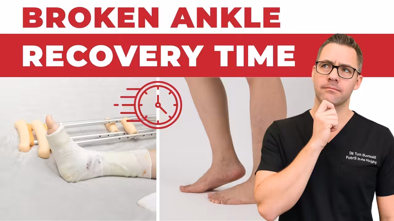
Peroneal Tendonitis 2026: Pain, Treatment & Recovery Gu relates to tendon injury — typically caused by overuse or sudden strain. Most patients improve in 6-12 weeks with conservative care. Same-week appointments in Howell + Bloomfield Twp: (810) 206-1402.

✅ Medically reviewed by Dr. Thomas Biernacki, DPM — Board-Certified Podiatrist · Last updated April 6, 2026

Medically Reviewed by Dr. Carl Jay, DPM — Board-Qualified Podiatric Physician
Balance Foot & Ankle, Michigan · Updated April 2026
⚡ Quick Answer
Peroneal tendonitis is inflammation of the peroneal tendons that run along the outer ankle and foot, causing pain behind and below the outer ankle bone (lateral malleolus). It’s most commonly triggered by overuse, sudden increases in activity, ankle sprains, or high-arched feet. Most cases respond well to conservative treatment within 4–8 weeks: rest and activity modification, ice, supportive footwear, ankle bracing, and a structured stretching and strengthening program. Surgery is rarely needed but may be considered for chronic tears or subluxation that doesn’t respond to 3–6 months of conservative care.
Pain along the outer edge of your ankle that started after you ramped up your running mileage, switched to a hillier route, or rolled your ankle — this is the classic story of peroneal tendonitis. The peroneal tendons are the hardest-working stabilizers of the outer ankle, and when they become inflamed or damaged, every step reminds you they exist.
At Balance Foot & Ankle, we see peroneal tendon injuries regularly — from weekend athletes who increased activity too quickly to patients with high arches who’ve been unknowingly overloading these tendons for years. The condition responds well to proper treatment, but misdiagnosis is common because the pain can mimic ankle sprains, stress fractures, or nerve issues. Here’s how to identify peroneal tendonitis, what’s happening inside the tendon, and how to fix it.
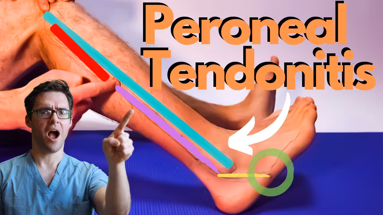
Anatomy: The Two Peroneal Tendons
Two peroneal muscles originate on the outer lower leg and send tendons behind the outer ankle bone (lateral malleolus), through a groove in the bone called the fibular groove, and into the foot.
Peroneus longus travels under the foot to attach on the inner side at the base of the first metatarsal and medial cuneiform. It’s responsible for everting the foot (turning the sole outward) and assists with plantarflexion. It also plays a critical role in stabilizing the first ray during push-off.
Peroneus brevis attaches to the base of the fifth metatarsal on the outer foot. It primarily everts the foot and is the key dynamic stabilizer against ankle inversion (rolling inward) — which is why peroneal tendon injuries are so common after ankle sprains.
Both tendons are held in place behind the ankle bone by the superior peroneal retinaculum — a band of tissue that acts like a seatbelt. When this retinaculum is damaged (often during ankle sprains), the tendons can sublux (snap over the bone), causing a snapping sensation and ongoing instability.
Types of Peroneal Tendon Injuries
| Injury Type | What’s Happening | Symptoms | Common Cause |
|---|---|---|---|
| Tendonitis (most common) | Inflammation of the tendon and its sheath | Aching pain behind/below outer ankle; worse with activity; swelling | Overuse, sudden activity increase, repetitive ankle motion |
| Tendinosis | Chronic degeneration; microtears without acute inflammation | Persistent ache; thickened tendon on exam; gradual onset over months | Chronic overload, inadequate healing from recurrent tendonitis |
| Peroneal Tear | Partial or full-thickness tear, often of the peroneus brevis | Sharp pain, weakness with eversion, ankle gives way | Ankle sprain, chronic subluxation, degenerative changes |
| Subluxation / Dislocation | Tendons snap over the lateral malleolus due to damaged retinaculum | Audible/palpable snapping behind ankle; instability; recurrent “giving way” | Ankle sprain damage to retinaculum, shallow fibular groove (anatomic variant) |
Causes and Risk Factors
Overuse and training errors are the leading cause. Running on uneven terrain (trails, beaches, crowned roads), suddenly increasing mileage or intensity, and repetitive lateral movements (basketball, tennis, soccer) all overload the peroneal tendons. The classic pattern is a runner who increases weekly mileage by more than 10% or adds hill training too quickly.
Ankle sprains — particularly inversion sprains — stretch and sometimes tear the peroneal retinaculum, destabilizing the tendons. Research shows that up to 40% of ankle sprains have associated peroneal tendon injury that goes undiagnosed because the attention focuses on the lateral ligaments.
High arches (pes cavus) tilt the heel into a varus (inward-leaning) position, placing the peroneal tendons on constant stretch and increasing the mechanical load with every step. This is one of the most overlooked risk factors — patients with high arches and chronic outer ankle pain often have undiagnosed peroneal tendinosis.
Improper footwear — shoes with worn-out lateral edges, inadequate ankle support, or too much lateral rigidity can all contribute. Minimalist shoes and racing flats provide minimal peroneal support.
Chronic ankle instability — recurrent ankle sprains leave the lateral ligaments lax, forcing the peroneal tendons to compensate as the primary stabilizers. This chronic overwork eventually leads to tendonitis or degeneration.
⚠️ When to See a Doctor Urgently
- Inability to bear weight after an ankle injury — may indicate fracture or complete tendon tear
- Visible snapping or popping of tendons behind the ankle bone — suggests subluxation requiring evaluation
- Significant bruising extending behind and below the outer ankle — may indicate tendon rupture
- Progressive weakness — difficulty lifting the outer edge of your foot or walking on uneven ground
- Pain and swelling that worsen despite 2 weeks of rest — suggests something beyond simple tendonitis
Diagnosis
Your podiatrist can typically diagnose peroneal tendonitis through a physical exam. Key tests include palpation along the tendon path (tenderness behind and below the lateral malleolus), resisted eversion (pain with turning the foot outward against resistance), and the peroneal compression test (pressing behind the ankle while dorsiflexing and everting the foot to check for subluxation).
Ultrasound is the best first-line imaging study for peroneal tendon injuries — it’s dynamic (performed while you move your foot), so it can visualize subluxation in real-time, identify tears, and assess tendon thickness and fluid in the sheath. MRI is used for complex cases, surgical planning, or when ultrasound is inconclusive. X-rays can rule out fractures (particularly a 5th metatarsal avulsion fracture, which can mimic peroneal tendonitis).
Conservative Treatment Protocol
Phase 1: Acute Phase (Weeks 1–2)
Relative rest — Reduce or eliminate the aggravating activity, but avoid complete immobilization unless pain is severe. Cross-train with swimming or cycling (no push-off component). Walking is usually fine if pain is mild.
Ice — Apply ice for 15–20 minutes, 3–4 times daily, along the outer ankle. A frozen water bottle rolled under the outer foot and ankle is effective.
Ankle brace or taping — A lace-up ankle brace or kinesiology tape supports the peroneal tendons, reduces the work they need to do, and prevents the ankle from rolling inward during daily activities.
NSAIDs — Short-term ibuprofen or naproxen (7–10 days) reduces inflammation and pain. Topical diclofenac gel applied directly over the tendons avoids systemic side effects.
Phase 2: Rehabilitation (Weeks 2–6)
Eccentric strengthening is the gold standard for tendon rehabilitation. For peroneal tendons: sit with your foot hanging, use a resistance band wrapped around the outside of the foot, slowly resist as the foot returns to a neutral position. 3 sets of 15 repetitions, twice daily. Eccentric loading stimulates tendon remodeling and has the strongest evidence base for tendonitis recovery.
Balance and proprioception training — Single-leg balance on a flat surface (work up to 60 seconds), progress to a wobble board or foam pad, then add eyes-closed challenges. Proprioception training retrains the neuromuscular control that’s often impaired after peroneal tendon injuries.
Calf stretching — Tight calves increase the load on the peroneal tendons by limiting ankle dorsiflexion. Wall stretches (knee straight for gastrocnemius, knee bent for soleus), 30 seconds each, 3 times per side, twice daily.
Phase 3: Return to Activity (Weeks 6–12)
Gradually reintroduce running or sport-specific activities. Start at 50% of your pre-injury volume and increase by no more than 10% per week. Continue the eccentric strengthening and balance exercises as maintenance. If pain returns, pull back to the previous comfortable level for one week before progressing again.
Footwear and Orthotics
Proper footwear is critical for both recovery and prevention. The ideal shoe for peroneal tendonitis provides lateral stability (prevents ankle rolling), moderate cushioning (reduces impact forces on the tendons), and a heel counter that keeps the heel upright rather than letting it tilt inward.
For patients with high arches — the foot type most prone to peroneal problems — a cushioned neutral shoe with a lateral posting or custom orthotic to reduce the varus heel tilt can be transformative. Over-the-counter orthotics with a deep heel cup help stabilize the rearfoot and reduce peroneal strain.
Products We Recommend
We may earn a commission through affiliate links below — but every product is independently selected and recommended based on clinical experience. This does not affect our recommendations.
🏆 #1 Pick — ASICS Gel-Kayano
The Gel-Kayano provides the combination of lateral stability and cushioning that peroneal tendonitis patients need. The Dynamic DuoMax support system and GEL technology in the rearfoot prevent excessive inversion, and the structured heel counter keeps the foot properly aligned. This is our top recommendation for runners recovering from peroneal tendon injuries — the stability prevents the ankle rolling that aggravates the tendons while the cushioning reduces impact forces.
PowerStep Pinnacle Maxx Orthotics
The Pinnacle Maxx’s deep heel cup stabilizes the rearfoot and controls the varus (inward) heel tilt that’s a primary driver of peroneal tendon overload, especially in high-arched feet. The semi-rigid arch support redistributes forces away from the lateral foot and ankle. For many patients with recurrent peroneal tendonitis, adding these to their shoes is the single change that prevents recurrence.
Hoka Bondi 8
For daily walking and standing, the Bondi’s maximum cushioning absorbs the repetitive forces that aggravate inflamed peroneal tendons. The oversized midsole and meta-rocker geometry provide smooth forward propulsion, reducing the lateral forces that stress the outer ankle. Wide toe box prevents forefoot compression that can alter gait mechanics.
More Podiatrist-Recommended Foot Health Essentials
Hoka Clifton 10

Max-cushion everyday shoe — podiatrist favorite for walking and running.
PowerStep Pinnacle Insole

- The Pinnacle Full length insoles for men & women provide maximum cushioning, from high activity to moderate support. The PowerStep arch support shape provides stability to the foot and ankle, helping to relieve foot pain.
- When you spend all day on your feet, every step counts. PowerStep insoles are a podiatrist-recommended orthotic to help relieve & prevent foot pain related to athletes, runners, Plantar Fasciitis, heel spurs & other common foot, ankle & knee injuries
- The Pinnacle plantar fasciitis insoles offer superior heel cushioning and arch support. The dual-layer cushioning is designed to reduce stress and fatigue, while PowerStep premium arch support is designed for plantar fasciitis relief.
- The PowerStep Pinnacle arch support inserts for men & women can be worn in a variety of shoe types such as; athletic, walking, running, work & some casual shoes. Orthotic Inserts are ordered by shoe size, no trimming required.
- Made in the USA & backed by a 30-day money-back guarantee. PowerStep orthotic inserts for men & women are designed for shoes where the factory insole can be removed. HSA & FSA Eligible
The podiatrist-recommended over-the-counter orthotic.
OOFOS Recovery Slide

- The Original Recovery Footwear.
- Finding Your Size - For your perfect fit, consult the “size chart” link above. Wear a half size? In general, we recommend that women who wear a ½ size size UP, and men who wear a ½ size size DOWN
- OOahh - An evolution of the OOriginal, the OOahh slide features our proven foundation of OOfoam technology + patented footbed design with a slide-style strap that has become a best-seller in the OOFOS line
- OOfoam Technology - Our revolutionary OOfoam technology absorbs 37% more impact than traditional footwear foams to reduce the stress on your feet, joints & back. Plus, the closed-cell foam is machine washable and designed to minimize odor
- Patented Footbed - Our patented footbed cradles and supports arches to reduce energy exertion in the ankles by up to 47% compared to competitors’ footwear. So walking is easier. Recovery is faster. And yOO feel better
Impact-absorbing recovery sandal — wear after long days on your feet.
As an Amazon Associate, Balance Foot & Ankle earns from qualifying purchases. Product recommendations are based on clinical experience; prices and availability shown above update live from Amazon.
When to See a Podiatrist
If foot or ankle pain has been bothering you for more than a few weeks, home care alone may not be enough. Balance Foot & Ankle offers same-week appointments at our Howell and Bloomfield Hills clinics — no referral needed in most cases. Bring your current shoes and a short list of symptoms and we’ll build you a treatment plan in one visit.
Call Balance Foot & Ankle: (810) 206-1402 · Book online · Offices in Howell & Bloomfield Hills
Frequently Asked Questions
Mild peroneal tendonitis typically improves within 2–4 weeks with rest, ice, and proper footwear. Moderate cases with significant tendon thickening or pain with daily activities usually take 6–8 weeks with a structured rehabilitation program. Chronic tendinosis (where the tendon has degenerated rather than just inflamed) can take 3–6 months to fully resolve. The most important factor in recovery time is catching it early — continuing to train through peroneal pain significantly extends healing time.
Usually yes — walking on flat, even surfaces in supportive shoes is generally fine and even encouraged during recovery. Walking maintains blood flow to the healing tendon and prevents deconditioning. However, avoid walking on uneven terrain, hills, or inclines (which increase peroneal demand), and stop if walking causes more than mild discomfort. Use an ankle brace for additional support during the acute phase. If walking causes significant pain, a walking boot may be needed temporarily.
The most common triggers are sudden increases in activity (running more miles, adding hill work, increasing sport intensity), wearing unsupportive or worn-out shoes, walking or running on uneven surfaces, ankle sprains that damage the peroneal retinaculum, and having high arches that place extra stress on the outer tendons. Cold weather can also cause flare-ups because it reduces blood flow to the tendons and increases tissue stiffness.
Simple tendonitis is not serious and responds well to conservative treatment. However, if left untreated, tendonitis can progress to tendinosis (chronic degeneration), partial tears, or chronic subluxation — which are more difficult to treat and may eventually require surgery. Persistent outer ankle pain that hasn’t improved after 2–3 weeks of home treatment deserves professional evaluation to ensure you’re not dealing with a tear, subluxation, or fracture that requires different management.
The Bottom Line
Peroneal tendonitis is a frustrating but highly treatable condition when approached correctly. The keys to successful recovery are reducing the aggravating activity early (not pushing through the pain), starting eccentric strengthening once acute pain subsides, addressing underlying biomechanical factors like high arches with proper footwear and orthotics, and returning to activity gradually. Most patients are back to full activity within 6–12 weeks — and with continued attention to footwear and ankle strengthening, recurrence is uncommon.
In-Office Treatment at Balance Foot & Ankle
When conservative care isn’t enough, Dr. Tom Biernacki and the team at Balance Foot & Ankle offer advanced, same-day options — including Peroneal Tendon Disorders Treatment in Michigan at our Howell and Bloomfield Hills clinics.
Same-day appointments available. Call (810) 206-1402 or book online.
Sources
- Davda K, et al. “Peroneal Tendon Disorders.” EFORT Open Reviews, 2017;2(6):281-292.
- Heckman DS, et al. “Tendon Disorders of the Foot and Ankle.” Journal of the American Academy of Orthopaedic Surgeons, 2009;17(4):231-238.
- van Dijk PAD, et al. “Peroneal Tendon Pathology.” Foot and Ankle Clinics, 2015;20(4):563-575.
- Alfredson H. “Eccentric Muscle Training in Sports and Tendinopathies.” Scandinavian Journal of Medicine & Science in Sports, 2015.
Outer Ankle Pain That Won’t Go Away?
Our podiatrists specialize in diagnosing peroneal tendon injuries using in-office ultrasound, developing targeted rehabilitation programs, and fitting custom orthotics for high-arched feet. Most patients see significant improvement within their first few weeks of treatment.
📞 (810) 206-1402 · Howell & Bloomfield Hills, Michigan
Outer Ankle Pain That Won’t Go Away?
Peroneal tendonitis is often misdiagnosed. Our podiatrists specialize in accurate diagnosis and targeted treatment of ankle tendon conditions.
Clinical References
- Dombek MF et al. Peroneal tendon tears: a retrospective review. Journal of Foot and Ankle Surgery. 2003;42(5):250-258.
- Heckman DS et al. Tendon disorders of the foot and ankle, part 2: achilles tendon disorders. American Journal of Sports Medicine. 2009;37(6):1223-1234.
- Davda K et al. Peroneal tendon disorders. EFORT Open Reviews. 2017;2(6):281-292.
Insurance Accepted
BCBS · Medicare · Aetna · Cigna · United Healthcare · HAP · Priority Health · Humana · View All →
👟 Dr. Tom Also Recommends
Podiatrist Recommended Shoes 2026: Dr. Tom’s Top Picks for Every Condition
The right footwear can make or break your recovery. Dr. Tom’s complete guide to the best shoes for plantar fasciitis, flat feet, neuropathy, bunions & more — with clinical picks for every foot type.
See Dr. Tom’s Top Shoe Picks →Howell Office
3980 E Grand River Ave, Suite 140
Howell, MI 48843
Get Directions →
Bloomfield Hills Office
43700 Woodward Ave, Suite 207
Bloomfield Hills, MI 48302
Get Directions →
Your Board-Certified Podiatrists


Ready to Get Back on Your Feet?
Same-week appointments available at both locations.
Book Your AppointmentPros & Cons of Conservative Care for foot care
Advantages
- ✓ Conservative care first
- ✓ Same-week appointments
- ✓ Multiple insurance accepted
Considerations
- ✗ Self-treatment can mask issues
- ✗ See a podiatrist if pain >2 weeks
Dr. Tom’s Recommended Products for foot care
Affiliate disclosure: As an Amazon Associate, Balance Foot & Ankle earns from qualifying purchases. We only recommend products we use with patients.


Footnanny Heel Cream Dr. Tom’s Pick
Best for: Daily moisturizer for cracked heels
Ready to Get Back on Your Feet?
Same-day appointments in Howell + Bloomfield Twp. Most insurance accepted. Dr. Tom Biernacki, DPM & team.
Book Today — Same-Day Appointments Available
Call Now: (810) 206-1402
About Your Care Team at Balance Foot & Ankle
Dr. Tom Biernacki, DPM · Board-Certified Foot & Ankle Surgeon. Specializes in conservative-first care, minimally invasive bunion surgery, and complex reconstruction.
Dr. Carl Jay, DPM · Accepting new patients. Specializes in sports medicine, athletic injuries, and routine podiatric care.
Dr. Daria Gutkin, DPM, AACFAS · Accepting new patients. Specializes in surgical reconstruction and pediatric podiatry.
Locations: 4330 E Grand River Ave, Howell, MI 48843 · 43494 Woodward Ave Suite 208, Bloomfield Twp, MI 48302
Hours: Mon–Fri 8:00 AM – 5:00 PM · (810) 206-1402
Ankle & Peroneal Tendon Conditions
Dr. Tom Biernacki, DPM is a double board-certified podiatrist and foot & ankle surgeon at Balance Foot & Ankle Specialists in Southeast Michigan. With over a decade of clinical experience, he specializes in heel pain, bunions, diabetic foot care, sports injuries, and minimally invasive surgery. Dr. Biernacki is a member of the APMA and ACFAS, and his patient education content on MichiganFootDoctors.com and YouTube has reached over one million views.
- Plantar Fasciitis: Diagnosis and Conservative Management (PubMed)
- Plantar Fasciitis (APMA)
- Diagnosis and Treatment of Plantar Fasciitis (PubMed / AAFP)
- Heel Pain (APMA)