
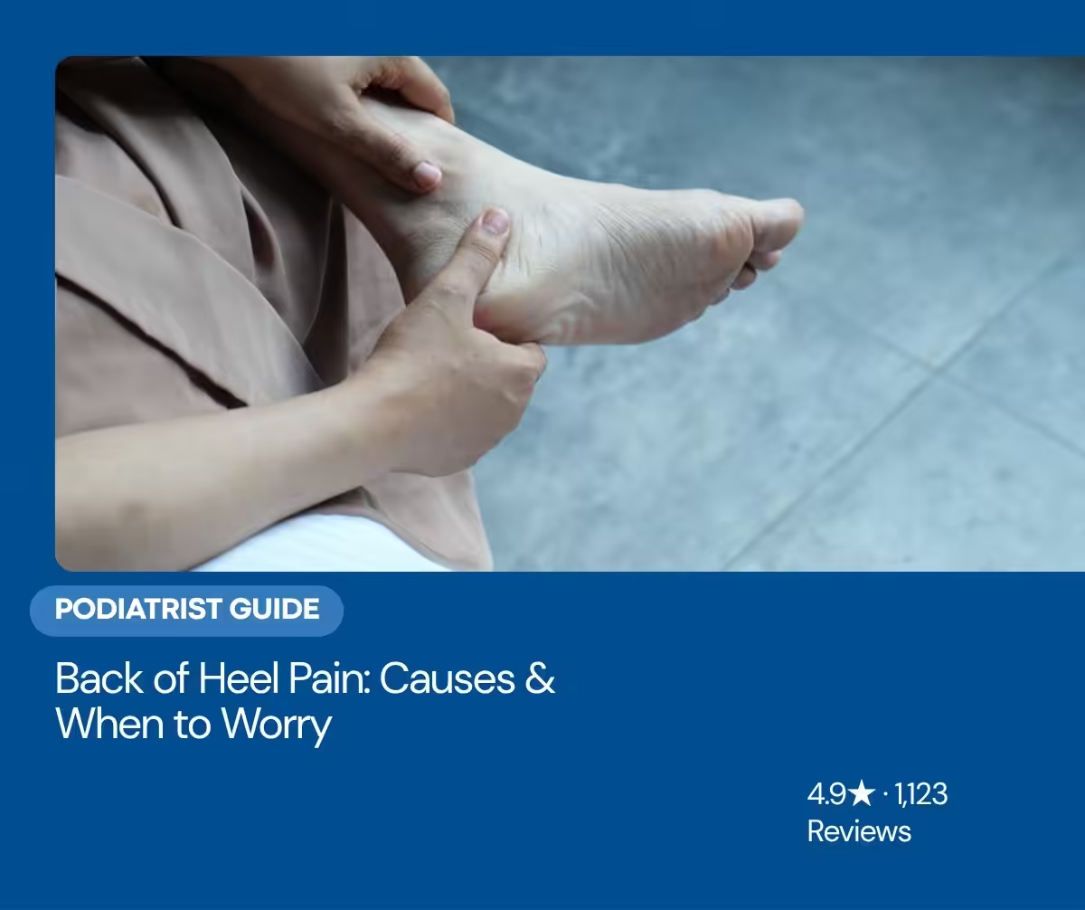
Back heel pain — pain on the back of the heel, not the bottom — is most often insertional Achilles tendonitis, retrocalcaneal bursitis, or Haglund’s deformity. Each has its own fix.
You’ve come to the right podiatry team. Dr. Tom Biernacki, DPM, FACFAS — board-certified foot & ankle surgeon with 3,000+ surgeries — explains exactly what back of heel pain means and what works. Call (810) 206-1402 for same-day appointment at Howell or Bloomfield Township.
Pain at the back of the heel is most often insertional Achilles tendinopathy, retrocalcaneal bursitis, or a Haglund deformity (and Sever disease in active kids). The right treatment depends on the exact cause. Below we explain how to tell them apart and the options that actually help. Call (810) 206-1402.
Quick Answer
Back of heel pain is most commonly caused by insertional Achilles tendonitis (where the tendon attaches to the heel bone), Haglund’s deformity (“pump bump” — bony enlargement of the heel), and retrocalcaneal bursitis (inflamed fluid sac between Achilles and heel bone). Less common causes include calcaneal stress fracture, Sever’s disease in kids, and rheumatoid enthesitis. Treatment depends on the cause — most cases respond to heel lifts, eccentric exercises, and supportive footwear within 8-12 weeks.
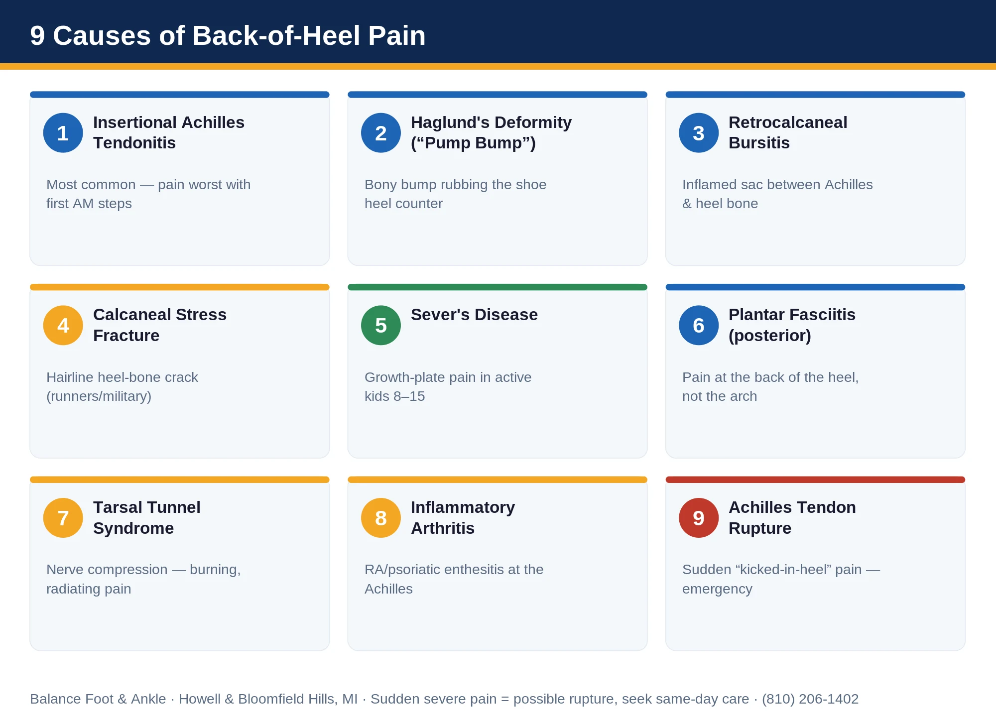
9 Most Common Causes of Back of Heel Pain
1. Insertional Achilles Tendonitis
The most common cause — chronic micro-tears where the Achilles tendon inserts into the calcaneus (heel bone). Pain is at the very back of the heel, worse with first steps in morning, and tender to touch. Treatment: supportive shoes with 8-12mm heel drop, eccentric heel raises, heel lifts.
2. Haglund’s Deformity (“Pump Bump”)
A bony enlargement of the back of the heel bone that rubs against shoe heel counters. Often visible as a hard bump. Treatment: shoe modification (open-back shoes), heel pads, ice. Surgery only for chronic cases.
3. Retrocalcaneal Bursitis
Inflammation of the fluid sac (bursa) between the Achilles tendon and the heel bone. Often coexists with Haglund’s. Treatment: ice, NSAIDs, ultrasound therapy, occasional cortisone injection.
4. Calcaneal Stress Fracture
Hairline fracture of the heel bone, usually from running or military training. Pain with weight-bearing, worse with jumping. Diagnosis requires MRI (X-ray often misses early). Treatment: 6-8 weeks in a CAM walker boot.
5. Sever’s Disease (children/teens)
Growth plate inflammation in active kids ages 8-15. Self-limiting once growth plate closes. Treatment: heel cups, cushioned shoes, activity modification.
6. Plantar Fasciitis (Posterior Variant)
Less common variant where pain is at the back of the heel rather than under it. Same fascial inflammation, different presentation. Treatment same as classic plantar fasciitis.
7. Tarsal Tunnel Syndrome
Compression of the posterior tibial nerve causes burning pain that can radiate to the back and bottom of the heel. Treatment: nerve gliding exercises, supportive footwear, occasional surgical release.
8. Inflammatory Arthritis (Rheumatoid, Psoriatic)
Autoimmune conditions cause enthesitis at the Achilles insertion. Often bilateral. Requires rheumatology workup and disease-modifying treatment.
9. Achilles Tendon Rupture (acute)
Sudden severe pain like being kicked in the back of the heel, often during sports. Inability to push off. Emergency — needs immediate evaluation.
At-Home Treatment Tools That Actually Work
- Heel cups / pads — silicone heel cups (e.g., Tuli’s Cheetahs) absorb impact and elevate the heel slightly to offload Achilles. Available on Amazon.
- Achilles tendon brace — Bauerfeind AchilloTrain or similar for daytime support.
- Strassburg Sock — overnight stretch device that prevents morning stiffness.
- Eccentric heel raises — 3 sets of 15, twice daily. The single most effective Achilles exercise per research.
- Foam roller / massage gun — calf release reduces tension on the Achilles insertion.
When to See a Podiatrist (Red Flags)
- Pain longer than 4-6 weeks despite home treatment
- Sharp pain with sudden onset (rule out rupture or stress fracture)
- Visible deformity or swelling that doesn’t resolve
- Pain that wakes you at night
- Bilateral heel pain (rule out inflammatory arthritis)
Schedule an evaluation: Howell or Bloomfield Township · (810) 206-1402 · Book online
✅ Medically Reviewed by Dr. Tom Biernacki, DPM
Board-certified podiatric physician & surgeon | Balance Foot & Ankle | Updated April 2026
★ DR. TOM BIERNACKI, DPM, FACFAS · BOARD-CERTIFIED PODIATRIST
Back of Heel Pain: Quick Answer
Pain at the back of your heel comes from one of six structures: (1) Achilles tendon insertion (insertional Achilles tendonitis), (2) Achilles tendon mid-substance (3-5 cm above the heel), (3) the calcaneus bone itself (Haglund’s deformity bony bump), (4) retrocalcaneal bursa (fluid sac between Achilles and bone), (5) the heel skin (cracked heels, blister), or (6) referred pain from sciatic nerve compression.
The pattern that helps diagnose: If pain hurts FIRST thing in the morning and improves with walking, suspect Achilles tendonitis or plantar fasciitis (heel-bottom). If a hard bump on the back of the heel hurts when shoes press on it, suspect Haglund’s deformity. If pain worsens during exercise and the area feels boggy/swollen, suspect retrocalcaneal bursitis. Most cases respond to heel lifts (reduce Achilles tension), a 2-4 week reduction in calf-loading exercise, and eccentric calf strengthening protocols. See a podiatrist if pain lasts >3 weeks.
Medically reviewed by Dr. Tom Biernacki, DPM — Board-certified foot & ankle surgeon, 3,000+ surgeries performed. Updated April 2026 with current clinical evidence. This article reflects real practice experience from Balance Foot & Ankle Specialists in Howell and Bloomfield Township, Michigan.
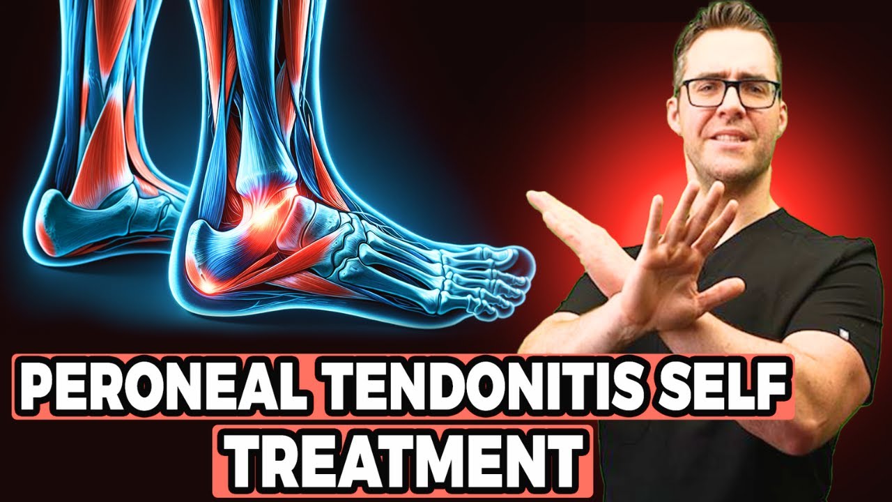
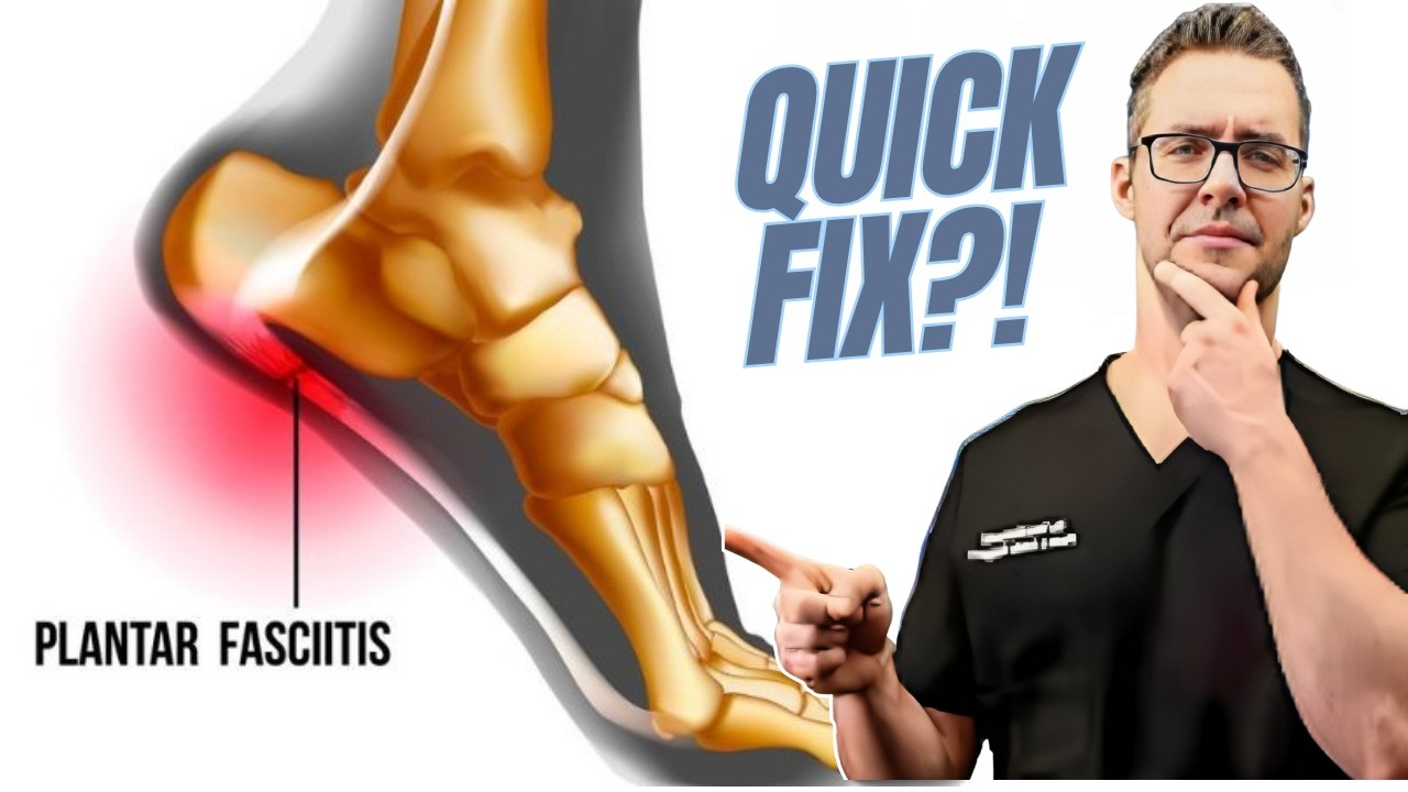
Watch: Dr. Tom Biernacki, DPM
![Retrocalcaneal Heel Bursitis [Pain in the back of the Heel TREATMENT!]](https://www.michiganfootdoctors.com/wp-content/cache/flying-press/b89b8ee4358be1bbe72667e9a51f7d5f.jpg)
What Causes Pain in the Back of the Heel?
The back of the heel is one of the highest-load regions in the entire lower extremity. The Achilles tendon — the largest and strongest tendon in the body — inserts here, transferring enormous forces from the calf muscles to the heel bone (calcaneus) with every step. When patients present with posterior heel pain, we systematically work through the most common diagnoses, because treatment differs significantly between them.
1. Achilles Tendonitis (Midportion)
Midportion Achilles tendonitis is inflammation or degeneration of the Achilles tendon 2–6 cm above the heel bone insertion — the classic “runner’s heel.” It develops from repetitive overloading: sudden increases in training mileage, insufficient recovery time, worn-out shoe cushioning, or calf muscle tightness that increases tension on the tendon.
Key signs: Tender, thickened segment of the tendon in the mid-portion (not at the heel bone), morning stiffness that warms up with activity, and pain that returns after activity. The tendon may feel “crunchy” or nodular to touch.
2. Insertional Achilles Tendinopathy
Insertional tendinopathy affects the point where the Achilles attaches to the heel bone. Unlike midportion Achilles issues, insertional disease involves calcification and bony changes at the attachment site. It’s more common in older, heavier, or less active patients. Standard eccentric calf exercises — effective for midportion tendonitis — often aggravate insertional disease and should be modified.
Key signs: Pain and tenderness directly at the back of the heel bone (not in the tendon belly), a visible or palpable bony prominence at the insertion, and pain with direct pressure from shoe counters. X-ray often shows calcification at the attachment.
3. Haglund’s Deformity (Pump Bump)
A Haglund’s deformity is an abnormal bony enlargement of the posterior-superior calcaneus — the upper back corner of the heel bone. It develops from a combination of genetics (high-arched, rigid foot) and shoe friction from stiff heel counters. Women who wear pumps frequently are disproportionately affected, giving rise to the term “pump bump.”
Key signs: A prominent bony bump at the upper back of the heel, redness and skin thickening overlying the bump, and severe pain with shoe wear. Pain is typically localized to the superficial bony prominence rather than the tendon itself.
4. Retrocalcaneal Bursitis
A small bursa (fluid-filled sac) sits between the Achilles tendon and the calcaneus, cushioning the tendon as it moves. Retrocalcaneal bursitis is inflammation of this bursa, often occurring alongside Haglund’s deformity or insertional tendinopathy. It presents as swelling on either side of the Achilles just above the heel bone, and is tender to lateral and medial compression at this level.
• A sudden “pop” in the back of the heel with inability to push up on your toes — possible Achilles rupture
• Severe swelling and bruising after an acute injury
• Pain so severe you cannot bear weight
• Numbness or weakness in the foot alongside heel pain
• Symptoms that are worsening despite 3–4 weeks of rest
How Is Back of Heel Pain Diagnosed?
Accurate diagnosis requires hands-on evaluation. In our clinic, we use a combination of physical examination (palpation of the tendon, squeeze test for bursitis, Thompson test for rupture), weight-bearing X-rays to assess bony pathology, and diagnostic ultrasound to evaluate tendon integrity and bursa fluid. MRI is reserved for cases where ultrasound is inconclusive or surgical planning is needed.
The distinction between midportion and insertional Achilles disease is clinically significant — the two conditions require different treatment protocols, and confusing them leads to prolonged recovery.
Treatment for Back of Heel Pain
For midportion Achilles tendonitis: The Alfredson eccentric calf raise protocol is the gold standard — 3 sets of 15 reps, twice daily, for 12 weeks. Heel lifts (1–2 cm) reduce tendon load during healing. Physical therapy, shockwave therapy (ESWT) for chronic cases, and activity modification are the mainstays. Most patients see significant improvement by 8–12 weeks.
For insertional Achilles tendinopathy: Eccentric exercises must be modified — perform them only to the horizontal (not below), avoiding the range that compresses the insertion. Heel lifts, cushioned heel counters, night splinting, and extracorporeal shockwave therapy (ESWT) are effective. Platelet-rich plasma (PRP) injection is an option for refractory cases. Surgery is reserved for calcific disease unresponsive to 6+ months of conservative care.
For Haglund’s deformity: Conservative management focuses on eliminating friction — open-back shoes, heel lifts to shift the calcaneus forward, and custom orthotics to unload the posterior heel. Surgical resection of the bony prominence is very effective when conservative care fails and delivers lasting relief.
For retrocalcaneal bursitis: Ice, NSAIDs, and heel lifts are first-line. Ultrasound-guided corticosteroid injection directly into the bursa (not the tendon) provides rapid relief in refractory cases. Injection into the Achilles tendon itself is contraindicated due to rupture risk.
Recommended Products for Back of Heel Pain

If home care isn’t resolving your heel pain, a visit with a board-certified podiatrist is the fastest path to accurate diagnosis and a personalized plan. At Balance Foot & Ankle Specialists, Dr. Tom Biernacki, Dr. Carl Jay, and Dr. Daria Gutkin offer same-day and next-day appointments at both our Howell and Bloomfield Township offices. We perform on-site diagnostic ultrasound, digital X-ray, conservative care, advanced regenerative treatments, and minimally invasive surgery when indicated.
Call (810) 206-1402 or request an appointment online. Most insurance plans accepted, including Medicare, Blue Cross Blue Shield, Aetna, Cigna, and United Healthcare.
More Podiatrist-Recommended Plantar Fasciitis Essentials
Best Night Splint
Keeps fascia stretched overnight — the #1 intervention for morning heel pain.
Top Podiatrist-Recommended Insole

Deep heel cup + arch support unloads the plantar fascia all day.
Plantar Fasciitis Compression Sock

Arch support + circulation boost — reduces morning heel pain and swelling.
As an Amazon Associate, Balance Foot & Ankle earns from qualifying purchases. Product recommendations are based on clinical experience; prices and availability shown above update live from Amazon.

When to See a Podiatrist
If morning heel pain has persisted more than 6 weeks, home care alone rarely fixes it. At Balance Foot & Ankle, we combine in-office ultrasound diagnostics, custom orthotics, and — when needed — shockwave or PRP to resolve plantar fasciitis that hasn’t responded to stretching and inserts. Most patients are walking pain-free within 4-8 weeks of starting a structured plan.
Call Balance Foot & Ankle: (810) 206-1402 · Book online · Offices in Howell & Bloomfield Township
⭐ 4.5★ | 15K+ Sold
Eliminates back-of-heel morning pain by stretching the Achilles overnight — the most effective conservative treatment for posterior heel pain.
⭐ 4.6★ | 30K+ Sold
Provides compression and support to the Achilles at the back of the heel — reduces pain with every step during recovery.
Ready to Get Relief? Same-Day Appointments Available
Board-certified podiatric surgeon | Howell & Bloomfield Township, MI | Most Insurance Accepted
4.9★ | 1,123 Reviews | 3,000+ Surgeries
Or call: (810) 206-1402
Frequently Asked Questions
Is back of heel pain the same as plantar fasciitis?
No — they are distinct conditions affecting different anatomical structures. Plantar fasciitis causes pain on the bottom of the heel (inferior calcaneus), typically worst with the first steps of the morning. Back of heel pain affects the posterior calcaneus and Achilles insertion. Treatment protocols differ significantly, which is why accurate diagnosis matters.
How long does back of heel pain take to heal?
Acute Achilles tendonitis in active patients: 6–8 weeks with a structured eccentric loading protocol. Chronic insertional tendinopathy or Haglund’s deformity: 3–6 months of consistent conservative care, with some patients requiring shockwave therapy or surgery. Early diagnosis and appropriate treatment dramatically shortens recovery time.
Can I run with back of heel pain?
For mild Achilles tendonitis, modified running (reduced volume, softer surfaces, slower pace) is generally acceptable as long as pain during and 24 hours after running stays below 4/10. For insertional disease, bursitis, or Haglund’s deformity with significant pain, a temporary running hiatus (2–4 weeks) while initiating a rehabilitation program produces better long-term outcomes than running through it.
The Bottom Line
Back of heel pain has four main causes — Achilles tendonitis (midportion), insertional tendinopathy, Haglund’s deformity, and retrocalcaneal bursitis — each with its own treatment approach. Getting the diagnosis right from the start is the single most important factor in a fast, complete recovery. Most patients do not need surgery. With the right protocol started early, 80–90% of posterior heel pain cases resolve with conservative care.
Heel Pain That Isn’t Getting Better?
Dr. Tom and Dr. Carl provide accurate diagnosis and personalized treatment for posterior heel pain at our Howell and Bloomfield Township offices. Same-week appointments available.
Book Your Evaluation →⭐⭐⭐⭐⭐ 4.9 Stars · 1,100+ Reviews · Howell & Bloomfield Township, MI
Differential Diagnosis: What Else Could It Be?
Several conditions share symptoms with Plantar Fasciitis and are commonly misdiagnosed in the first office visit. Considering these alternatives is part of every Balance Foot & Ankle exam:
- Baxter’s neuropathy. Compressed first branch of lateral plantar nerve — burning medial heel pain rather than first-step sharpness.
- Calcaneal stress fracture. Squeeze test of the heel reproduces pain anywhere; PF is reproduced only at the medial-plantar attachment.
- Heel spur (incidental). Spurs show on X-ray but rarely cause pain on their own — treat the fascia, not the spur.
If your symptoms don’t fit the textbook pattern, ask your podiatrist which differentials they ruled out — that conversation often shortcuts months of trial-and-error treatment.
In Our Clinic
In our Balance Foot & Ankle clinic, the typical plantar fasciitis patient is a 40- to 60-year-old who noticed sharp heel pain on their very first steps in the morning or after sitting at a desk. Many arrive having already tried cheap shoe-store inserts and a week of ice without relief. On exam, we palpate the medial calcaneal tubercle, check for a positive windlass test, and rule out Baxter’s neuropathy and calcaneal stress fractures. Most of our plantar fasciitis patients respond to a custom orthotic + eccentric calf loading + night splinting protocol within 6–12 weeks — without injections or surgery.
Most Common Mistake We See
The most common mistake we see is: Stretching aggressively before the fascia warms up. Fix: apply heat or move the foot through gentle circles for 3-5 minutes before your first morning steps, then stretch.
Warning Signs That Need Same-Day Care
Seek immediate evaluation at Balance Foot & Ankle if you experience any of the following:
- Unable to bear weight on the heel
- Bruising or visible swelling around the heel
- Constant rest or night pain in the heel
- No improvement after 6 weeks of home care
Call (810) 206-1402 — same-day and next-day appointments at our Howell and Bloomfield Township offices.
What is Heel pain?
Heel pain is a common foot/ankle condition that affects mobility and quality of life. Understanding the underlying cause is the first step in successful treatment. Our podiatrists at Balance Foot & Ankle perform a hands-on biomechanical exam, review your activity history, and use diagnostic imaging when appropriate to identify the root cause—not just treat the symptom. Many patients have been told to “rest and ice” without a deeper diagnostic workup; our approach is different.
Symptoms and warning signs
Common signs of heel pain include pain that worsens with activity, morning stiffness, swelling, tenderness when palpated, and difficulty bearing weight. If you experience sudden severe pain, inability to walk, visible deformity, numbness or color change, contact our office the same day or visit urgent care—these can signal a more serious injury such as a fracture, tendon rupture, or vascular compromise. Diabetics with any foot wound should seek same-day care.
Conservative treatment options
Most cases of heel pain respond to non-surgical care: structured rest, supportive footwear changes, custom orthotics, targeted stretching and strengthening protocols, anti-inflammatory medications when medically appropriate, and in-office procedures such as ultrasound-guided injections. We also offer advanced therapies including MLS laser therapy, EPAT/shockwave, regenerative injections, and image-guided procedures. Treatment is sequenced from least invasive to most invasive, and we explain the rationale at every step.
When is surgery considered?
Surgery is reserved for cases that fail 3-6 months of well-structured conservative care, when there is structural pathology (severe deformity, complete tear, advanced arthritis), or when imaging shows damage that will not heal without intervention. Our surgeons have performed 3,000+ foot and ankle procedures and prioritize minimally-invasive techniques whenever appropriate. We discuss recovery timelines, return-to-activity milestones, and realistic outcome expectations before any procedure is scheduled.
Recovery timeline and prevention
Recovery from heel pain varies based on severity and chosen treatment path. Conservative cases often improve within 4-8 weeks with consistent adherence to the protocol. Post-procedural recovery may range from a few days (in-office procedures) to several months (reconstructive surgery). Long-term prevention involves footwear assessment, activity modification, structured strengthening, and regular check-ins with your podiatrist if you have a history of recurrence. We provide written home-exercise plans and digital follow-up support.
Ready to feel better?
Same-week appointments available in Howell and Bloomfield Township, Michigan.
Book Your VisitReady to fix this for good?
Reading goes only so far. The fastest path to relief is a 30-minute office visit with Dr. Biernacki — same-day Howell or Bloomfield Township. Call (810) 206-1402 or use our online booking.
Doctor Hoy’s Natural Pain Relief Gel
Natural topical pain relief I use in our clinic. Arnica + camphor formula — apply directly to the area 3–4x daily. ($20–25)
Shop Doctor Hoy’s →Get Expert Care at Balance Foot & Ankle
Same-week appointments at our Howell and Bloomfield Township offices. Board-certified podiatric surgeons. Most insurance accepted.

Dr. Tom Biernacki, DPM is a board-certified foot & ankle surgeon (ABFAS & ABPM) at Balance Foot & Ankle Specialists in Southeast Michigan. With over a decade of clinical experience, he specializes in heel pain, bunions, diabetic foot care, sports injuries, and minimally invasive surgery. Dr. Biernacki is a member of the APMA and ACFAS, and his patient education content on MichiganFootDoctors.com and YouTube has made him one of the most-followed foot & ankle educators on YouTube.
More questions patients ask
What causes heel pain?
Heel pain most commonly results from plantar fasciitis, accounting for ~80% of cases. Other causes include Achilles tendinitis, heel spurs, stress fractures, and Baxter's nerve entrapment. Dr. Biernacki performs thorough biomechanical evaluations. Call 810-206-1402 for a same-day appointment.
How long does plantar fasciitis take to heal?
With proper treatment, most cases improve within 6-12 weeks. Options include custom orthotics, stretching, physical therapy, and injections. For persistent cases, Dr. Biernacki offers shockwave therapy and minimally invasive procedures with 90%+ success rates.
Should I see a podiatrist for heel pain?
Yes. A board-certified podiatrist can accurately diagnose heel pain using X-rays and diagnostic ultrasound, ruling out stress fractures. Early intervention leads to faster recovery. Call 810-206-1402.
Still have a question about coverage or cost? Call (810) 206-1402 and we will check your benefits before you come in — or book online: Book in Howell · Book in Bloomfield Township