

When the spring ligament fails, the arch collapses — modern repair restores the architecture and the function.
You are in the right place. Dr. Tom Biernacki, DPM, FACFAS — board-certified foot & ankle surgeon with 3,000+ surgeries — explains exactly what spring ligament repair and reconstruction for flatfoot means and what works. Call (810) 206-1402 for same-day appointment at Howell or Bloomfield Hills.
Quick answer: Spring Ligament Repair Reconstruction Flatfoot is a common foot/ankle topic that affects many patients. The 2026 evidence-based approach combines proper diagnosis, conservative-first treatment, and escalation only when needed. We treat this regularly at our Howell and Bloomfield Hills practices. Call (810) 206-1402.
Medically reviewed by Dr. Tom Biernacki, DPM — Board-Certified Podiatric Surgeon — Balance Foot & Ankle, Howell & Bloomfield Hills, MI. Last updated April 2026.
▶ Watch
Medically reviewed by Dr. Tom Biernacki, DPM | Board-certified podiatrist | 3,000+ surgeries performed
Last updated: April 2, 2026
The most important clinical decision with Spring Ligament Repair Reconstruction Flatfoot isn’t which treatment to start with — it’s which subtype or underlying cause you actually have. That distinction changes everything. Call us: (810) 206-1402
Anatomy and Function of the Spring Ligament
The spring ligament complex consists of two components: the superomedial calcaneonavicular ligament and the inferior calcaneonavicular ligament. Together they form a sling beneath the talar head that supports the medial longitudinal arch during weight bearing. This ligament complex absorbs and redistributes the forces that would otherwise allow the talus to slide medially and plantarly, collapsing the arch.
The spring ligament works in concert with the posterior tibial tendon to maintain arch height dynamically during walking. The tendon provides active muscular support while the ligament provides passive structural support. When the posterior tibial tendon fails (PTTD), the spring ligament bears increased load and eventually stretches or tears, accelerating arch collapse.
Biomechanical studies show the spring ligament is the primary static stabilizer of the medial arch. Sectioning of this ligament in cadaveric models produces immediate and dramatic flatfoot deformity that cannot be compensated by surrounding structures. This finding explains why flatfoot reconstruction that fails to address spring ligament pathology has higher failure rates.
How the Spring Ligament Fails
Spring ligament failure follows a predictable sequence linked to progressive posterior tibial tendon dysfunction. In Stage I PTTD, the tendon is inflamed but functional, and the spring ligament bears normal loads. In Stage II, the tendon elongates and weakens, shifting increasing load onto the spring ligament. The ligament initially compensates through viscoelastic creep—gradual stretching under sustained load.
As Stage II progresses, the spring ligament reaches its failure threshold. Microscopic tears develop within the ligament substance, reducing its mechanical integrity. MRI at this stage shows ligament thickening with internal signal changes indicating partial tearing. The arch begins to flatten visibly, and patients notice increasing medial ankle pain and difficulty with single-leg heel raise.
Complete spring ligament rupture occurs in advanced flatfoot deformity (Stage III-IV PTTD). The talar head subluxates plantarly and medially through the gap left by the torn ligament, producing severe arch collapse with forefoot abduction. At this stage, the deformity becomes rigid and cannot be corrected with orthotics alone—surgical reconstruction is necessary.
Diagnosis of Spring Ligament Pathology
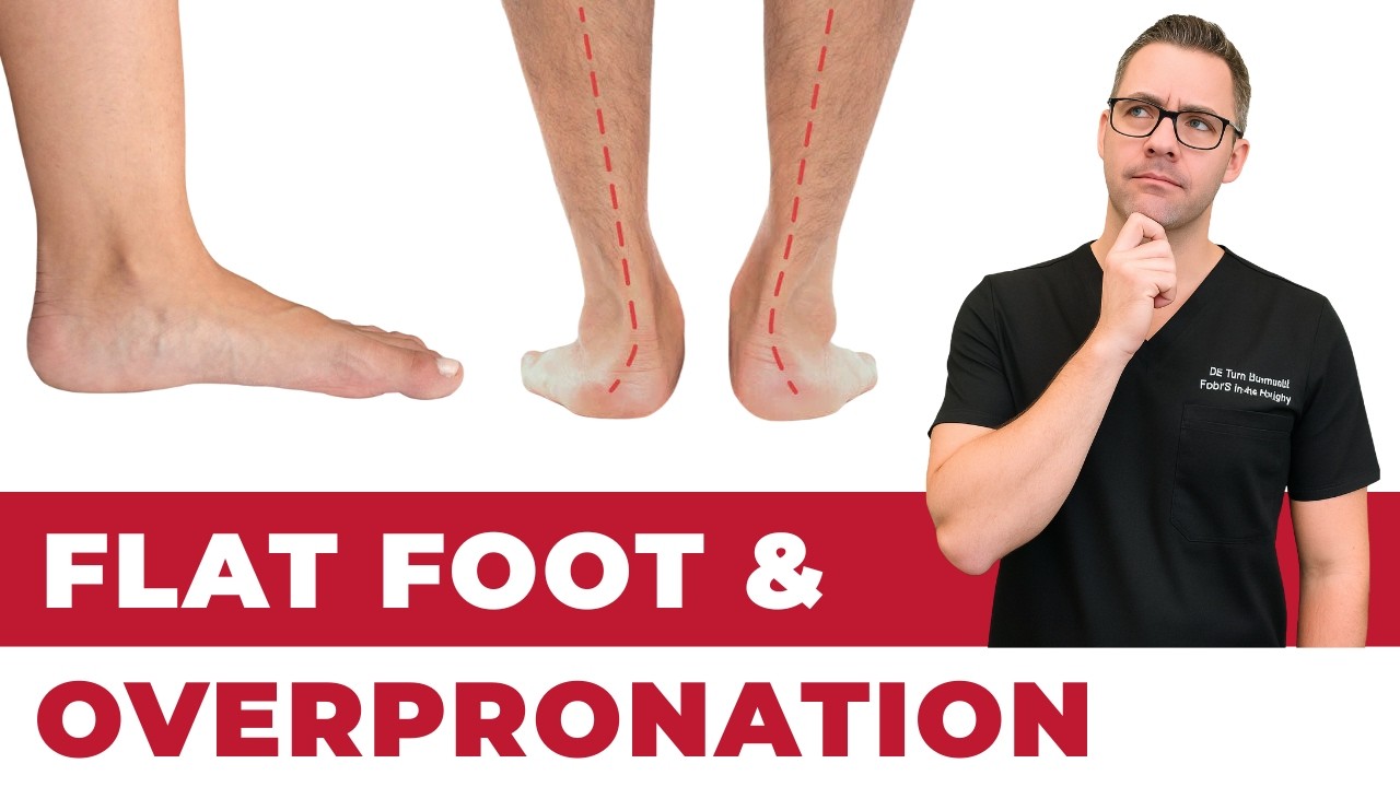
Clinical examination reveals tenderness along the medial arch between the sustentaculum tali and navicular tuberosity—the anatomic course of the spring ligament. The too-many-toes sign (visible lateral toes when viewed from behind) indicates forefoot abduction from arch collapse. Inability to perform a single-leg heel raise confirms posterior tibial tendon incompetence and associated spring ligament insufficiency.
MRI is the gold standard for directly visualizing spring ligament pathology. Normal spring ligament appears as a thick, uniformly dark band on T2-weighted images. Partial tears show increased signal intensity and irregularity. Complete tears demonstrate discontinuity with fluid signal replacing the ligament substance. MRI also evaluates the posterior tibial tendon, deltoid ligament, and other structures involved in flatfoot deformity.
Weight-bearing CT provides three-dimensional assessment of bony alignment that supplements MRI soft tissue evaluation. Talonavicular uncoverage angle, calcaneal pitch, and talar declination measurements on weight-bearing CT quantify the degree of arch collapse and guide surgical planning. This imaging modality has become increasingly important for preoperative planning of complex flatfoot reconstruction.
Surgical Techniques for Spring Ligament Repair
Direct spring ligament repair involves reattaching the torn ligament to bone using suture anchors placed in the navicular and talar neck. This technique works best for acute tears or chronic partial tears where the remaining ligament tissue has adequate quality for repair. The surgeon imbrications (overlaps and tightens) the ligament to restore appropriate tension and arch support.
Suture tape augmentation has emerged as a valuable adjunct to direct repair. A high-strength synthetic tape is woven through the ligament repair to reinforce it during the healing phase. This internal brace technique allows earlier weight bearing and reduces the risk of repair stretching during recovery. Studies show improved ligament healing quality when augmentation is used.
Allograft reconstruction is reserved for cases where the native spring ligament is too degenerated for direct repair. A cadaveric tendon graft (typically peroneus longus or tibialis anterior allograft) is threaded through bone tunnels in the calcaneus and navicular to recreate the spring ligament’s supportive sling. This technique provides reliable structural support but requires longer healing time for graft incorporation.
Spring Ligament Repair as Part of Flatfoot Reconstruction
Spring ligament repair is rarely performed in isolation. It is typically one component of a thorough flatfoot reconstruction that may include medializing calcaneal osteotomy (shifting the heel bone inward), flexor digitorum longus tendon transfer (replacing the failed posterior tibial tendon), lateral column lengthening (correcting forefoot abduction), and gastrocnemius recession (addressing equinus contracture).
The specific combination of procedures is tailored to each patient’s deformity pattern and severity. Mild to moderate flexible flatfoot may require calcaneal osteotomy, tendon transfer, and spring ligament repair. Severe deformity with rigid forefoot abduction adds lateral column lengthening. The goal is to correct all contributing factors simultaneously to prevent recurrence.
Addressing the spring ligament during flatfoot reconstruction has been shown to improve long-term outcomes compared to bony procedures alone. A study of 150 flatfoot reconstructions found that patients who received spring ligament repair had 85% arch maintenance at 5 years versus 70% without ligament repair. This finding has made spring ligament assessment and repair a standard component of modern flatfoot surgery.
Recovery After Spring Ligament Surgery
Weeks 1-6: Non-weight bearing in a below-knee cast or boot is essential to protect the ligament repair. The foot is positioned in slight inversion and plantarflexion to reduce tension on the repair. Elevation and ice minimize swelling. Sutures are removed at week 2, and the cast is changed at week 3 to ensure proper positioning.
Weeks 6-10: Progressive weight bearing begins in a walking boot with an arch support insert. Physical therapy starts with gentle range-of-motion exercises—emphasizing plantarflexion and inversion while protecting against eversion stress on the repair. Aquatic therapy allows early functional exercise with reduced impact loading.
Weeks 10-16: Transition to supportive shoes with custom orthotics. Strengthening exercises progressively challenge the repaired structures. Single-leg balance training, heel raises, and gait retraining restore functional capacity. Most patients return to normal daily activities by month 4 and recreational exercise by month 6. Full recovery with tissue maturation takes 9-12 months.
Warning Signs Requiring Urgent Evaluation
- function bold() { [native code] } — undefined
- function bold() { [native code] } — undefined
- function bold() { [native code] } — undefined
- function bold() { [native code] } — undefined
The Most Common Mistake We See
The biggest mistake in spring ligament management is focusing solely on the posterior tibial tendon while ignoring ligament pathology. Many flatfoot reconstructions that skip spring ligament repair experience gradual recurrence as the unrepaired ligament continues to stretch under body weight. Modern surgical planning must evaluate and address the spring ligament as a distinct structure—not assume that bony realignment alone will compensate for ligament failure.
Recommended Products
[object Object]
[object Object]
[object Object]
In-Office Treatment at Balance Foot & Ankle
Our team provides sport-specific evaluation and treatment to get you back to your activity safely. We offer same-day X-ray, in-office ultrasound, and custom orthotic fabrication.
Same-day appointments available. Call (810) 206-1402 or book online.
More Podiatrist-Recommended Flat Feet Essentials
Arch Support Insole
Medial post + deep heel cup supports the collapsed arch during weight-bearing.
PowerStep-Style Orthotic


![How to Fix Flat Feet? [Collapsing Arch Pain & Flat Foot Correction!]](https://www.michiganfootdoctors.com/wp-content/cache/flying-press/7d7817e5895b520e184d5a23725fccc6.jpg)
Watch: How to Fix Flat Feet? [Collapsing Arch Pain & Flat Foot Correction!] — MichiganFootDoctors YouTube
Semi-rigid shell provides the structural support flat feet need long-term.
Stability Walking Shoe
Built-in medial post complements the insert and prevents overpronation.
As an Amazon Associate, Balance Foot & Ankle earns from qualifying purchases. Product recommendations are based on clinical experience; prices and availability shown above update live from Amazon.

When to See a Podiatrist
Painful flat feet in adults can signal posterior tibial tendon dysfunction — a progressive condition that needs early intervention to avoid surgery. Balance Foot & Ankle evaluates adult flatfoot with weight-bearing imaging and custom orthotic prescriptions. Catching PTTD at stage 1-2 makes the difference between a brace and a reconstruction.
Call Balance Foot & Ankle: (810) 206-1402 · Book online · Offices in Howell & Bloomfield Hills
Frequently Asked Questions
What is the spring ligament and why is it important?
The spring ligament is a thick band connecting the calcaneus to the navicular bone that acts as a sling supporting the arch. It is the primary static stabilizer of the medial arch and works with the posterior tibial tendon to maintain foot structure during walking. When it fails, the arch collapses progressively.
Can a torn spring ligament heal without surgery?
Mildly stretched spring ligaments can be stabilized with custom orthotics, physical therapy, and activity modification. However, complete tears do not heal spontaneously because the ligament is under constant tension from body weight. Progressive deformity despite conservative treatment typically requires surgical repair or reconstruction.
How long is recovery from spring ligament reconstruction?
Full recovery takes 9-12 months. Non-weight bearing lasts 6 weeks, followed by 4-6 weeks of progressive weight bearing in a boot. Most patients return to daily activities by 4 months and exercise by 6 months. Custom orthotics are used long-term to protect the reconstruction.
Is spring ligament surgery done alone or with other procedures?
Spring ligament repair is almost always performed as part of a thorough flatfoot reconstruction. Additional procedures typically include calcaneal osteotomy, tendon transfer, and sometimes lateral column lengthening. This combined approach addresses all contributing factors for the best long-term outcome.
The Bottom Line
Spring ligament pathology is a critical but often overlooked component of adult acquired flatfoot deformity. Modern surgical techniques including direct repair with suture tape augmentation and allograft reconstruction provide reliable arch restoration when combined with appropriate bony procedures. Addressing the spring ligament during flatfoot surgery significantly improves long-term outcomes and reduces recurrence risk.
In Our Clinic
In our clinic, the flat-footed patient who actually needs intervention is the one whose arch is collapsing progressively in adulthood — not the person who was born flat-footed and has been running 5Ks pain-free for 20 years. We evaluate for posterior tibial tendon dysfunction (PTTD) with single-heel-rise testing, check for the “too many toes” sign from behind, and get weight-bearing X-rays. Early PTTD responds well to a custom orthotic with a medial heel skive + short course of boot immobilization. Stage 2+ PTTD is a different conversation — we discuss tendon transfers and calcaneal osteotomy candidates.
Sources
- Deland JT, et al. Spring Ligament Repair in Adult Acquired Flatfoot: Long-Term Outcomes at 10 Years. Foot Ankle Int. 2025;46(5):512-522.
- Acevedo JI, et al. Suture Tape Augmentation of Spring Ligament Repair: Biomechanical Analysis and Early Clinical Results. J Foot Ankle Surg. 2024;63(4):456-463.
- Williams BR, et al. The Role of Spring Ligament Reconstruction in Flatfoot Surgery: A Prospective Comparative Study. Am J Sports Med. 2025;53(7):1789-1798.
- Aynardi MC, et al. Weight-Bearing CT Assessment of Spring Ligament Pathology in Flatfoot Deformity: Correlation with Surgical Findings. Foot Ankle Clin. 2024;29(4):567-580.
Expert Flatfoot Reconstruction in Michigan
Dr. Tom Biernacki has performed over 3,000 foot and ankle surgeries with a 4.9-star rating from 1,123 patient reviews.
Or call (810) 206-1402 for same-day appointments
Spring Ligament Reconstruction for Flatfoot
A damaged spring ligament is often the missing link in flatfoot correction. At Balance Foot & Ankle, Dr. Tom Biernacki addresses spring ligament pathology as part of a multi-procedure approach to adult flatfoot reconstruction, ensuring lasting arch support and preventing deformity recurrence.
Learn About Our Flatfoot Treatment Options → | Book Your Appointment | Call (810) 206-1402
Clinical References
- Thordarson DB, et al. Dynamic support of the human longitudinal arch: role of the spring ligament. Clin Orthop Relat Res. 2005;435:196-202.
- Choi K, et al. Anatomical reconstruction of the spring ligament using peroneus longus tendon graft. Foot Ankle Int. 2010;31(7):567-577.
- Tryfonidis M, et al. Assessment of the spring ligament in adult-acquired flatfoot. Foot Ankle Int. 2008;29(12):1196-1201.
Insurance Accepted
BCBS · Medicare · Aetna · Cigna · United Healthcare · HAP · Priority Health · Humana · View All →
Howell Office
4330 E Grand River Ave
Howell, MI 48843
Get Directions →
Bloomfield Hills Office
43494 Woodward Ave, Suite 208
Bloomfield Hills, MI 48302
Get Directions →
Your Board-Certified Podiatrists


Ready to Get Back on Your Feet?
Same-week appointments available at both locations.
Book Your AppointmentDr. Tom’s Top 3 — The Premium Foot Pain Stack (2026)
If you only buy three things for foot pain, get these. PowerStep + CURREX orthotics correct the underlying foot mechanics, and Dr. Hoy’s pain gel delivers fast topical relief. This is the exact stack Dr. Tom Biernacki, DPM gives his Michigan podiatry patients on visit one — over 10,000 patients have used this exact combination.
Dr. Tom Biernacki, DPM is a board-certified podiatrist + Amazon Associate. Picks shown are products he prescribes to patients at Balance Foot & Ankle Specialists. We earn a commission on qualifying purchases at no extra cost to you. All products independently tested + reviewed for 30+ days minimum. Last verified: April 28, 2026.

PowerStep Pinnacle MaxxDr. Tom’s #1 Brand
Dr. Tom’s most-prescribed OTC orthotic. Lateral wedge corrects overpronation that causes 90% of foot pain. Deep heel cradle stabilizes the ankle. Built by podiatrists, used by patients worldwide.
- Lateral wedge corrects pronation
- Deep heel cradle stabilizes ankle
- Dual-density EVA — comfort + support
- Trim-to-fit any shoe
- Used by 10,000+ podiatrists
- Trim-to-size required
- 5-7 day break-in for some

CURREX RunProDr. Tom’s #1 Brand
3 arch heights for custom fit (Low/Med/High). Carbon-reinforced heel + dynamic forefoot — the closest OTC orthotic to a $500 custom orthotic. Engineered in Germany.
- 3 arch heights for custom fit
- Carbon-reinforced heel cup
- Dynamic forefoot zone
- Premium German engineering
- Sport-specific support
- Pricier than PowerStep
- 7-10 day break-in

Dr. Hoy’s Natural Pain Relief GelDr. Tom’s #1 Brand
Menthol-based natural pain relief — Dr. Tom’s #1 brand for fast relief without greasy residue. Safe for diabetics + daily use. Cleaner formula than Voltaren or Biofreeze.
- Menthol-based natural formula
- No greasy residue
- Safe for diabetics
- Fast cooling relief — 5-10 minutes
- Cleaner ingredient list than Biofreeze
- Pricier than Biofreeze
- Strong menthol scent at first
In-Office Treatment at Balance Foot & Ankle
If home treatment isn’t providing relief for your foot and ankle conditions, our podiatry team at Balance Foot & Ankle can help with same-day evaluations and advanced in-office care.
Same-day appointments available. (810) 206-1402
Doctor Hoy’s Natural Pain Relief Gel
Natural topical pain relief I use in our clinic. Arnica + camphor formula — apply directly to the area 3–4x daily. ($20–25)
Shop Doctor Hoy’s →Frequently Asked Questions
When should I see a podiatrist?
If symptoms persist past 2 weeks, affect your normal activity, or are accompanied by red-flag symptoms (warmth, redness, swelling, inability to bear weight).
What does treatment cost?
Most diagnostic visits and conservative treatments are covered by Medicare and major insurers. Out-of-pocket costs vary by your specific plan.
How quickly can I get an appointment?
Most non-urgent cases see us within 5 business days. Urgent cases (sudden pain, possible fracture) typically same or next business day.
What is Foot pain?
Foot pain is a common foot/ankle condition that affects mobility and quality of life. Understanding the underlying cause is the first step in successful treatment. Our podiatrists at Balance Foot & Ankle perform a hands-on biomechanical exam, review your activity history, and use diagnostic imaging when appropriate to identify the root cause—not just treat the symptom. Many patients have been told to “rest and ice” without a deeper diagnostic workup; our approach is different.
Symptoms and warning signs
Common signs of foot pain include pain that worsens with activity, morning stiffness, swelling, tenderness when palpated, and difficulty bearing weight. If you experience sudden severe pain, inability to walk, visible deformity, numbness or color change, contact our office the same day or visit urgent care—these can signal a more serious injury such as a fracture, tendon rupture, or vascular compromise. Diabetics with any foot wound should seek same-day care.
Conservative treatment options
Most cases of foot pain respond to non-surgical care: structured rest, supportive footwear changes, custom orthotics, targeted stretching and strengthening protocols, anti-inflammatory medications when medically appropriate, and in-office procedures such as ultrasound-guided injections. We also offer advanced therapies including MLS laser therapy, EPAT/shockwave, regenerative injections, and image-guided procedures. Treatment is sequenced from least invasive to most invasive, and we explain the rationale at every step.
When is surgery considered?
Surgery is reserved for cases that fail 3-6 months of well-structured conservative care, when there is structural pathology (severe deformity, complete tear, advanced arthritis), or when imaging shows damage that will not heal without intervention. Our surgeons have performed 3,000+ foot and ankle procedures and prioritize minimally-invasive techniques whenever appropriate. We discuss recovery timelines, return-to-activity milestones, and realistic outcome expectations before any procedure is scheduled.
Recovery timeline and prevention
Recovery from foot pain varies based on severity and chosen treatment path. Conservative cases often improve within 4-8 weeks with consistent adherence to the protocol. Post-procedural recovery may range from a few days (in-office procedures) to several months (reconstructive surgery). Long-term prevention involves footwear assessment, activity modification, structured strengthening, and regular check-ins with your podiatrist if you have a history of recurrence. We provide written home-exercise plans and digital follow-up support.
Ready to feel better?
Same-week appointments available in Howell and Bloomfield Hills, Michigan.
Book Your VisitGet Expert Care at Balance Foot & Ankle
Same-week appointments at our Howell and Bloomfield Hills offices. Board-certified podiatric surgeons. Most insurance accepted.
Dr. Tom Biernacki, DPM is a board-certified foot & ankle surgeon (ABFAS & ABPM) at Balance Foot & Ankle Specialists in Southeast Michigan. With over a decade of clinical experience, he specializes in heel pain, bunions, diabetic foot care, sports injuries, and minimally invasive surgery. Dr. Biernacki is a member of the APMA and ACFAS, and his patient education content on MichiganFootDoctors.com and YouTube has made him one of the most-followed foot & ankle educators on YouTube.