

Sudden foot drop after a fall or strain may be a tib anterior rupture — quick repair restores the lift.
You are in the right place. Dr. Tom Biernacki, DPM, FACFAS — board-certified foot & ankle surgeon with 3,000+ surgeries — explains exactly what tibialis anterior tendon rupture — surgical repair means and what works. Call (810) 206-1402 for same-day appointment at Howell or Bloomfield Hills.
Quick answer: Tibialis Anterior Tendon Rupture Surgical Repair is a common foot/ankle topic that affects many patients. The 2026 evidence-based approach combines proper diagnosis, conservative-first treatment, and escalation only when needed. We treat this regularly at our Howell and Bloomfield Hills practices. Call (810) 206-1402.
Watch: Torn Achilles Tendon Rupture — MichiganFootDoctors YouTube

Medically Reviewed by Dr. Tom Biernacki, DPM, FACFAS — Board-certified podiatrist & foot surgeon | Balance Foot & Ankle | Last updated: May 2026
Tibialis anterior tendon rupture causes sudden foot drop and inability to lift the foot—often mistaken for a simple ankle sprain. Surgical repair within 3–6 weeks gives the best outcome; delayed repair requires tendon graft reconstruction. Most patients return to full activity in 4–6 months with surgery. Non-surgical treatment in older, low-demand patients is an option but results in permanent weakness.
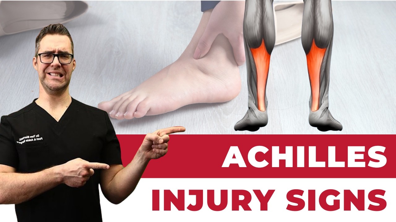
What Is the Tibialis Anterior Tendon?
The tibialis anterior is the primary muscle responsible for dorsiflexion—lifting the front of the foot during walking. Its tendon runs down the front of the shin and inserts at the inner base of the first metatarsal and medial cuneiform. Rupture of this tendon is relatively rare compared to Achilles or posterior tibial tendon injuries, but when it occurs—typically in men over 50 from a single traumatic misstep or in athletes—it causes immediate and profound functional loss. The characteristic presentation is a visible or palpable gap on the front of the ankle, inability to clear the ground during walking, and a “floppy foot” gait identical to peroneal nerve palsy, which is a common diagnostic confusion point.
Tibialis Anterior Rupture: Treatment Options Compared
| Treatment | Best For | Recovery | Expected Outcome |
|---|---|---|---|
| Primary Surgical Repair | Acute rupture (<3–6 weeks), active patients | 4–6 months | Excellent — near-full strength restoration |
| Reconstruction with Tendon Graft | Delayed presentation (>6 weeks), tendon retraction | 6–9 months | Good — functional but some residual weakness |
| EHL Tendon Transfer | Massive tendon loss, poor tissue quality | 6–8 months | Good functional outcome; EHL deficit usually tolerable |
| AFO Brace (Non-Surgical) | Low-demand, elderly, medical comorbidities | Immediate but permanent bracing | Compensatory function; no strength recovery |
How Surgical Repair Works
Primary repair is performed through a longitudinal incision over the dorsal ankle and foot. I identify the proximal stump (which retracts toward the shin) and the distal tendon end attached near the first metatarsal, then freshen the ends and perform an end-to-end repair under appropriate tension using non-absorbable sutures. If the rupture is at the bony insertion, suture anchors fix the tendon directly back to the bone. The foot is held in slight dorsiflexion in a cast for 4 weeks, followed by a boot with progressive range-of-motion exercises and physical therapy. Athletes typically return to sport-level activity by month 5 or 6.
Waiting too long for a diagnosis. Tibialis anterior rupture is frequently misdiagnosed as an ankle sprain in the emergency department because both produce anterior ankle swelling and pain. The critical difference is functional: a sprain patient can still lift the foot, a tibialis anterior rupture patient cannot. Every week of delay shrinks the primary repair window—after 6 weeks, tendon retraction and muscle atrophy typically require a more complex graft reconstruction. If you suspect foot drop after an ankle injury, insist on an MRI before accepting a sprain diagnosis.
Frequently Asked Questions
How do I know if my tibialis anterior is ruptured?
The hallmark sign is sudden inability to lift the front of the foot (foot drop) combined with a palpable gap or fullness on the front of the ankle after an injury. Unlike peroneal nerve damage, the skin sensation remains normal. An MRI confirms the diagnosis and shows the degree of tendon retraction, which guides the surgical approach. If you can’t clear your toes during walking after an ankle injury, seek immediate evaluation.
Can a tibialis anterior rupture heal without surgery?
Partial tears in low-demand patients can sometimes be managed conservatively with casting and physical therapy, with acceptable functional outcomes for activities of daily living. Complete ruptures in active adults almost always benefit from surgical repair—non-surgical management results in permanent dorsiflexion weakness and requires lifelong AFO bracing. For anyone who wants to walk normally, hike, or participate in sport, surgery is the standard of care.
What is the recovery timeline after tibialis anterior repair?
Week 0–4: non-weight-bearing cast with foot in dorsiflexion. Week 4–8: walking boot with progressive weight-bearing. Week 8–12: physical therapy for range of motion, strength, and gait. Month 3–4: return to low-impact activity. Month 5–6: return to sport or demanding physical work. Full strength recovery typically takes 6–9 months but most patients feel functionally normal well before that.
Will I lose strength permanently after tendon repair?
Primary repair within the first 3–6 weeks typically restores 90–95% of original dorsiflexion strength. Delayed reconstruction with a tendon graft achieves approximately 80–85% strength. Even with some residual weakness, most patients cannot functionally distinguish the repaired side from the healthy side during everyday activities. Competitive athletes at the elite level may notice subtle differences in explosive acceleration.
Does insurance cover tibialis anterior tendon repair?
Yes—tendon rupture repair is a covered surgical procedure under most PPO plans and Medicare Part B when documented with MRI and clinical examination. CPT codes 27691/27659 apply depending on whether primary repair or reconstruction is performed. Our team at Balance Foot & Ankle handles pre-authorization from both the Howell and Bloomfield Hills offices. Call (810) 206-1402 for a same-day surgical evaluation.
Sudden Foot Drop After an Injury? Don’t Wait.
Dr. Tom Biernacki performs tibialis anterior tendon repair and reconstruction at Balance Foot & Ankle — Howell and Bloomfield Hills, MI. The repair window closes fast — same-day surgical evaluations available.
Book a Same-Day Visit (810) 206-1402Related Resources
- Foot Drop Surgery: Peroneal Nerve & Tendon Transfer
- Ankle Fractures: Types & Recovery
- What Is a Podiatrist? When to See One
- Custom Orthotics in Michigan
In-Office Treatment at Balance Foot & Ankle
If home treatment isn’t providing relief for your Achilles tendinitis, our podiatry team at Balance Foot & Ankle can help with same-day evaluations and advanced in-office care.
Same-day appointments available. (810) 206-1402
Doctor Hoy’s Natural Pain Relief Gel
Natural topical pain relief I use in our clinic. Arnica + camphor formula — apply directly to the area 3–4x daily. ($20–25)
Shop Doctor Hoy’s →Frequently Asked Questions

When should I see a podiatrist?
If symptoms persist past 2 weeks, affect your normal activity, or are accompanied by red-flag symptoms (warmth, redness, swelling, inability to bear weight).
What does treatment cost?
Most diagnostic visits and conservative treatments are covered by Medicare and major insurers. Out-of-pocket costs vary by your specific plan.
How quickly can I get an appointment?
Most non-urgent cases see us within 5 business days. Urgent cases (sudden pain, possible fracture) typically same or next business day.
What is Foot pain?
Foot pain is a common foot/ankle condition that affects mobility and quality of life. Understanding the underlying cause is the first step in successful treatment. Our podiatrists at Balance Foot & Ankle perform a hands-on biomechanical exam, review your activity history, and use diagnostic imaging when appropriate to identify the root cause—not just treat the symptom. Many patients have been told to “rest and ice” without a deeper diagnostic workup; our approach is different.
Symptoms and warning signs
Common signs of foot pain include pain that worsens with activity, morning stiffness, swelling, tenderness when palpated, and difficulty bearing weight. If you experience sudden severe pain, inability to walk, visible deformity, numbness or color change, contact our office the same day or visit urgent care—these can signal a more serious injury such as a fracture, tendon rupture, or vascular compromise. Diabetics with any foot wound should seek same-day care.
Conservative treatment options
Most cases of foot pain respond to non-surgical care: structured rest, supportive footwear changes, custom orthotics, targeted stretching and strengthening protocols, anti-inflammatory medications when medically appropriate, and in-office procedures such as ultrasound-guided injections. We also offer advanced therapies including MLS laser therapy, EPAT/shockwave, regenerative injections, and image-guided procedures. Treatment is sequenced from least invasive to most invasive, and we explain the rationale at every step.
When is surgery considered?
Surgery is reserved for cases that fail 3-6 months of well-structured conservative care, when there is structural pathology (severe deformity, complete tear, advanced arthritis), or when imaging shows damage that will not heal without intervention. Our surgeons have performed 3,000+ foot and ankle procedures and prioritize minimally-invasive techniques whenever appropriate. We discuss recovery timelines, return-to-activity milestones, and realistic outcome expectations before any procedure is scheduled.
Recovery timeline and prevention
Recovery from foot pain varies based on severity and chosen treatment path. Conservative cases often improve within 4-8 weeks with consistent adherence to the protocol. Post-procedural recovery may range from a few days (in-office procedures) to several months (reconstructive surgery). Long-term prevention involves footwear assessment, activity modification, structured strengthening, and regular check-ins with your podiatrist if you have a history of recurrence. We provide written home-exercise plans and digital follow-up support.
Ready to feel better?
Same-week appointments available in Howell and Bloomfield Hills, Michigan.
Book Your VisitDr. Tom Biernacki, DPM is a board-certified foot & ankle surgeon (ABFAS & ABPM) at Balance Foot & Ankle Specialists in Southeast Michigan. With over a decade of clinical experience, he specializes in heel pain, bunions, diabetic foot care, sports injuries, and minimally invasive surgery. Dr. Biernacki is a member of the APMA and ACFAS, and his patient education content on MichiganFootDoctors.com and YouTube has made him one of the most-followed foot & ankle educators on YouTube.